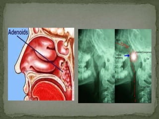
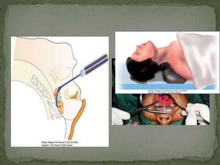
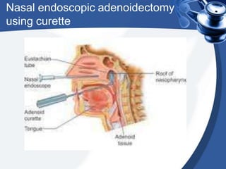
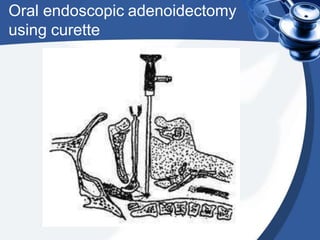
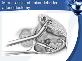
Dr Rajesh Kar
MBBS, MS, DNB
Synonym: Nasopharyngeal
tonsil
• Adenoids is the
hypertrophied mass of
lymphoid tissue situated at
the junction of roof & post.
wall of nasopharynx.
• The mass of lymphoid
tissue is termed as
Adenoids only when it is
hypertrophied.
• It usually undergoes atrophy
by puberty (13-14 yrs)
• Pink, globular mass
• Vertical ridges on its
surface
• No crypts
• Lined by ciliated
columnar epithelium
• No capsule
• The formation of the adenoids begins in the 3rd month
of fetal development. This starts with glandular
primordia in the posterior nasopharynx becoming
associated with infiltrating lymphocytes.
• In the 5th month sagittal folds are formed which are
the beginnings of pharyngeal crypts. The surface is
covered with pseudostratified ciliated epithelium.
• By the 7th month of development the adenoids are
fully formed.
• Ascending palatine
branch of facial artery
• Ascending pharyngeal
artery
• Pharyngeal branch of 3rd
part of maxillary artery
Venous drainage is through
the pharyngeal plexus and
the pterygoid plexus
flowing ultimately into the
facial and internal jugular
veins.
•Innervation is derived from
the glossopharyngeal and
vagus nerves.
LYMPHATIC DRAINAGE :
Upper deep jugular nodes
• Retropharyngeal nodes
• Parapharyngeal nodes
• The tonsils and adenoids are part of the secondary
immune system.
• Without afferent lymphatics the lymphoid nodules
in these structures are exposed to antigen only in
the crypts of the palatine tonsils and the folds of
the adenoids where it is transported through the
epithelial layer.
• These are involved in the production of mostly
secretory IgA, which is transported to the surface
providing local immune protection.
Common Diseases of the Tonsils and
Adenoids
1. Acute adenoiditis/tonsillitis
2. Recurrent/chronic adenoiditis/tonsillitis
3. Obstructive hyperplasia
4. Malignancy
LOCAL
• B/L nasal obstruction
• Snoring
• Mouth breathing
• Rhinolalia clausa
• Frequent rhinorrhoea
• Epistaxis
• Feeding problems in
children
• Conductive deafness due to
ET block
• Cervical lymphadenopathy
• Otitis media
• Adenoids facies
• Bronchitis
GENERAL
• Anorexia
• Lethargy
• Poor physical & mental
development
• Bed-wetting
• Pigeon chest
• Protuberent abdomen
Recurrent acute adenoiditis is 4 or more episodes of
acute adenoiditis in a 6-month period with
intervening periods of wellness.
•Chronic adenoiditis symptoms include
• persistent rhinorrhea,
• postnasal drip,
•malodorous breath, and
• associated otitis media or extra esophageal reflux
lasting at least 3 months.
•Obstructive adenoid hyperplasia includes symptoms
of chronic nasal obstruction, rhinorrhea, snoring,
mouth breathing, and a hyponasal voice.
•Obstructive sleep apnea in children is clinically
marked by loud snoring, apneic episodes while
sleeping, daytime somnolence, behavioral problems,
and enuresis
• It is the long, open
mouthed, face of children
with adenoid hypertrophy.
• The mouth is always open
because upper airway
congestion has made
patients obligatory mouth
breathers.
• The most common
presenting symptoms are
chronic mouth breathing
and snoring.
• The most dangerous
symptom is sleep apnea
•The characteristic facial appearance
consists of:
• Underdeveloped thin nostrils
• Short upper lip
• Prominent upper teeth
•Crowded teeth
• Narrow upper alveolus• High-arched
palate
• Hypoplastic maxilla
• Eustachian blockage causing glue ear
/deafness
• The deafness and inattentiveness
interferes with the learning
• Child grows with lowered intelligence
and understanding
• Sunken eyes
• Narrow pinched nostrils
• Open mouth
• High-arched palate
• Crowded teeth
• Dull mask-like face
• Protruding teeth
• Drooling saliva
• Everted upper lip
• Rhinorrhoea
• Loss of nasolabial fold
•Endoscopy
•Posterior rhinoscopy
•Otoscopy
•Radiological examination can also help : CT scan/x ray
•Posterior rhinoscopy is
done to look for lesions in
the post nasal space – for
example, adenoids,
tumors of the
nasopharynx, etc.
Uses:
• Examination of the post nasal space by a
procedure called posterior rhinoscopy, an
outpatient procedure.
• The mirror is warmed and introduced into
the oral cavity while the tongue is depressed
with a tongue depressor.
• The mirror is turned upwards in
order to examine the post nasal
space.
• The shaft of the instrument is bent
to achieve a bayonet shape, a
feature that helps differentiate it
from the indirect laryngoscopy
mirror.
• The mirror is available in 5 sizes.
• Nasopharyngoscopy is a
procedure which enables the
doctor to examine the
internal
surfaces of the nose and
throat (nasopharynx).
• Nasopharyngoscopy
provides a direct view of
every part of the upper
respiratory tract from the
nasal passages down the
throat to the larynx
• The main imaging study
to evaluate the adenoid is
a lateral neck
radiograph, as in the
images.
• CT scan is not normally used to
evaluate the adenoids.
However, when a CT scan is
performed to evaluate the
sinuses, the choana and
nasopharynx are occasionally
imaged, providing information
on the size of the adenoids
• If the adenoids look abnormal
or if a mass is present in the
nasopharynx in an older child
or in an adult, an imaging
study (eg, CT scan, MRI) is
obtained to rule out a lesion
other than an adenoid
MANAGEMENT
•Management options include
•wait until they involutes
•surgical removal (ADENOIDECTOMY)
•Non surgical management include intranasal
corticosteroids
Medical Management
• No good evidence supports any curative medical
therapy for chronic infection of the adenoids.
• Systemic antibiotics have been used long-term (ie,
6 wk) for lymphoid tissue infection, but eradication
of the bacteria failed.
• In fact, with the current trend of resistant bacteria,
the use of prophylactic or long-term antibiotics has
been decreased to prevent the formation of
resistant bacteria.
• Some studies indicate a benefit with using topical nasal
steroids{Fluticasone, Mometasone, Budesonide} in
children with adenoid hypertrophy.
• Studies indicate that while using the medication, the
adenoid may shrink slightly (ie, up to 10%), which may help
relieve some nasal obstruction.
• However, once the topical nasal steroid is discontinued, the
adenoid can again hypertrophy and continue to cause
symptoms.
• In a child with nasal obstructive symptoms with or without
presumed allergic rhinitis, a trial of topical nasal steroid
spray and saline spray may be considered for effective
control of symptoms.
• Four or more episodes of recurrent purulent rhinorrhea in prior 12
months in a child <12. One episode documented by intranasal
examination or diagnostic imaging.
• Persisting symptoms of adenoiditis after 2 courses of antibiotic
therapy.
• Sleep disturbance with nasal airway obstruction persisting for at
least 3 months.
• Hyponasal or nasal speech
• Otitis media with effusion >3 months
• Dental malocclusion or orofacial growth disturbance documented
by orthodontist.
• Cardiopulmonary complications including cor pulmonale,
pulmonary hypertension, right ventricular hypertrophy
associated with upper airway obstruction.
• A submucous cleft palate which may lead to
velopharyngeal insufficiency after surgery. If the adenoid
obstruction is severe enough, then only superior half
adenoidectomy is performed.
• Avoid surgery in patients with hemoglobin less than 10.
• Perform surgery at least 2 weeks after the last attack of
acute tonsillitis.
• Wait at least 6 weeks after polio vaccination.
• Avoid surgery in patients with uncontrolled systemic
diseases (ie. leukemia).
• The adenoid curette is
used in adenoidectomy
operations.
• The instrument has a
strong handle, a shaft and
a curette at the tip. The
curette itself is a curved,
square window that allows
for the tissue to engage in
it.
With guard Without guard
How the adenoid curette is used
• For the adenoidectomy operation, the patient lies supine in
the neutral position.
• The mouth is held open with a mouth gag.
• The curette is held at the handle like a dagger.
• The curette is then introduced into the oral cavity, all the
way above and behind the soft palate.
• The adenoid tissue is caught in the curette and removed
with a smooth, shaving movement.
• Adenoidectomy was earlier performed as a blind procedure.
A nasal endoscope can now be used to visualize the
procedure.
Nasal endoscopic adenoidectomy
using curette
Oral endoscopic adenoidectomy
using curette
Micro debrider
Micro debrider Micro debrider-tip blade
Mirror assisted microdebrider
adenoidectomy
Coblation adenoidectomy
• It is also other wise known as cold
abalation. This technique utilises a field
of plasma, or ionised sodium
molecules, to ablate tissues. The heat
generated varies from 40 - 80 degrees
centigrade, much lower than that of
electro cautery. The major advantage of
this procedure is reduced bleeding and
reduced post operative pain.
Coblation adenoidectomy
• The incidence of mortality from adenotonsillar surgery
ranges from 1 in 16,000 to 1 in 35,000 cases.
• Anesthetic complications and hemorrhage cause the
majority of deaths.
• The prevalence of hemorrhage ranges from 0.1% to 8.1%.
• It is divided into primary bleeding, in the first 24 hours, and
secondary bleeding, around 7-10 days post operatively.
Griesel syndrome. Patient complains of neck pain and
develops torticollis. Mostly it is due to spasm of
paraspinal muscles, but can be due to atlanto-axial
dislocation requiring cervical collar and even traction.
• Vomiting
• Dehydration
• Airway obstruction due to edema
• Pulmonary edema
• Velopharyngeal insufficiency
• Dental injury
• Burns
• Nasopharyngeal stenosis
Adenoids

Adenoids

  • 1.
  • 2.
    Synonym: Nasopharyngeal tonsil • Adenoidsis the hypertrophied mass of lymphoid tissue situated at the junction of roof & post. wall of nasopharynx. • The mass of lymphoid tissue is termed as Adenoids only when it is hypertrophied. • It usually undergoes atrophy by puberty (13-14 yrs)
  • 3.
    • Pink, globularmass • Vertical ridges on its surface • No crypts • Lined by ciliated columnar epithelium • No capsule
  • 4.
    • The formationof the adenoids begins in the 3rd month of fetal development. This starts with glandular primordia in the posterior nasopharynx becoming associated with infiltrating lymphocytes. • In the 5th month sagittal folds are formed which are the beginnings of pharyngeal crypts. The surface is covered with pseudostratified ciliated epithelium. • By the 7th month of development the adenoids are fully formed.
  • 6.
    • Ascending palatine branchof facial artery • Ascending pharyngeal artery • Pharyngeal branch of 3rd part of maxillary artery
  • 7.
    Venous drainage isthrough the pharyngeal plexus and the pterygoid plexus flowing ultimately into the facial and internal jugular veins. •Innervation is derived from the glossopharyngeal and vagus nerves. LYMPHATIC DRAINAGE : Upper deep jugular nodes • Retropharyngeal nodes • Parapharyngeal nodes
  • 8.
    • The tonsilsand adenoids are part of the secondary immune system. • Without afferent lymphatics the lymphoid nodules in these structures are exposed to antigen only in the crypts of the palatine tonsils and the folds of the adenoids where it is transported through the epithelial layer. • These are involved in the production of mostly secretory IgA, which is transported to the surface providing local immune protection.
  • 9.
    Common Diseases ofthe Tonsils and Adenoids 1. Acute adenoiditis/tonsillitis 2. Recurrent/chronic adenoiditis/tonsillitis 3. Obstructive hyperplasia 4. Malignancy
  • 10.
    LOCAL • B/L nasalobstruction • Snoring • Mouth breathing • Rhinolalia clausa • Frequent rhinorrhoea • Epistaxis • Feeding problems in children • Conductive deafness due to ET block • Cervical lymphadenopathy • Otitis media • Adenoids facies • Bronchitis GENERAL • Anorexia • Lethargy • Poor physical & mental development • Bed-wetting • Pigeon chest • Protuberent abdomen
  • 11.
    Recurrent acute adenoiditisis 4 or more episodes of acute adenoiditis in a 6-month period with intervening periods of wellness. •Chronic adenoiditis symptoms include • persistent rhinorrhea, • postnasal drip, •malodorous breath, and • associated otitis media or extra esophageal reflux lasting at least 3 months.
  • 12.
    •Obstructive adenoid hyperplasiaincludes symptoms of chronic nasal obstruction, rhinorrhea, snoring, mouth breathing, and a hyponasal voice. •Obstructive sleep apnea in children is clinically marked by loud snoring, apneic episodes while sleeping, daytime somnolence, behavioral problems, and enuresis
  • 13.
    • It isthe long, open mouthed, face of children with adenoid hypertrophy. • The mouth is always open because upper airway congestion has made patients obligatory mouth breathers. • The most common presenting symptoms are chronic mouth breathing and snoring. • The most dangerous symptom is sleep apnea
  • 14.
    •The characteristic facialappearance consists of: • Underdeveloped thin nostrils • Short upper lip • Prominent upper teeth •Crowded teeth • Narrow upper alveolus• High-arched palate • Hypoplastic maxilla • Eustachian blockage causing glue ear /deafness • The deafness and inattentiveness interferes with the learning • Child grows with lowered intelligence and understanding
  • 15.
    • Sunken eyes •Narrow pinched nostrils • Open mouth • High-arched palate • Crowded teeth • Dull mask-like face • Protruding teeth • Drooling saliva • Everted upper lip • Rhinorrhoea • Loss of nasolabial fold
  • 16.
  • 17.
    •Posterior rhinoscopy is doneto look for lesions in the post nasal space – for example, adenoids, tumors of the nasopharynx, etc.
  • 18.
    Uses: • Examination ofthe post nasal space by a procedure called posterior rhinoscopy, an outpatient procedure. • The mirror is warmed and introduced into the oral cavity while the tongue is depressed with a tongue depressor. • The mirror is turned upwards in order to examine the post nasal space. • The shaft of the instrument is bent to achieve a bayonet shape, a feature that helps differentiate it from the indirect laryngoscopy mirror. • The mirror is available in 5 sizes.
  • 19.
    • Nasopharyngoscopy isa procedure which enables the doctor to examine the internal surfaces of the nose and throat (nasopharynx). • Nasopharyngoscopy provides a direct view of every part of the upper respiratory tract from the nasal passages down the throat to the larynx
  • 20.
    • The mainimaging study to evaluate the adenoid is a lateral neck radiograph, as in the images.
  • 22.
    • CT scanis not normally used to evaluate the adenoids. However, when a CT scan is performed to evaluate the sinuses, the choana and nasopharynx are occasionally imaged, providing information on the size of the adenoids • If the adenoids look abnormal or if a mass is present in the nasopharynx in an older child or in an adult, an imaging study (eg, CT scan, MRI) is obtained to rule out a lesion other than an adenoid
  • 23.
    MANAGEMENT •Management options include •waituntil they involutes •surgical removal (ADENOIDECTOMY) •Non surgical management include intranasal corticosteroids
  • 24.
    Medical Management • Nogood evidence supports any curative medical therapy for chronic infection of the adenoids. • Systemic antibiotics have been used long-term (ie, 6 wk) for lymphoid tissue infection, but eradication of the bacteria failed. • In fact, with the current trend of resistant bacteria, the use of prophylactic or long-term antibiotics has been decreased to prevent the formation of resistant bacteria.
  • 25.
    • Some studiesindicate a benefit with using topical nasal steroids{Fluticasone, Mometasone, Budesonide} in children with adenoid hypertrophy. • Studies indicate that while using the medication, the adenoid may shrink slightly (ie, up to 10%), which may help relieve some nasal obstruction. • However, once the topical nasal steroid is discontinued, the adenoid can again hypertrophy and continue to cause symptoms. • In a child with nasal obstructive symptoms with or without presumed allergic rhinitis, a trial of topical nasal steroid spray and saline spray may be considered for effective control of symptoms.
  • 26.
    • Four ormore episodes of recurrent purulent rhinorrhea in prior 12 months in a child <12. One episode documented by intranasal examination or diagnostic imaging. • Persisting symptoms of adenoiditis after 2 courses of antibiotic therapy. • Sleep disturbance with nasal airway obstruction persisting for at least 3 months. • Hyponasal or nasal speech • Otitis media with effusion >3 months • Dental malocclusion or orofacial growth disturbance documented by orthodontist. • Cardiopulmonary complications including cor pulmonale, pulmonary hypertension, right ventricular hypertrophy associated with upper airway obstruction.
  • 27.
    • A submucouscleft palate which may lead to velopharyngeal insufficiency after surgery. If the adenoid obstruction is severe enough, then only superior half adenoidectomy is performed. • Avoid surgery in patients with hemoglobin less than 10. • Perform surgery at least 2 weeks after the last attack of acute tonsillitis. • Wait at least 6 weeks after polio vaccination. • Avoid surgery in patients with uncontrolled systemic diseases (ie. leukemia).
  • 28.
    • The adenoidcurette is used in adenoidectomy operations. • The instrument has a strong handle, a shaft and a curette at the tip. The curette itself is a curved, square window that allows for the tissue to engage in it.
  • 29.
  • 30.
    How the adenoidcurette is used • For the adenoidectomy operation, the patient lies supine in the neutral position. • The mouth is held open with a mouth gag. • The curette is held at the handle like a dagger. • The curette is then introduced into the oral cavity, all the way above and behind the soft palate. • The adenoid tissue is caught in the curette and removed with a smooth, shaving movement. • Adenoidectomy was earlier performed as a blind procedure. A nasal endoscope can now be used to visualize the procedure.
  • 32.
  • 33.
  • 34.
    Micro debrider Micro debriderMicro debrider-tip blade
  • 35.
  • 36.
    Coblation adenoidectomy • Itis also other wise known as cold abalation. This technique utilises a field of plasma, or ionised sodium molecules, to ablate tissues. The heat generated varies from 40 - 80 degrees centigrade, much lower than that of electro cautery. The major advantage of this procedure is reduced bleeding and reduced post operative pain.
  • 37.
  • 38.
    • The incidenceof mortality from adenotonsillar surgery ranges from 1 in 16,000 to 1 in 35,000 cases. • Anesthetic complications and hemorrhage cause the majority of deaths. • The prevalence of hemorrhage ranges from 0.1% to 8.1%. • It is divided into primary bleeding, in the first 24 hours, and secondary bleeding, around 7-10 days post operatively. Griesel syndrome. Patient complains of neck pain and develops torticollis. Mostly it is due to spasm of paraspinal muscles, but can be due to atlanto-axial dislocation requiring cervical collar and even traction.
  • 39.
    • Vomiting • Dehydration •Airway obstruction due to edema • Pulmonary edema • Velopharyngeal insufficiency • Dental injury • Burns • Nasopharyngeal stenosis