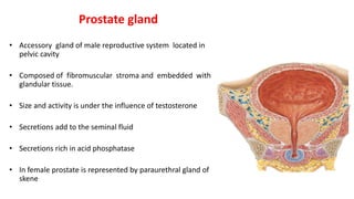
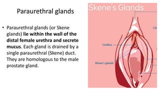
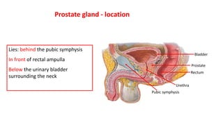
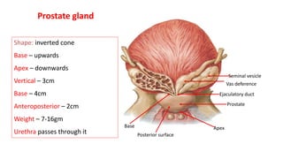
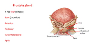
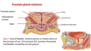
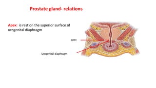
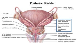
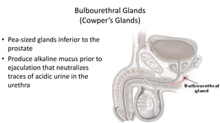
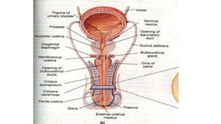
Prostate gland
Prostate gland
• Accessory gland of male reproductive system located in
pelvic cavity
• Composed of fibromuscular stroma and embedded with
glandular tissue.
• Size and activity is under the influence of testosterone
• Secretions add to the seminal fluid
• Secretions rich in acid phosphatase
• In female prostate is represented by paraurethral gland of
skene
Paraurethral glands
• Paraurethral glands (or Skene
glands) lie within the wall of the
distal female urethra and secrete
mucus. Each gland is drained by a
single paraurethral (Skene) duct.
They are homologous to the male
prostate gland.
Bladder
Prostate
Rectum
Pubic symphysis
Urethra
Prostate gland - location
Lies: behind the pubic symphysis
In front of rectal ampulla
Below the urinary bladder
surrounding the neck
Prostate
Ejaculatory duct
Vas deference
Seminal vesicle
Apex
Base
Posterior surface
Prostate gland
Shape: inverted cone
Base – upwards
Apex – downwards
Vertical – 3cm
Base – 4cm
Anteroposterior – 2cm
Weight – 7-16gm
Urethra passes through it
Prostate
Apex
Posterior surface
Prostate gland
Inferolateral
surface
Base
It has four surfaces:
Base (superior)
Anterior
Posterior
Two inferolateral
Apex
Prostate gland-relations
Levator ani
Urogenital diaphragm
Base
Apex
Inferolateral
surface
Prostatic plexus
Base – neck of bladder. Urethra pierces in median plane at
the junction of ant. 1/3 and post 2/3. junction of prostate
and bladder marked by circular groove.
Prostate gland- relations
Apex: is rest on the superior surface of
urogenital diaphragm
apex
Urogenital diaphragm
Prostate - relations
Posterior:
Rectal ampulla
Rectovesical fascia
Pubic symphysis
Retropubic space
Prosatatic venous plexus
Anterior
Urethra pierces the anterior surface slightly above the apex
Prostate – posterior surface
It is pierced by the ejaculatory ducts which
divide this surface in to a smaller upper and
a lower larger part.
Upper part belongs to the median lobe
Ejaculatory duct
Ampulla of rectum is seprated by the fascia of Denonvilliers
Inferolateral surfaces:
Related to the anterior fibers of levator ani muscle
Relations of prostate gland
Levator ani
True and false capsules
Between the two there is prostatic
venous.
It is absent posteriorly
True capsule – peripheral
condensation of connective tissue
stroma
Prostate gland - capsule
Capsules and ligaments of Prostate
• True Capsule It is formed
by condensation of the
peripheral part of the
gland. It is fibromuscular in
structure and is continuous
with stroma of the gland.
The lies between true and
false capsules venous
plexus.
 False Capsule It lies outside the true capsule
and is derived from the endopelvic fascia.
Anteriorly, it is continuous with the
puboprostatic ligaments. On each side, the
prostatic venous plexus is embedded in between
false and true capsules. Posterioily, it is
avascular, and is formed by the rectovesical
fascia of Denonvilliers. A pair of medial
puboprostatic and a pair of lateral puboprostatic
ligaments extend from the false capsule to the
back of pubic bone. The medial pair lie near the
apex while lateral pair is close to the base. These
four Iigaments support the gland.
Prostate gland - capsule
• False capsule – part of pelvic fascia
• Puboprostatic ligaments are attached to it
• Puboprostatic ligaments – medial and lateral connect prostate to pubic symphysis
and pubic bone
• Form the floor of retropubic space
Prostate gland - lobes
Anterior
Lateral
Posterior
Urethra
Ejaculatory duct
Median
Prostatic utricle
Prostate gland - lobes
• Divided incompletely in to 5 lobes
• Anterior lobe or isthmus – lies in front of urethra connects the lateral lobes . It is
made up off fibromuscular tissue. No glandular tissue
• Posterior lobe: lies behind the urethra and connects the posterior ends of the lateral
lobes. It contains glandular tissue and site of primary carcinoma
Lies behind urethra, above the ejaculatory
ducts.
It produces a bulge called uvula vesicae at the
lower part of the trigone of bladder.
It contains more number of glandular tissue
and is prone for benign hypertrophy of the
prostate.
Median/middle lobe:
Prostate gland - lobes
Lateral lobes: lie on either side of the urethra
It contains numerous glands
May give rise to adenoma
Prostate can be divided into 3 zones
Inner- mucosal
Middle – submucosal
Outer – contain main glands. The ducts of which
open in the prostatic sinus
Prostate gland
Prostate gland – blood supply
• Arterial supply: inferior vesical, middle rectal and internal pudendal arteries
• Venous drainage: prostatic venous plexus which lies between the true and false
capsules
• It receives deep dorsal vein of penis
Prostate gland – blood supply
• Prostatic venous plexus drains in to vesical venous plexus
• This drains in to internal iliac veins
• These plexus of veins are connected to internal vertebral plexus of veins ( do not
have valves)
• These connections form important route for depositing malignant cells from
prostate to the vertebral bodies and skull
Lymphatic drainage of prostate
External iliac group
Preaortic group
Presacral group
Internal iliac group
Age changes in prostate gland
• Newborn and child: fibromuscular stroma and rudiments of the duct
system
• Puberty: sudden in growth under the influence testosterone
follicles develop and proliferate and begin secreting acid phosphatase,
prostate specific antigen and prostaglandins
Age changes in prostate gland
• From 20 to 30 years: epithelium grows by multiplications with infoldings
in the lumen of the follicles, making them irregular.
• After 31 -45: epithelium infoldings disappears and amyloid concretions
start forming inside the follicles . years may show benign hypertrophy or
progressively atrophy.
THANK YOU
SEMINAL VESICLES
The seminal vesicles are two coiled sacculated tubes about 2
inches (5 cm) long which can be unraveled to three times of this
length. They lie extraperitoneally on each side, at the base of the
urinary bladder, lateral to the termination of the vas deferens and
in front of the rectum. The lower narrow end of seminal vesicle
(duct of seminal vesicle) joins the ductus deferens to form the
ejaculatory duct. It do not form a reservoir for sperms. Their
secretions form a large amount of the seminal fluid. The secretion
of seminal vesicles is slightly alkaline, containing fructose, choline,
and a coagulating enzyme called vesiculos.
• The secretion of seminal vesicle contains fructose, which is not
produced anywhere else in the body. This provides a forensic
evidence of the occurrence of rape. However, choline crystals
provide the preferred basis for determination of the presence
of seminal fluid (Florence test).
Applied aspects
• It is easily palpable per rectum
• Median lobe undergoes benign hypertrophy
• Carcinoma most common malignant tumor in males over 65 years of age
• Surgical removal: retropubic approach
• Suprapubic transvesical approach
• Transurethral approach – most accepted. It is known as transurethral resection of
prostate (TURP)
Symptoms of Prostate Cancer?
• Difficulty starting urination.
• Weak or interrupted flow of urine.
• Frequent urination, especially at night.
• Difficulty emptying the bladder completely.
• Pain or burning during urination.
• Blood in the urine or semen.
• Pain in the back, hips, or pelvis that doesn’t go away.
• Painful ejaculation.
Rectal examination of
Prostate
• It is easily palpable per
rectum.
• The discussion about
screening should take place
at: Age 50 for men who are at
average risk of prostate
cancer and are expected to
live at least 10 more years.
Age 45 for men at high risk of
developing prostate cancer.
Fibromuscular tube. 18 – 20 cm length
Common passage for urine and semen
Begins at the neck of bladder passes through
prostate, pelvic diaphragm, perineal pouches
and penis
Opens at the external urethral orifice at the tip
of glans
It shows double curve when penis is flaccid state
Male urethra
urethra
Prostatic
membranous
Spongy
External urethral meatus
Preprostatic
Male urethra – parts
Preprostatic part
Extends from the neck of the bladder to the base
of prostate
Length:1cm
Surrounded by smooth muscle which is part of
sphincter vesicae.
Richly innervated by sympathetic fibers
Prevents retrograde ejaculation of semen
Preprostatic part
Length: 2.5cm
Most dilatable part of the urethra
Lumen crescentic in shape
Begins at the base of the prostate
Emerges out on the anterior surface of the
prostate
Has a median crest in the posterior wall called
urethral crest
Prostatic urethra
Colliculus seminalis
Urethral crest
Prostatic utricle
Ejaculatory duct
Prostatic ducts
Prostatic urethra -features
The depression on either side of the
crest is the prostatic sinus
It receives the openings of the ducts
of the gland
Median crest shows a rounded swelling
called colliculus seminalis
It has the openings of the prostatic utricle
opening of ejaculatory ducts
Prostatic urethra - features
Membranous urethra
Length : 1.5cm
Lies in the deep perineal pouch
Narrowest and least dilatable part
Cross section – stellate shaped
Bulbourethral glands lie on either side
Surrounded by sphincter urethrae
Memembranous urethra
Bulbourethral galnd
Sphincter urethrae
Length : 15cm
Passes through the bulb in the superficial
perineal pouch, corpus spongiosum and
glans penis
Part in the bulb is dilated to form intrabulbar
fossa
Spongy or penile part of the urethra
In this part it receives the opening of
bulbourethral gland
In the bulbar fossa the anterior wall is
shorter than the floor and sides
Cross section – trapezoid shape
Bulbar fossa Bulbourethral gland
Spongy or penile part of the urethra
It passes through the corpus spongiosum of the
penis
Transverse section – transverse slit
Part in the glans is called – navicular fossa or
terminal fossa which is a vertical slit
It opens at the tip as external urethral opening or
meatus
Navicular fossa
Bulbar fossa
Spongy or penile part of the urethra
Urethral glands (glands of Litter) open in the entire
spongy urethra in mucous out pouching called lacunae
Largest lacuna is present in the roof of the navicular
fossa (lacuna magna)
Spongy or penile part of the urethra
Female urethra
4cm long
Related to the anterior wall of
vagina
Paraurethral glands open here
It is equivalent to the prostatic
urtethra upto the colliculus
seminalis
57
Posterior Bladder
Bulbourethral Glands (Cowper's)
• small, about the size of a pea, and located near the base of the
penis. A short duct from each enters the proximal end of the
penile urethra.
• In response to sexual stimulation, the bulbourethral glands
secrete an alkaline mucus-like fluid
59
Bulbourethral Glands
(Cowper’s Glands)
• Pea-sized glands inferior to the
prostate
• Produce alkaline mucus prior to
ejaculation that neutralizes
traces of acidic urine in the
urethra
prostateAnatomyand lobeswith agechanges.pptx

prostateAnatomyand lobeswith agechanges.pptx

  • 1.
  • 3.
    Prostate gland • Accessorygland of male reproductive system located in pelvic cavity • Composed of fibromuscular stroma and embedded with glandular tissue. • Size and activity is under the influence of testosterone • Secretions add to the seminal fluid • Secretions rich in acid phosphatase • In female prostate is represented by paraurethral gland of skene
  • 4.
    Paraurethral glands • Paraurethralglands (or Skene glands) lie within the wall of the distal female urethra and secrete mucus. Each gland is drained by a single paraurethral (Skene) duct. They are homologous to the male prostate gland.
  • 5.
    Bladder Prostate Rectum Pubic symphysis Urethra Prostate gland- location Lies: behind the pubic symphysis In front of rectal ampulla Below the urinary bladder surrounding the neck
  • 6.
    Prostate Ejaculatory duct Vas deference Seminalvesicle Apex Base Posterior surface Prostate gland Shape: inverted cone Base – upwards Apex – downwards Vertical – 3cm Base – 4cm Anteroposterior – 2cm Weight – 7-16gm Urethra passes through it
  • 7.
    Prostate Apex Posterior surface Prostate gland Inferolateral surface Base Ithas four surfaces: Base (superior) Anterior Posterior Two inferolateral Apex
  • 8.
    Prostate gland-relations Levator ani Urogenitaldiaphragm Base Apex Inferolateral surface Prostatic plexus Base – neck of bladder. Urethra pierces in median plane at the junction of ant. 1/3 and post 2/3. junction of prostate and bladder marked by circular groove.
  • 9.
    Prostate gland- relations Apex:is rest on the superior surface of urogenital diaphragm apex Urogenital diaphragm
  • 10.
    Prostate - relations Posterior: Rectalampulla Rectovesical fascia Pubic symphysis Retropubic space Prosatatic venous plexus Anterior Urethra pierces the anterior surface slightly above the apex
  • 11.
    Prostate – posteriorsurface It is pierced by the ejaculatory ducts which divide this surface in to a smaller upper and a lower larger part. Upper part belongs to the median lobe Ejaculatory duct Ampulla of rectum is seprated by the fascia of Denonvilliers
  • 12.
    Inferolateral surfaces: Related tothe anterior fibers of levator ani muscle Relations of prostate gland Levator ani
  • 13.
    True and falsecapsules Between the two there is prostatic venous. It is absent posteriorly True capsule – peripheral condensation of connective tissue stroma Prostate gland - capsule
  • 16.
    Capsules and ligamentsof Prostate • True Capsule It is formed by condensation of the peripheral part of the gland. It is fibromuscular in structure and is continuous with stroma of the gland. The lies between true and false capsules venous plexus.
  • 17.
     False CapsuleIt lies outside the true capsule and is derived from the endopelvic fascia. Anteriorly, it is continuous with the puboprostatic ligaments. On each side, the prostatic venous plexus is embedded in between false and true capsules. Posterioily, it is avascular, and is formed by the rectovesical fascia of Denonvilliers. A pair of medial puboprostatic and a pair of lateral puboprostatic ligaments extend from the false capsule to the back of pubic bone. The medial pair lie near the apex while lateral pair is close to the base. These four Iigaments support the gland.
  • 18.
    Prostate gland -capsule • False capsule – part of pelvic fascia • Puboprostatic ligaments are attached to it • Puboprostatic ligaments – medial and lateral connect prostate to pubic symphysis and pubic bone • Form the floor of retropubic space
  • 22.
    Prostate gland -lobes Anterior Lateral Posterior Urethra Ejaculatory duct Median Prostatic utricle
  • 23.
    Prostate gland -lobes • Divided incompletely in to 5 lobes • Anterior lobe or isthmus – lies in front of urethra connects the lateral lobes . It is made up off fibromuscular tissue. No glandular tissue • Posterior lobe: lies behind the urethra and connects the posterior ends of the lateral lobes. It contains glandular tissue and site of primary carcinoma
  • 24.
    Lies behind urethra,above the ejaculatory ducts. It produces a bulge called uvula vesicae at the lower part of the trigone of bladder. It contains more number of glandular tissue and is prone for benign hypertrophy of the prostate. Median/middle lobe:
  • 25.
    Prostate gland -lobes Lateral lobes: lie on either side of the urethra It contains numerous glands May give rise to adenoma
  • 26.
    Prostate can bedivided into 3 zones Inner- mucosal Middle – submucosal Outer – contain main glands. The ducts of which open in the prostatic sinus Prostate gland
  • 28.
    Prostate gland –blood supply • Arterial supply: inferior vesical, middle rectal and internal pudendal arteries • Venous drainage: prostatic venous plexus which lies between the true and false capsules • It receives deep dorsal vein of penis
  • 29.
    Prostate gland –blood supply • Prostatic venous plexus drains in to vesical venous plexus • This drains in to internal iliac veins • These plexus of veins are connected to internal vertebral plexus of veins ( do not have valves) • These connections form important route for depositing malignant cells from prostate to the vertebral bodies and skull
  • 30.
    Lymphatic drainage ofprostate External iliac group Preaortic group Presacral group Internal iliac group
  • 31.
    Age changes inprostate gland • Newborn and child: fibromuscular stroma and rudiments of the duct system • Puberty: sudden in growth under the influence testosterone follicles develop and proliferate and begin secreting acid phosphatase, prostate specific antigen and prostaglandins
  • 32.
    Age changes inprostate gland • From 20 to 30 years: epithelium grows by multiplications with infoldings in the lumen of the follicles, making them irregular. • After 31 -45: epithelium infoldings disappears and amyloid concretions start forming inside the follicles . years may show benign hypertrophy or progressively atrophy.
  • 33.
  • 34.
    SEMINAL VESICLES The seminalvesicles are two coiled sacculated tubes about 2 inches (5 cm) long which can be unraveled to three times of this length. They lie extraperitoneally on each side, at the base of the urinary bladder, lateral to the termination of the vas deferens and in front of the rectum. The lower narrow end of seminal vesicle (duct of seminal vesicle) joins the ductus deferens to form the ejaculatory duct. It do not form a reservoir for sperms. Their secretions form a large amount of the seminal fluid. The secretion of seminal vesicles is slightly alkaline, containing fructose, choline, and a coagulating enzyme called vesiculos.
  • 37.
    • The secretionof seminal vesicle contains fructose, which is not produced anywhere else in the body. This provides a forensic evidence of the occurrence of rape. However, choline crystals provide the preferred basis for determination of the presence of seminal fluid (Florence test).
  • 38.
    Applied aspects • Itis easily palpable per rectum • Median lobe undergoes benign hypertrophy • Carcinoma most common malignant tumor in males over 65 years of age • Surgical removal: retropubic approach • Suprapubic transvesical approach • Transurethral approach – most accepted. It is known as transurethral resection of prostate (TURP)
  • 39.
    Symptoms of ProstateCancer? • Difficulty starting urination. • Weak or interrupted flow of urine. • Frequent urination, especially at night. • Difficulty emptying the bladder completely. • Pain or burning during urination. • Blood in the urine or semen. • Pain in the back, hips, or pelvis that doesn’t go away. • Painful ejaculation.
  • 40.
    Rectal examination of Prostate •It is easily palpable per rectum. • The discussion about screening should take place at: Age 50 for men who are at average risk of prostate cancer and are expected to live at least 10 more years. Age 45 for men at high risk of developing prostate cancer.
  • 41.
    Fibromuscular tube. 18– 20 cm length Common passage for urine and semen Begins at the neck of bladder passes through prostate, pelvic diaphragm, perineal pouches and penis Opens at the external urethral orifice at the tip of glans It shows double curve when penis is flaccid state Male urethra urethra
  • 42.
  • 43.
    Preprostatic part Extends fromthe neck of the bladder to the base of prostate Length:1cm Surrounded by smooth muscle which is part of sphincter vesicae. Richly innervated by sympathetic fibers Prevents retrograde ejaculation of semen Preprostatic part
  • 44.
    Length: 2.5cm Most dilatablepart of the urethra Lumen crescentic in shape Begins at the base of the prostate Emerges out on the anterior surface of the prostate Has a median crest in the posterior wall called urethral crest Prostatic urethra
  • 45.
    Colliculus seminalis Urethral crest Prostaticutricle Ejaculatory duct Prostatic ducts Prostatic urethra -features The depression on either side of the crest is the prostatic sinus It receives the openings of the ducts of the gland
  • 46.
    Median crest showsa rounded swelling called colliculus seminalis It has the openings of the prostatic utricle opening of ejaculatory ducts Prostatic urethra - features
  • 47.
    Membranous urethra Length :1.5cm Lies in the deep perineal pouch Narrowest and least dilatable part Cross section – stellate shaped Bulbourethral glands lie on either side Surrounded by sphincter urethrae Memembranous urethra Bulbourethral galnd Sphincter urethrae
  • 48.
    Length : 15cm Passesthrough the bulb in the superficial perineal pouch, corpus spongiosum and glans penis Part in the bulb is dilated to form intrabulbar fossa Spongy or penile part of the urethra
  • 49.
    In this partit receives the opening of bulbourethral gland In the bulbar fossa the anterior wall is shorter than the floor and sides Cross section – trapezoid shape Bulbar fossa Bulbourethral gland Spongy or penile part of the urethra
  • 50.
    It passes throughthe corpus spongiosum of the penis Transverse section – transverse slit Part in the glans is called – navicular fossa or terminal fossa which is a vertical slit It opens at the tip as external urethral opening or meatus Navicular fossa Bulbar fossa Spongy or penile part of the urethra
  • 51.
    Urethral glands (glandsof Litter) open in the entire spongy urethra in mucous out pouching called lacunae Largest lacuna is present in the roof of the navicular fossa (lacuna magna) Spongy or penile part of the urethra
  • 53.
    Female urethra 4cm long Relatedto the anterior wall of vagina Paraurethral glands open here It is equivalent to the prostatic urtethra upto the colliculus seminalis
  • 57.
  • 58.
    Bulbourethral Glands (Cowper's) •small, about the size of a pea, and located near the base of the penis. A short duct from each enters the proximal end of the penile urethra. • In response to sexual stimulation, the bulbourethral glands secrete an alkaline mucus-like fluid
  • 59.
    59 Bulbourethral Glands (Cowper’s Glands) •Pea-sized glands inferior to the prostate • Produce alkaline mucus prior to ejaculation that neutralizes traces of acidic urine in the urethra