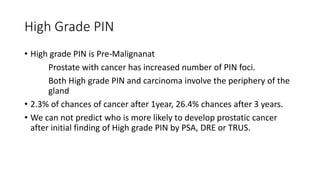
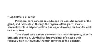
1. Carcinoma prostate
Speaker : Dr. Ajai Sasidhar
PG Student
KIMS Hubli
Chair Person : Dr. Ravikumar Jadhav
Associate professor
Department of Urology
2. Prostate carcinoma
• Anatomy of the Prostate gland
• Epidemiology and aetiology of prostate cancer
• Pathological types
• Diagnosis and staging of prostate cancer
• Treatment
6. Anatomy
• Pyramidal Fibro Muscular gland, Surrounds the 1st 3cm of urethra
• Consists of Glandular tissue in fibro muscular stroma
• Embryologically develop as a protrusion from the dorsal wall of
prostatic urethra at 12th week of gestation
• Has no true capsule, but enclosed by visceral fascia which sends
numerous fibro muscular septa, which divide the gland into different
lobes.
14. Base
• The base is directed upwards and continuous with the neck of
bladder.
• The junction is marked by circular groove which lodges veins of the
vesical and prostate plexuses.
15.
16. Apex
• The apex is directed downwards and rests on the upper surface of
urogenital diaphragm.
18. Anterior surface
• Convex from side to side.
• Lies in the arch of pubis, being separated by it by dorsal venous
complex.
• Anterior surface is connected to pubic bone by pubo prostatic
ligament.
• Urethra emerges from this surface
19.
20.
21. Inferio Lateral Surface
Are related to the anterior fibers of the levator ani , and they are
separated from the muscles by a plexus of veins embedded in its
sheath.
22.
23. Posterior surface
• It is flattened from side to side and convex from above downwards .
• Near its upper border it is pierced on each side of the median plane
by the ejaculatory ducts.
• It is separated from the rectum by the fascia of denonvilliers.
24.
25.
26. Anatomy
The prostate has 5 lobes.
• Anterior
• Posterior
• Middle
• 2 lateral lobes (Right and Left)
27.
28. • ANTERIOR LOBE- lies in front of the urethra and contains little or no
glandular tissue and therefore seldom forms an adenoma.
• POSTERIOR LOBE – lies behind the middle lobe and ejaculatory ducts ,
adenoma never occurs here but primary carcinoma of the prostate is said
to begin in this part.
29. • MIDDLE/MEDIAN LOBE- This is a wedge of tissue situated behind the
urethra and in front of ejaculatory ducts , just below the neck of bladder
and contains much glandular tissue.
• It produces an elevation in the lower part of trigone of bladder , known
as uvula vesicae .
• Middle lobe projects into urethra raising a prominence verumontanum
• It is common site for adenoma.
30.
31. • LATERAL LOBES – Lies on each side of urethra . Each lobe contains
glandular tissue from which an adenoma may arise in old age.
32. • 5 lobes can be distinguished in foetal gland prior to 20 weeks of
gestation
• After 20 weeks only 3 lobes are recognizable , Two lateral lobes and a
median lobe.
So, The latest concept is Prostate can be subdivided into 3 distinct
zones
33. The zonal anatomy of the prostate by Mc Neil
• Transitional zone (5%)
• Central zone (25%)
• Peripheral zone (70%)
• Anterior fibro muscular stroma.
34.
35. Transitional zone (5%)
• The transitional zone lies around the distal part of prostatic urethra,
proximal to the apex of central zone and ejaculatory ducts.
• This zone is typically prone to benign hypertrophy (BPH).
36.
37. Central zone (25%)
• The central zone surround the ejaculatory ducts posterior to the
prostatic urethra.
• It is the inner small zone is composed of sub mucosal glands opening
in prostatic sinuses.
38.
39. Peripheral zone (70%)
• The peripheral zone is cup shaped and encloses the central,
transitional zones and prostatic urethra anteriorily.
• This zone is frequently the site of carcinoma 70% carcinomas arises
here.
40.
41. • The anterior portion is filled by non glandular tissue also called
fibomuscular stroma.
42.
43.
44.
45. Vascular Supply
• The prostate gland is supplied by branches from
• Inferior vesical
• Middle rectal&
• Internal pudendal arteries
46. • The majority of blood supply comes from inferior vesical artery, A
branch of internal iliac artery.
• The urethral and capsular groups.
47.
48. • The capsular arteries run with neuro vascular bundles (Walsch) and it
penetrate the prostate perpendicularly.
• These are important as these capsular vessels provide the
macroscopic landmark to aid in the identification of the microscopic
branches of the pelvic plexus that innervate the corpora cavernosa.
49.
50. Venous drainage
• The veins form a rich plexus around the sides and base of the gland,
the plexus receives the deep dorsal vein of the penis.
• The plexus communicate with the vesical plexus and the internal
pudendal vein and drains into the vesical and internal iliac veins.
53. • Vas deference drains to external iliac nodes.
• Seminal vesicle drains to external and internal iliac nodes
54. Nerve Supply
• Nerve supply is from the pelvic plexus.
• Pelvic plexus is formed by parasympathetic, visceral, efferent,
preganglionic fibers that arise from the sacral center (S2 to S4) and
sympathetic fibers via the hypogastric nerve from the thoracolumbar
center .
• The branches from the plexus run in neuro vascular bundle.
55.
56. • The pelvic plexus provides visceral branches that innervate the
bladder, ureter, seminal vesicles, prostate, rectum, membranous
urethra, and corpora cavernosa.
• The somatic motor axons also travel through the pelvic plexus to
supply the levator ani, coccygeus, and striated urethral musculature.
57. • These nerves are microscopic but the capsular vessels which
penetrate prostate serves as a landmark during dissection of prostate
58. The capsules of prostate
• The prostate doesn’t have a true capsule.
• The false capsule of prostate is formed by condensation of prostatic
tissue all around which forms a surgical capsule.
59. The prostate is covered by 3 different facial layers
• The Denonvillers fascia
• Prostatic fascia
• Levator fascia
60. The Denonvillers fascia
• Located between anterior wall of rectum and prostate.
• Formed from peritoneal pouch.
• Covers the posterior surface of seminal vesicle
61. Prostatic fascia
• Anteriorly continuous with prostatic capsule.
• Dorsal vein of penis and Santorini’s plexus travel within anterior
prostatic fascia
62. Levator fascia
• Laterally the prostatic fascia fuses with levator fascia to form lateral
pelvic fascia
• The prostate receives innervation and blood supply between the
layers of the levator fascia and prostatic fascia.
63.
64. Prostatic fluid - functions
• The average volume of the normal human ejaculate is approximately
3 mL. Range 2-6mL
• Spermatozoa, which represent less than 1% of the total ejaculate, are
present in the range of 100 million per millilitre.
• The major contribution to the volume of seminal plasma (average 3
mL) comes from the seminal vesicles (1.5 to 2 mL), from the prostate
(0.5 mL), and from Cowper's gland and glands of Littre (0.1 to 0.2 mL).
65. Prostatic fluid - functions
• The prostatic fluid provides optimum pH for the motility of the
sperms by neutralizing the acidity of vaginal secretions and maintains
a ph of 6.0 to 6.5.
• The clotting enzymes in prostatic secretions cause conversion of
fibrinogen into coagulum.
• It is essential in holding the sperms to uterine cervix.
66. Lysis of coagulum :
• The coagulum is dissolved by fibrinolysin of prostatic secretions so
the sperms become motile.
68. History
• Adams J. The case of scirrhous of the prostate gland with
corresponding affliction of the lymphatic glands in the lumbar region
and in the pelvis. Lancet. 1853;1:393.
• Described prostate cancer as “very rare disease”
69. Incidence
• Most common visceral malignant neoplasm in US men.
• Prostate cancer is the 4th most common male malignant neoplasm
worldwide.
• Lowest incidence in Asians, and highest in African Americans
70. Age at diagnosis
• Rarely diagnosed below the age of 50
• Median diagnosis at the age of 68 years
• Peak incidence between ages 70 and 74
• 20% risk of having clinically palpable carcinoma, and 75% chance of
histopathological carcinoma at age 80.
71. Stage at diagnosis
• PSA testing
• The incidence of loco regional disease increased but that of
metastatic disease is decreased.
• Non palpable tumors account for 75% of newly diagnosed cases.
72. Mortality
• Now because of widespread use of PSA screening, 5 year survival and
10 year survival are 100% and 92% respectively.
73. Effect of screening on mortality
• PLCO(prostate, lung, colorectal, ovary) trial
• ERSPC(European randomised screening for prostate cancer) reduction
of rate of death by 20%. But to prevent one death one would need to
scan 1410 men and treat additional 48 cases of prostate cancer.
74. Risk Factors for Ca Prostate
• Family history
• Genetic Factors
• Chronic inflammation
• Hormonal Factors- Androgen and Estrogen
• Diet
• Oesity
• Leptins
• Lack of vitamin D
• Alcohol
78. Family History
• Sporadic
• Familial
• Hereditory – A subset of familial cancer where,
Nuclear family with 3 or more affected members
Prostate cancer in 3 successive generations
2 affected individuals with cancer onset before 55 years
79. The relative risks can be summarised as.
Family History Relative Risk Absolute Risk (%)
None 1 8
Father or brother 2 15
Father or brother affected < 60 years 3 20
Father and brother 4 30
Hereditary prostate cancer 5 35-45
80. Genetic factors
Many genes are associated with carcinoma prostate
HPC 1, OGG1, CHEK2, BRCA2,PON1,and MIC1
• Most important is HPC1
Prostate cancer is more common in African Americans and less
common in Asians.
81. Presence of chronic inflammation
• Chronic inflammation leads to hyper proliferation
• Significant association with STDs and prostatic cancer
82. Hormonal Factors
• Androgen :Absence will protect against prostate cancer.But There is
no dose response relationship of testosterone with prostate cancer
• Estrogen: Initially thought to be protective. Estrogen also promote
epithelial growth
83. • Diet – High level of fat in diet will promote carcinogenesis.
• Obesity
• Vitamin D found to be protective
• Smoking and alcohol – No definite role.
84. Prevention of Carcinoma prostate
• “Prevention is better than cure, because it saves the labor of being
sick.”
85. 5 Alpha reductase inhibitors
• The main findings of the PCPT are as follows:
1. The prevalence of prostate cancer was reduced by 24.8%
2. The prevalence of Gleason grade 7 to 10 tumors was higher in
the finasteride group than in the placebo
3. The risk reductions associated with finasteride among risk groups
defined by age, family history, race, and PSA level were of the
same general magnitude.
4. Sexual side effects were more common with finasteride, whereas
urinary symptoms were more common with placebo.
87. Summary Epidemiology
• In the United States, prostate cancer is the most common visceral
malignant neoplasm in men and the second leading cause of cancer-
related deaths.
• Mortality has declined since 1991 and for white men is now lower
than before PSA was introduced.
• Worldwide, prostate cancer incidence and mortality rates vary
significantly between countries and regions and are highest in African
American men.
• PSA testing has induced a significant downward migration in age and
stage (both clinical and pathologic) at diagnosis
88. • Both genetics and environment are important in the origin and
evolution of prostate cancer.
• Most prostate cancer is polygenic, but some hereditary forms may be
determined by variants in a single gene or relatively few genes.
• HPC1 is an autosomal dominant gene with high penetrance.
• HPC1 encodes the enzyme RNase L, which has both antiviral and
proapoptotic activity.
89. • Androgen exposure of the prostate plays an important but
incompletely defined role in prostate carcinogenesis.
• Long-term absence of androgen exposure to the prostate appears to
protect against the development of cancer, but a dose-response
relationship between androgen levels and cancer risk has not been
established .
• Estrogen is also important in prostate cancer development and may
have varying effects.
• Dietary factors, including saturated fat consumption, are also
important in prostate cancer risk.
90. • The Prostate Cancer Prevention Trial demonstrated that finasteride
reduces the period prevalence of prostate cancer by 25%, albeit with
a slightly higher risk of high-grade disease. Whether the risk of higher
grade disease is real or an artifact of the study design is currently
under study.
• Other 5α-reductase inhibitors, the antioxidants selenium and vitamin
E, and other agents are currently under study as potential
chemopreventive agents.
91. Patology of Prostatic cancer
• Pathology of adenocarcinoma of the prostate from its precursor lesions to
invasive carcinomas.
• Precursor Lesions
Prostatic intraepithelial neoplasia.
Low Grade
High Grade
Adeno carcinoma
Mucinous adenocarcinoma, prostatic duct
adenocarcinomas, - Rarer forms
Rare malignancies
Mesenchymal tumors, Lymphoma
92. Prostatic intraepithelial neoplasia
• Prostatic intraepithelial neoplasia (PIN) consists of architecturally
benign prostatic acini or ducts lined by cytologically atypical cells and
is classified into low grade and high grade lesions.
94. High Grade PIN
• High grade PIN is Pre-Malignanat
Prostate with cancer has increased number of PIN foci.
Both High grade PIN and carcinoma involve the periphery of the
gland
• 2.3% of chances of cancer after 1year, 26.4% chances after 3 years.
• We can not predict who is more likely to develop prostatic cancer
after initial finding of High grade PIN by PSA, DRE or TRUS.
95. Adeno Carcinoma Prostate
• Location
85% in peripheral zone. 15% in transitional zone.
Adenocarcinoma of the prostate is multifocal in more than 85%
of cases.
96. • Local spread of tumor
Peripheral zone cancers spread along the capsular surface of the
gland, and may extend through the capsule of the gland, invade
seminal vesicles and periprostatic tissues, and involve the bladder neck
or the rectum.
Transitional zone tumors demonstrate a lower frequency of extra
prostatic extension. May harbor large volumes of disease with
relatively high PSA levels but remain confined to the prostate.
97. • Metastatic spread
Most commonly through lymphatics – to iliac, presacral,
common iliac inguinal and para aortic nodes.
Secondly through blood – Bone followed by lung , liver and
adrenals
98. Histological grading
• The gleason score
• Based on the pattern of gland under relatively low
magnifications.
• Histologic variation in tumor, so two predominant patterns are
recorded for each case.
• Primary or predominant and Secondary or second most
prevalent patterns are identified and assaigned a score from 1
to 5.
99. • Final score is the sum of both the primary and secondary scores.
• >50% ca contain ≥2 patters
• One of the strongest predictors of biological behaviour, invasiveness
and metastatic potential.
100.
101. • Gleason pattern 1 and pattern 2 tumors are composed of relatively
circumscribed nodules of uniform, single, separate, closely packed,
medium-sized glands.
102. • Gleason pattern 3 tumor infiltrates the non-neoplastic prostate, and
the glands have marked variation in size and shape, with smaller
glands than in Gleason pattern 1 or pattern 2.
103. • Gleason pattern 4 glands are no longer single and separate as in
patterns 1 to 3. In Gleason pattern 4, one may also see large,
irregular, cribriform glands as opposed to the smoothly
circumscribed smaller nodules of cribriform Gleason pattern 3.
104. • It is important to recognize Gleason pattern 4 tumor because tumors
with this pattern have a significantly worse prognosis than those with
pure Gleason pattern 3.
• It has also been demonstrated in radical prostatectomy specimens
that tumors with Gleason score 4 + 3 = 7 have a worse prognosis than
those with Gleason score 3 + 4 = 7.
105. • Gleason pattern 5 tumor shows no glandular differentiation and is
composed of solid sheets, cords, single cells, or tumor with central
comedonecrosis.
109. Mucinous adeno carcinoma
• Mucinous adenocarcinoma of the prostate gland is one of the least
common morphologic variants of prostatic carcinoma.
• Has an aggressive biologic behaviour.
• Has a propensity to develop bone metastases and increased serum
acid phosphatase and PSA levels with advanced disease.
110. Small Cell carcinoma
• Small cell carcinomas of the prostate are identical to small cell
carcinomas of the lung.
• Shows neuro endocrine differentiation.
• Produce adrenocorticotropic hormone or antidiuretic hormone.
• The average survival of patients with small cell carcinoma of the
prostate is less than a year.
111. Prostatic duct adenocarcinoma
• Between 0.4% and 0.8% of prostatic adenocarcinomas arise from
prostatic ducts.
• They may grow as an exophytic lesion into the urethra, most
commonly in and around the verumontanum.
112. Mesenchymal tumors
Sarcomas
• Account for 0.1% to 0.2% of all malignant prostatic tumors.
Rhabdomyosarcoma is the most frequent mesenchymal tumor within
the prostate and is seen almost exclusively in childhood.
• Leiomyosarcomas are the most common sarcomas involving the
prostate in adults.
113. Transitional cell carcinomas
• Primary transitional cell carcinoma of the prostate without bladder
involvement accounts for 1% to 4% of all prostate carcinomas.
• Primary transitional cell carcinomas of the prostate show a propensity
to infiltrate the bladder neck and the surrounding soft tissue such
that more than 50% of the patients present with stage T3 or T4
tumors.
• 5-year survival of approximately 34%.
114. Summary
• Low-grade PIN should not be commented on in diagnostic reports
because the diagnosis is not reproducible among pathologists and
lacks clinical significance.
• For patients diagnosed with high-grade PIN on extended initial core
sampling, repeat biopsy within the first year is unnecessary in the
absence of other clinical indicators of cancer.
• Regardless of the serum PSA level, all patients with an initial atypical
diagnosis on needle biopsy should undergo repeat biopsy; the risk of
cancer is approximately 40%, and clinical findings are not helpful in
predicting who is more likely to have cancer .
115. • Tumor volume measured in the radical prostatectomy specimen does
not independently predict postsurgical progression once grade,
pathologic stage, and margins are accounted for and should not be
required for routine pathologic analysis.
• Gleason grade, whether it is assessed on needle biopsy, transurethral
resection, or radical prostatectomy specimens, remains one of the
most influential prognostic factors.
116. Tumor markers
• The discovery and utilization of tumor markers have positively
affected early detection, diagnosis, and staging for prostatic
malignancies.
• By improving early detection, tumor markers contribute to improved
curative success rates.
117. Tumor markers in prostate cancer
• PSA
• PROSTATIC ACID PHOSPHATASE
NEWER TUMOR MARKERS
• Human kallikrein 2 (hK2)
• Insulin-like growth factor 1
• PCA3 – detected in urine.
• CDKN1B (P27)
• Ki-67
119. PSA
• A product kallikerin gene family
• Also known as hk3, identified in 1970
• PSA is normally found in low concentration in sera (ng/mL). Within
sera, PSA circulates in both bound and unbound forms. Most PSA in
sera is bound or complexed to the antiproteases ACT and
macroglobulin (MG)
• PSA expression is strongly influenced by androgens.
• In the absence of prostate cancer, serum PSA levels vary with age,
race, and prostate volume.
120. • Elevated serum PSA levels are probably a product of disruption of
cellular architecture within the prostate gland.
• This can occur in the setting of prostate disease (BPH, prostatitis,
prostate cancer) and with prostate manipulation (prostate massage,
prostate biopsy).
• So, it is organ specific and not disease specific.
• The serum half-life of PSA is 2 to 3 days.
• Prostatic trauma such as occurs after prostatic biopsy can result in a
leak of PSA into the circulation that may require more than 4 weeks
for return to baseline values.
121. PSA DERIVATIVES
• PSAD (PSA Density)
• Cancers produce less PSA per cell than nonmalignant
prostatic tissues
• Calculated by dividing the serum PSA concentration
by the volume of the prostate gland measured by
TRUS.
• PSAD greater than 0.15 has been suggested as
discriminatory for the presence of cancer.
• Important in elderly.
122. • PSAV (PSA Velocity)
• Serial PSA measurements and calculate the rate of rise in PSA
• A rate of rise of >0.75 ng/ml/yr is associated with a higher
frequency of cancer
• Important for young patients
123. • Age specific PSA
• Age PSA Range %
40-50 0-2.5 4-10 25
60-70 0-4.5 >10 60 (localized 50%)
high suspicion of carcinoma
70-80 0-6.5 >50 ↑ LN mets/ bone mets
disseminated disease
124. • Rule out the possibility of ca
• Gives idea about intraprostatic volume of disease
• Decision regarding Biopsy and treatment
• Monitoring response to therapy
• Base line for follow up and detecting recurrence after therapy
• Detection yield
• DRE+PSA = 4.7-5.8%
• PSA+DRE+TRUS = 60%
• DRE+Bx = 22%
• PSA+DRE+Bx = 48%
125. • PROSTATIC ACID PHOSPHATASE (PAP) (N- 0-5.5 u/l)
• Sensitivity- 10% Specificity-90%
• Abnormal PAP correlates with higher tumor stage T3, T4, higher
grade,high pre therapy serum PSA
• Affected by circadian rhythm, prostatic message, Sx manipulation and
BHP.
126. Summary PSA
• Today, most prostate cancer arises as clinically nonpalpable (stage
T1c) disease with PSA between 2.5 and 10 ng/mL.
• Screening by PSA have resulted in stage migration to nonpalpable,
clinically localized (stage T1c) disease and a parallel reduction in
mortality.
• Although PSA is widely accepted as a prostate cancer tumor marker,
it is organ specific and not disease specific.
127. • PSA is normally found in low concentration in serum (ng/mL). In
serum, PSA circulates in both bound and unbound forms. Three
proteins that are recognized to bind to PSA within the blood are α1-
antichymotrypsin (ACT), α2-macroglobulin (A2M), and α1-protease
inhibitor (API).
• The serum half-life of PSA, calculated after removal of all prostate
tissue, is 2 to 3 days.
• Finasteride (5 mg) and other 5α-reductase inhibitors for treatment of
BPH have been shown to lower PSA levels by an average of 50%.
128. • Interpretation of PSA values should always take into account the
presence of prostate disease, previous diagnostic procedures, and
prostate-directed treatments.
129. Detection of prostatic cancer
• Long latent phase of disease.
• Disease progresses without symptoms.
• Screening will provide opportunity for early detection
130. Screening for prostate cancer
• Routine population based screening is not advised, As it is not cost
effective and leads to over diagnosis of the disease.
• Also false positive and borderline reports cause psychological stress in
the subject.
• Although the value of PSA screening remains controversial, men who
present for periodic health examinations should be made aware of
the availability of the PSA test so that they can make an informed
decision about the need for routine screening.
131. • ERSPC(European randomised screening for prostate cancer) reduction
of rate of death by 20%. But to prevent one death one would need to
scan 1410 men and treat additional 48 cases of prostate cancer.
132. Screening recommendations
• Using a computer generated (Markov) model of the natural history of
prostate cancer, Ross and colleagues (2000) found that a strategy of
PSA testing at age 40 years, age 45 years, and biennially (every other
year) after age 50 years with a PSA threshold of 4 ng/mL used fewer
resources and saved more lives than a strategy that tested annually
starting at age 50 years.
• Evidence based models not available.
• Can be stopped at 65 years if PSA is below 1.5ng/ml.
133. Detection of prostatic cancer
• Clinical Methods
• History-
• Urinary status
• sexual function
• Skeletal symptoms
• co-morbidities
134. • Extra prostatic spread- often asymptomatic
• Rectal involvement-
• Renal impairment due to prolonged BOO
• Fluid retention/ electrolyte imbalance
141. • Once gland volume has been obtained, one can calculate derivatives
such as the PSA density (PSAD = serum PSA/gland volume)
• All hypoechoic lesions within the PZ should be noted and included in the
biopsy material
• Hypoechoic-14-39%(~40%)
• Isoechoic- 14-29%
• Hyperechoic-1%
• TZ BPH nodules are typically hypoechoic but may contain isoechoic or
even hyperechoic foci
• A hypoechoic lesion is malignant in 17% to 57% of cases.
147. CT SCAN
• Primary role
• Size determination of the gland
• Assess pelvic LN metastasis
• Treatment planning in RT
• Extra Prostatic Extension:
• LN involvement
148. • Limitation of CT
• Can not detect intraprotatic anatomic changes
• Can't detect microscopic disease
• False Positive
149. MRI
• Superior to CT in defining prostate apex, NVB and anterior rectal wall
• Better delineation of periprostatic fat involvement
• Ca Prostate: A focal, peripheral region of decreased signal intensity
surrounded by a normal(high intensity) peripheral zone
• BHP: centrally located nodules of similar signal
• Primary staging sensitivity- 69%
• Endorectal surface coil MRI- accuracy of 54-72% staging the primary
and detects SVI and ECE
150. • Indications: High likelihood of capsular invasion and LN metastasis
• Abnormal DRE
• PSA>20
• Poorly differentiated ca
• Sensitivity to locate gland tumor- 79% and specificity- 55%
• LN detection- Low sensitivity but high specificity
151. MR Spectroscopy
• Spectroscopy principle: The electron cloud surrounding different chemical
compounds shields the resonant atoms of interest to varying degrees
depending on the specific atomic structure of the compound
• Glandular Citrate concentrations 240 to 1,300 times greater than blood
plasma concentrations. High levels of citrate -Peripheral zone and lower
levels in the transition and central zones.
• Tumor: A significant reduction in prostate citrate and a significant increase
in prostate choline levels relative to the normal peripheral zone
Metabolic map of the prostate corresponding to normal and abnormal
metabolic activity that can be used to pinpoint the location of prostatic
tumors
152. • Improved diagnostic accuracy of MRI both in localizing and staging and risk-
stratifying patients
• Specificity for tumor location (MRI + MRSpectroscopy) ~ 91%.
• Accurate localization of prostate tumors and improved guided biopsy
• Combined MRI/MRSI enhances the assessment of both ECE and SVI and
capsular breech
• Predict tumor aggressiveness
• Distinguishing b/w tumor and post biopsy hemorrhage
• Detect residual cancer following t/t and follow-up
• Development of more focused therapy
153. 99Tc BONE SCAN
• Clinically apparent metastatic disease limited to bone in 80-85% of patients of
metastatic ca prostate
• A close correlation exists between pretreatment PSA level and incidence of
abnormal bone scan results
• Osteoblastic secondaries
• MC sites of metastasis
• Vertebral column- 74%
• Ribs- 70%
• Pelvis- 60%
• Femora- 44%
• Shoulder girdle-41%
154. • Indications: Pretherapy
• Post therapy: Skeletal symptoms in associate with ↑PSA level
• False +ve
• Fractures/ Arthritis / Paget’s disease
156. Carcinoma prostate
Speaker : Dr. Ajai Sasidhar
PG Student
KIMS Hubli
Chair Person : Dr. Ravikumar Jadhav
Associate professor
Department of Urology
157. Clinical methods Case 1
• Vinayak a 52 year old male patient presented to the OPD for an
annual health check up. He is the 2nd male child in his family. His
father had history of prostate cancer. It is his 4th visit to the urologist.
His initial visit was at his 40th age. At his 1st visit he was made aware
of the optional PSA testing and its importance as he has a family
history of prostate cancer. His subsequent visits at the age of 45 and
50, he was evaluated with PSA, PSA was less than 1.5ng/ml in all3
occasions.
158. • He is healthy, sexually active with no urinary complaints.No bone pain
or tenderness. His bowel and bladder habits are normal. Not a
diabetic or hypertensive.
On further evaluation, he has no pallor, icterus or
Lymphadenopathy.
CVS, RS within normal Limits
P/A no organomegaly
159. • On Digital rectal examination , There is no prostatomegaly, or any
palpable lesions in prostate.
• His biochemical parameters are normal except PSA .
• PSA was 4.5ng/ml.
• His PSA value was at previous visit 2 years back was 1.5ng/ml.
• PSA velocity 1ng/ml/year
160. • TRUS showing hypo echoic lesion in transitional zone, No extra
prostatic extension.
• Biopsy showing adeno carcinoma with Gleason score 5
• CT showing no lymph node involvement
• Bone scan –No hot spots(can be omitted).
• Diagnosis - Early prostatic carcinoma.
161. Clinical methods Case 2
• Krishna , a 65 year old male patient presented to the OPD with
difficulty in passing urine. He is having Urinary hesitancy,diminished
force of urine stream, intermittency,sense of incomplete voiding for
the last 2 months, the symptoms are gradually progressing for the last
2 months.He has no other complaints.There is no bone pain or
tenderness. His bowel habits are normal. Not a diabetic or
hypertensive.
162. On further evaluation, he has no pallor, icterus or
Lymphadenopathy.
CVS, RS within normal Limits
P/A no organomegaly
• On Digital rectal examination , he has a Hard, nodular, asymmetrical
swelling in the prostate gland raised above the surface of gland and is
surrounded by compressible prostatic tissue
• His biochemical parameters are normal except PSA .
• PSA was 14ng/ml.
163. • TRUS showing hypo echoic lesion with seminal vescicle and bladder
base involvement.
• Biopsy showing adeno carcinoma with gleason score 7
• CT scan no lymphadenopathy
• Bone scan – No hot spots
• Diagnosis – Locally advanced prostatic carcinoma
164.
165.
166. Clinical methods Case 3
• Shiva a 70 year old male patient presented to the OPD with
complaints of difficulty in passing urine. He is having Urinary
hesitancy, diminished force of urine stream, intermittency, sense of
incomplete voiding for the last 1 year, the symptoms are gradually
progressing for the last 1 year. He is also having Urgency,frequency,
and dribbling for last 6 months. He is having Hematochezia,
Constipation, Intermittent diarrhoea for the last 1 month. He is also
complaining of low back ache and pelvic pain for last 1 month.
167. • O/E
• He is thin emaciated, has pallor, and inguinal lymphadenopathy .
CVS, RS within normal Limits
P/A a firm 3x3 cm mass palpable in the abdomen below umbilicus
• On Digital rectal examination , he has a Hard, nodular, asymmetrical
swelling in the prostate gland Firmness extending superior to the
prostate gland. There was a mass palpable in the anal canal at 12 o
clock position and the finger stained with blood and mucus.
• He had tenderness at L1 vertebra region.
168. • His PSA was 30ng/ml.
• Prostatic acid phosphatase was 100 U/L
• He had hyponatremia.
169. • TRUS showing hypo echoic lesion in transitional zone, with extension
to the seminal vesicle, bladder base, and rectum
• Biopsy showing adeno carcinoma with Gleason score 7
• CT showing internal iliac, para and pre aortic lymph node involvement
• Bone scan –Hot spots at vertebral column, ribs and pelvis.
• Diagnosis - Metastatic prostatic carcinoma.
170.
171.
172. Clinical methods Case 4
• Vijay , a 60 year old male patient presented to the OPD with difficulty
in passing urine. He is having Urinary hesitancy,diminished force of
urine stream, intermittency,sense of incomplete voiding for the last 2
months, the symptoms are gradually progressing for the last 2
months.He has no other complaints.There is no bone pain or
tenderness. His bowel habits are normal. Not a diabetic or
hypertensive.
173. On further evaluation, he has no pallor, icterus or
Lymphadenopathy.
CVS, RS within normal Limits
P/A no organomegaly
• On Digital rectal examination , prostatomegaly
• His biochemical parameters are normal except PSA .
• PSA was 4ng/ml.
174. • He is diagnosed as having BPH
• Posted for TURP
• The tissue obtained after TURP sent for histopathological examination
• The HPR came as adeno carcinoma. Gleason score 3
175. • In TURP, the tissue is resected from transitional zone
• Tumors of the transitional area are more often low grade and less
prone to penetrate capsule and metastasize, and there is a fair chance
of cure even if the PSA is well above 20 ng/ml.
• Diagnosis - Early prostatic carcinoma.
176. Clinical methods Case 5
• Shankar a 70 year old male patient presented to the OPD with
complaints of difficulty in passing urine. He is having Urinary
hesitancy, diminished force of urine stream, intermittency, sense of
incomplete voiding for the last 1 year, the symptoms are gradually
progressing for the last 1 year. He is also having Urgency,frequency,
and dribbling for last 6 months.. He is also complaining of low back
ache and pelvic pain for last 1 month.
177. • O/E
• He is thin emaciated, has pallor, and inguinal lymphadenopathy .
CVS, RS within normal Limits
P/A no mass palpable
• On Digital rectal examination , he has a Hard, nodular, asymmetrical
swelling in the prostate gland Firmness extending superior to the
prostate gland. There was a mass palpable in the anal canal at 12 o
clock position and the finger stained with blood and mucus.
• He had tenderness at L1 vertebra region.
178. • He underwent extended prostate biopsy
• HPR Report came negative for adeno carcinoma
179. • Should be subjected to saturation biopsy or fusion biopsy.
• Diagnosis - Metastatic prostatic carcinoma.
181. TNM STAGING SYSTEM
• PRIMARY TUMOR (T)
• TX -Primary tumor cannot be assessed.
• T0 -No evidence of primary tumor
• T1 -Clinically inapparent tumor neither palpable nor visible by imaging
• T1a -Tumor incidental histologic findings in ≤5% of tissue resected
• T1b -Tumor incidental histologic finding in >5% of tissue resected
• T1c -Tumor identified by needle biopsy (e.g., because of elevated PSA)
• T2 -Tumor is confined within prostate.
• T2a -Tumor involves one half of a lobe or less
• T2b -Tumor involves more than one half of lobe, but not both lobes
• T2c -Tumor involves both lobes
• T3 Tumor extends through the prostate capsule.
• T3a -Unilateral extracapsular extension
• T3b -Bilateral extracapsular extension
• T3c -Tumor invades seminal vesicle(s)
• T4 -Tumor is fixed or invades adjacent structures other than seminal vesicles.
• T4a -Tumor invades bladder neck, external sphincter, or rectum.
• T4b -Tumor invades levator muscles or is fixed to pelvic wall, or both.
182.
183.
184.
185.
186. NODE (N)
• NX -Regional lymph nodes cannot be assessed.
• N0 -No regional node metastasis
• N1 -Metastasis in single lymph node, ≤2 cm
• N2 -Metastasis in a single node, >2 cm but ≤5 cm
• N3 -Metastasis in a node >5 cm
187.
188. METASTASIS (M)
• MX -Presence of metastasis can't be assessed
• M0 -No distant metastasis
• M1 -Distant metastasis
M1a -Nonregional LNs
M1b -Metastasis in bone(s)
M1c -Metastasis in other site(s)
189. STAGE GROUPING
• I T1a N0 M0 G1
• II T1a N0 M0 G2,3–4
T1b N0 M0 Any G
T1c N0 M0 Any G
T1 N0 M0 Any G
T2 N0 M0 Any G
• III T3 N0 M0 Any G
• IV T4 N0 M0 Any G
Any T N1 M0 Any G
Any T Any N M1 Any G
190. • Case 1 Vinayak – T1cN0M0 Stage II
• Case 2 Krishna – T3cN0M0 Stage III
• Case 3 Shiva – T4aN2M1 Stage IV
• Case 4 vijay – T1bN0MO Stage II
• Case 5 Shankar - T4aN2M1 Stage IV
191. Treatment
• Depends on stage, patient's age and Gleason score
• EARLY
• LOCALLY ADVANCED
• METASTATIC
• 3 risk group
Stage Initial PSA Gleason gr.
• LOW RISK T1 –T2a <10 ng/ml ≤ 6
• INTERMEDIATE RISK Bulky T2b 10- 20 7
• HIGH RISK ≥ T2c >20 8-10
196. Conservative Management
• Watchful Waiting
Watchful waiting is monitoring the patient until he develops
metastatic disease that requires palliative treatment.
197. Conservative Management
• Watchful Waiting
• Active Surveillance
Active monitoring allows delayed primary treatment if there is
evidence of cancer progression.
198. • Low risk cases
• Life expectancy < 10 yrs
• Elderly patients
• Low volume disease (<2.5cc) with PSA <10 and GS 2-6
• Probability of progression of disease <2%/yr.
• Palliative, not curative
• As disease progresses palliative measures initiated
• If a man lives long enough he will die from prostate cancer
Watchful Waiting
199. Active monitoring
• Active monitoring is now being studied in younger patients with low-
volume, low- or intermediate-grade tumors to avoid or to delay
treatment that might not be immediately necessary.
• Those with curable disease destined to progress would be
monitored, often with multiple extended biopsy procedures and
imaging studies.
• The patients who opt for active monitoring should be evaluated with
DRE and PSA testing quarterly or semiannually and consider having
repeated prostate biopsy procedures yearly or biennially.
200. Active Monitoring - Disadvantages
• Multiple extended biopsy procedures may complicate subsequent
attempts at nerve-sparing surgery or delay treatment until the
window of opportunity for cure is closed.
• Treatment is more likely to be successful if it is given earlier while the
tumor is smaller and the prospects for potency-sparing surgery are
greater.
• It can avoid or delay treatment for some patients, but there will
inevitably be those who will miss their opportunity for cure and,
ultimately progress to metastases and death from prostate cancer.
201. Surgical Management
•Radical Prostatectomy
It has been performed for more than 100 years
No treatment has supplanted radical prostatectomy, and it still
remains the “gold standard” because of the realization that hormone
therapy and chemotherapy are never curative, and not all cancer cells
can be eradicated consistently by radiation or other physical forms of
energy.
202. RADICAL PROSTATECTOMY
• Selection of patients
• <60 years
• Good General Condition.
• Life expectancy >10yrs
• Completely resectable tumor. T2 or less
• No life threatening ancillary disease.
• Usual upper age is 75 years.
204. Approaches
• Perineal.
Advantage - less blood loss and a shorter operative time than the
retropubic approach.
Disadvantages - it does not provide access for a pelvic lymph
node dissection, there is a higher rate of rectal injury, and there is
occasional postoperative fecal incontinence that does not occur
commonly with other approaches. It is more difficult to spare the
cavernous nerves through the perineal approach.
205.
206. Approaches
• Perineal.
• Retropubic.
Preferred by most urologists because of their familiar surgical
anatomy; the lower risk for rectal injury and postoperative fecal
incontinence; the wide exposure and access for pelvic
lymphadenectomy and prostate excision, with preservation of the
neurovascular bundles; and the lower risk for positive surgical margins.
207. Surgical technique
• Radical prostatectomy involves complete removal of the prostate
gland and seminal vesicles and usually includes a modified pelvic
lymph node dissection as well.
• Surgical steps.
• 1. pelvic lymphadenectomy;
• 2. opening of the endopelvic fascia and limited incision of the
puboprostatic ligaments;
• 3. suture ligation and transection of Santorini's dorsal venous
complex;
208. • 4. Dissection of the urethra at the apex of the prostate and
transection of the urethra (sometimes the anastomotic sutures
are placed at this point in the operation);
• 5. Dissection of the prostate from the neurovascular bundles;
• 6. Securing and transection of the prostatic pedicles;
• 7. Transection and reconstruction of the bladder neck;
• 8. Dissection of the seminal vesicles and ampullary portions of the
vasa deferentia; and
• 9. Performance of the vesicourethral anastomosis.
209. Approaches
• Perineal.
• Retropubic.
• Laparoscopic.
Associated with less bleeding, better visualization, less
postoperative pain, and shorter convalescence than the standard open
approach.
223. Approaches
• Perineal.
• Retropubic.
• Laparoscopic.
• Robotic.
Greater technical ease for the surgeon, especially for tying
sutures and performing the vesicourethral anastomosis.
The availability of three-dimensional visualization is an
advantage over standard laparoscopic techniques.
225. • Neo adjuvant chemotherapy is not advised as it does not enhance the
resectability of prostate cancer and often increases the difficulty of
performing nerve-sparing surgery.
226. • Advantages
• Curative
• Removal of entire prostate and seminal vesicle
• Pelvic lymphadenectomy for staging
• Preservation of distal sphincter
• Preservation of cavernosal nerves-to prevent impotence
• Definitive pathologic information
• Less chance of biochemical failure
227. Return of urinary continence.
• The return of urinary continence is associated with the patient's age:
more than 95% of men younger than 50 years are continent after
surgery; 85% of men older than 70 years regain continence
228. Erectile function after surgery
• The return of erectile function after radical retropubic prostatectomy
correlates with the age of the patient, preoperative potency status,
and the extent of nerve-sparing surgery.
• In most patients, erections usually begin to return as partial erections
3 to 6 months after surgery and may continue to improve for up to 3
years or more .
229. Complications
• Rate of complication is less than 10%
• Early Complications
• Hemorrhage
• Rectal, vascular, ureteral, or nerve injury; urinary leak or
fistula.
• Thromboembolic and cardiovascular events; urinary
tract infection;
• lymphocele
• wound problems
231. Prognosis after surgery
• STAGE LOCAL CONTROL at 5 yrs
• T1a- T2a > 95%
• T1b - 2b > 92%
• T3a > 82%
• Overall biochemical relapse rate 17%
• PSA relapse free survival rates 84% - 5 year, 74% -10 year and 66%
for 15 years
• Factors predictive of high chances of local recurrence
233. Forms of radiotherapy
• Conventional Radiotherapy
Uses beams of gamma radiation (usually photons) directed at
the prostate and surrounding tissues through multiple fields.
234. Forms of radiotherapy
• Conventional Radiotherapy
• Conformational Radiotherapy
The radiation beams conform to the shape of the treatment target
236. Forms of radiotherapy
• Conventional Radiotherapy
• Conformational Radiotherapy
• Intensity Modulated radiotherapy
Intensity-modulated radiation therapy (IMRT),is a form of three-
dimensional conformal radiotherapy (3D-CRT) and can provide
localization of the radiation dose geometrically and minimize injury to
surrounding structures.
237. Intensity modulated radiotherapy
• Three dimensional imaging and treatment planning
• CT-based images generate high-resolution 3D reconstructions of the
patient
• Computer controlled beam shaping can create arbitrary beam shapes
238.
239.
240. Radical external beam radiotherapy
• Indication:
• Age<70 yrs
• Stage T1b, T2, T3
• LN involvement detected by nodal sampling
• Risk of LN involvement ≥ 15%
• Documented SVI
• Gleason score ≥6
• PSA ≥ 20 ng/ml
• Treatment Volume- Prostate, Seminal Vesicle, prostatic urethra and
margin of 1-1.5cm for T1-T2 lesion and pelvic LNs to be included if the
lesion is T3.
241. Side effects of radiotherapy
• Conventional:
• Acute – 60% in 3rd week of RT
• Rectal - discomfort, tenesmus, diarrhoea
• Urinary- frequency, urgency, nocturia
• Urinary incontinence
• Late – 6 months/ later
• Chronic diarrhoea , proctitis, rectal-anal stricture
• Bleeding PR- 3.3%, bowel obstruction. perforation- 0.6%
• Erectile dysfunction (10–85%)
242. Forms of radiotherapy
• Conventional Radiotherapy.
• Conformational Radiotherapy.
• Intensity Modulated radiotherapy
• Brachytherapy
With brachytherapy, radioactive sources (seeds or needles) are implanted
directly into the prostate gland, and sometimes into the surrounding tissues, to
deliver a high dose of radiation to the tumor while sparing, to the extent
possible, the bladder and the rectum.
243. Brachy therapy
• Types : Permanent/ Temporary
• Isotopes
• Why-
• Prostate has slow growth which remains localized for a long period
• Small t/t volume
• Potency well maintained with minimal complications
• Older patients>60 yr, less tolerace to high dose XRT
244. • Selection:
• Life expectancy >10 yrs
• Biopsy proven adenocarcinoma
• Stage T1 ,T2a
• Grade : Gleason sum 2-6
• PSA ≤ 10 ng/ml
• Prostate volume < 60 cc
• No e/o pelvic LAP/ Negative bone scan/ No prior TURP
• Brachy+ XRT: T2b , T2c, GS = 7-10, PSA > 10 ng/ml
• Brachy+ XRT+ Hormonal t/t : Initial large prostate > 60 cc
245. Relative contra indications brachy therapy
• Large median lobe
• High GS
• H/o multiple pelvic surgeries
• DM with healing problems
• SVI, ECE
• Apical lesion
• Gland size > 60 cc or pubic arch interference
• Prior pelvic RT
251. Hormone Therapy
• Male sex hormones (testosterone, androgens) are critical to growth of
prostate cancer.
• Primary androgen deprivation therapy may be appropriate for older
men, those with significant medical comorbidities precluding the use
of curative therapy.
252. MECHANISMS OF ANDROGEN AXIS BLOCKADE
• Hormonal therapy is 1st line therapy
• Normalization of PSA < 4ng/ml - 60-70%
• Tumor masses will decrease by half or more in 30-50%
• Improvement in symptoms (bone pain, urinary obstruction)- 60%
• There are four general forms of ADT:
Ablation of androgen
source
Inhibition of LHRH
or LH
Inhibition of androgen
synthesis
Antiandrogens
Orchiectomy DES
Leuprolide
Aminogluthemide Cyprotene acetate
Goserelin Ketoconazole Flutamide
Triptorelin Biclutamide
Histrelin Nilutamide
Cetrorelix
Abarelix
255. Effects of androgen deprivation
• Bilateral orchiectomy - testosterone ↓ by 90% within 24 hrs
of surgery
• One year survival rate of 73 % and a five-year survival rate of 35 % in Stage IV
256. Combined Androgen Blockade
• Blocks androgen production from the testes (95%) and the adrenals (5%)
• GnRH agonists + antiandrogen (flutamide, bicalutamide, nilutamide)
• Have not been shown to be superior to GnRH therapy alone
• Higher cost and more side effects than GnRH therapy alone
257. Side-effects of hormonal therapy
• Castration
• Loss of libido
• Erectile dysfunction
• Hot flashes (55–80% )
• Gynaecomastia and breast pain (49–80% DES, 10–20% castration)
• Increase in body fat
• Decrease in bone mineral density
• Osteoporosis
• Muscle wasting
• Anaemia
• Cognitive decline
• Oestrogens
• Cardiovascular toxic effects (Acute MI, CHF, CVA, DVT, pulmonary embolism)
• LHRH agonists
• Flare phenomenon due to initial rise of testosterone
• Might worsen symptoms
• Costly
258. • Antiandrogens
• Steroidal
• Pharmacological S/E-loss of libido, erectile dysfunction, but rarely
gynaecomastia
• Non-steroidal
• Pharmacological S/E- gynaecomastia (49–66%), breast pain (40–72%), hot
flushes (9–13%)
• Non-pharmacological S/E related to individual drugs
• Androgen deprivation is one of the most effective therapies against any solid tumor;
unfortunately, with time, almost all prostate cancers will become androgen refractory
259. Hormone refractory prostate cancer
• Virtually all patients eventually develop clinical evidence of resistance
to treatment.
• Progression-free and overall survival figures of patients with
metastatic disease with various methods of Androgen Deprivation
Therapy have ranged from 12 to 20 months and 24 to 36 months.
260. Mechanism of hormone resistant prostate
cancer
• Somatic alterations of the androgen receptor.
• a molecularly altered androgen receptor can still undergo ligand-
dependent activation by other hormones such as estrogens and
progestational agents and non–ligand-dependent activation by
growth factors and cytokines.
• Androgen ablation affects primarily the cell death rate by inducing
apoptosis. As the tumor progresses, the threshold of apoptosis
progressively rises to a point that cell proliferation exceeds cell death.
• This results in the accumulation of endocrine-independent cells
261. Treatment of Hormone resistant prostate
cancer
• Discontinuation of antiandrogens (both steroidal and nonsteroidal)
can result in short-term clinical responses.
• Second-line hormonal manipulation
• Aminoglutethimide
• ketoconazole
• corticosteroids
• cytotoxic chemotherapy
262. CHEMOTHERAPY
• Response rates vary from 20-60%
• Improvement is usually temporary
• Patients who progress after further hormonal manipulation may be candidates for
chemotherapy
• FDA-approved agents prior to 2004:
• Mitoxantrone
• Estramustine
• Main benefit is improvement in pain with limited objective responses and NO
Survival benefit
263. Treatment of metastatic carcinoma
Localized bone pain Pharmacologic pain management Surgical stabilization of pathologic
fractures or extensive bone erosions
Localized radiotherapy (special
attention to weight-bearing areas,
lytic metastasis, and extremities)
Epidural metastasis and cord
compression should be evaluated in
patients with focal back pain
Radiopharmaceuticals should be
considered if local radiation therapy
fails
Diffuse bone pain Pharmacologic pain management Corticosteroids
“Multi-spot” or wide-field
radiotherapy
Bisphosphonates
Radiopharmaceuticals Calcitonin
Chemotherapy
264. Epidural metastasis and cord
compression
High-dose corticosteroids Pharmacologic pain management
Radiotherapy
Surgical decompression and
stabilization should be indicated in
high-grade epidural blocks, extensive
bone involvement, or recurrence
after irradiation
Plexopathies caused by direct tumor
extension or prior therapy (rare)
Pharmacologic pain management Tricyclics (amitriptyline)
Radiation therapy (if not previously
employed)
Anticonvulsants
Neurolytic procedures (nerve blocks)
265. Miscellaneous neurogenic causes:
post-herpetic neuralgia, peripheral
neuropathies
Careful neurologic evaluation Tricyclics (amitriptyline)
Pharmacologic pain management Anticonvulsants
Discontinuation of neurotoxic drugs:
paclitaxel, docetaxel, vinca alkaloids,
platinum compounds, suramin
Other uncommon pain syndromes:
extensive skull metastasis with
cranial nerve involvement, extensive
painful liver metastasis or pelvic
masses
Radiotherapy Chemotherapy
Pharmacologic pain management Intrathecal chemotherapy may
ameliorate symptoms of meningeal
involvement; regional infusions may
be considered
266. ADJUNCTIVE TREATMENT
• Bisphosphonates:
• Aledronate (Fosamax)
• Zoledronate (Zometa)
• Pamidronate (Aredia)
• Strong affinity for bone
• Directly inhibit osteoclast activity: prevent bone resorption
induce osteoclast apoptosis
• Rationale
• Bone resorption increaed in metastasis which contributes to skeletal morbidity
• ADT further increases bone resorption
267. TARGETED CRYOABLATION OF PROSTATE
• Cryoablation: cancer treatment by freezing to -40º
Centigrade
• Induces apoptosis
268. Newer approaches - Immunotherapy
• Sipuleucel-T (Provenge®)
• dendritic cell-based therapeutic cancer vaccine. It is designed to induce an
immune response targeted against the prostatic acid phosphatase (PAP)
antigen.
• PROSTVAC-VF
• PROSTVAC-VF uses viruses (vaccinia and fowlpox) as vectors to deliver the PSA
antigen. The virus vectors stimulate an immune response against the PSA
antigen, which directs the immune system to attack cancer in the prostate.
270. Relapse risk Expected survival Initial T/t
LOW:
T1, T2a
GS 2-6
PSA< 10
<10yr
Very high:
T3b-T4
Intermediate:
T2b, T2c
GS 7
PSA-10-20
Expectant m/m or RT or
RP±PLND
High:
T3a
GS 8-10
PSA>20
>10yr
Expectant m/m or RT
<10yr
>10yr
Expectant m/m or RT or
RP±PLND
RP±PLND or RT
Androgen ablation +RT or RT
or RP±PLND
Androgen ablation or
RT+Androgen ablation
Any T, N1
Any T, Any N, M1 Androgen ablation
Androgen ablation or
RT+Androgen ablation
271. Cockroach Analogy
• Prostate cancer is similar to finding a cockroach in the middle of your
kitchen table.
• You panic, knowing that where there is one there are probably more
and they do multiply.
• You call several exterminators.
272. • The surgeon recommends removal.
• He'll use a chain saw and remove the kitchen from the rest of the
house and repair the plumbing as best he can with what remains.
273. • The external beam radiation exterminator wants to stand out side the
kitchen and blast away with a twelve gauge shot gun hoping he will
miss the plumbing.
274. • The seed implant exterminator is really slick.
• He just wants to drill holes in the wall and toss in grenades.
275. • The cryosurgery exterminator wants to drill holes in the walls and
pump in liquid nitrogen, hoping he doesn't freeze the plumbing.
276. • The hormone guys.. well they just want to pump in sleeping gas.
Knowing all too well that in a couple of years the cockroaches will
wake up pissed off and hungry.
277. • Chemotherapy boys will offer to poison everything in the kitchen and
will promise you that if you eat the poison they will give you an
antidote which may or may not work.
278. • Then there are the watchful waiting folks, some of whom are not real
sure that there was a cockroach and some of whom think it may have
been old with no kids that they saw.
279. • The active surveillance men are a little different - they set up their
equipment color dopplers, infra-red cameras - ready to pounce on
those pesky cockroaches if they ever show themselves again.
280. • Now if there is only the one cockroach the odds are good - you can
get rid of the infestation.
• However if the little cockroach laid eggs elsewhere or more of his
buddies are lurking about in other places...
It is composed of large branched glands ,the ducts of which curve backwards and open mainly into the prostatic sinuses.
After the inferior vesical artery provides small branches to the seminal vesicle and the base of the bladder and prostate, the artery terminates in two large groups of prostatic vessels: the urethral and capsular groups.
Prostate cancer makes up 11.7% of all cancers world wide
Low-grade PIN should not be commented on in diagnostic reports. - First, pathologists cannot reproducibly distinguish between low-grade PIN and benign prostate tissue.
- Second, when low-grade PIN is diagnosed on needle biopsy, these patients are at no greater risk of having carcinoma on repeated biopsy than are men with a benign biopsy finding.
Human kallikrein 2 (hK2) Shares 80% amino acid sequence with PSA and is produced by prostatic epithelium at concentrations 50-100 times less than
PSA. Generally elevated in prostate cancer vs BPH, and is more sensitive than PSA at detecting extracapsular extension
IGF 1 serum concentrations elevated in prostate cancer; discriminates between cancer and BPH or no disease
PCA3 Prostate-specific gene highly expressed in prostate cancer compared to other genitourinary tissues and non-neoplastic tissue.
CDKN1B (P27) Cyclin-dependent kinase inhibitor. Protein decreased in prostate tumor cells and levels correlated with worse outcome.
Ki-67 Marker of cellular proliferation. Fractions of cells staining positive by IHC associated with worse outcome.
epigenetics is the study of cellular and physiological trait variations that are not caused by changes in the DNA sequence
Alpha-methylacyl-CoA racemase (AMACR)
Mitochondrial and peroxisomal enzyme involved in oxidation over-expressed in prostate cancer; detected in tissue by IHC
Glutathione S-transferase-pi Protects cells from oxidative damage; reduced expression in prostate cancer due to hypermethylation of its promoter region; in a clinical trial
distinguishes between BPH and cancer
OBTRUCTIVE SYMPTOMS: PROSTATISM
Symptom complex- initially
Urinary hesitancy
Diminished force of urine stream
Intermittancy
Sense of incomplete voiding
Loss of bladder detrusor compliance
Urgency
Frequency
Nocturia
Incontinence
Acute retention may occur
OBTRUCTIVE SYMPTOMS: PROSTATISM
Symptom complex- initially
Urinary hesitancy
Diminished force of urine stream
Intermittancy
Sense of incomplete voiding
Loss of bladder detrusor compliance
Urgency
Frequency
Nocturia
Incontinence
Acute retention may occur
Hematuria- prostatic urethra/ trigone involvement
Hematospermia
cornerstone of the physical examination/ instrumental in staging
Sim’s lateral prone/ Lithotomy/ Knee-chest/ Standing
Most commonly in dorsal position.
Organ palpation:
Craniocaudal and transverse dimension
Consistency/ Sensitivity/ Mobility
Any firm/ elevated area and its size
Typical finding ca prostate- Hard, nodular, asymmetrical may or may not be raised above the surface of gland and is surrounded by compressible prostatic tissue
Prostatic induration- BHP nodule/ calculi/ infection/ granulomatous prostatitis/ infarction
Seminal Vesicle involvement- Firmness extending superior to the prostate gland
CXR
Pulmonary metastasis
Miliary pattern
Axial skeletal survey : Specific sites of bony pain
Osteoblastic secondaries
USG abdomen-pelvis: HDUN/ large PVR/ RPLAP/ Liver mets.
Normal adult prostate : Symmetric, triangular, relatively homogenous structure with an echogenic capsule
The paired seminal vesicles are positioned posteriorly at the base of the prostate. They have a smooth, saccular appearance and should be symmetrical. Normal SV measures 4.5 to 5.5 cm(l) and 2 cm (w)
Evaluation of an abnormal DRE and/or abnormal PSA
Measurement of prostate volume for treatment planning and/or monitoring
TRUS-directed prostate needle biopsy remains the gold standard for diagnosis of prostate cancer
Guided biopsy of the prostate
Recommendation: TRUS guided Bx in patients with PSA> 4 ng/ml
(ACR & AUA)
To establish the diagnosis
To report extent and grade of each core
To document presence of Pelvic LN involvement and ECE
Staging of clinically localized prostate cancer
Guidance during the seed/interstitial brachytherapy
Monitoring prostate cryotherapy
Evaluation and aspiration of prostate abscess
Monitoring the response to prostate cancer treatment
All anticoagulant therapy (warfarin, clopidogrel, aspirin/NSAIDs, herbal supplements) should be stopped 7 to 10 days before prostate biopsy
International Normalized Ratio has been corrected below 1.5
a dose of an oral fluoroquinolone 30 to 60 minutes before biopsy and continue therapy for 2 to 3 days.
This practice decreases the amount of feces in the rectum, thereby producing a superior acoustic window for prostate imaging.
infiltration anesthesia around the nerve bundles with local anesthetic may provide excellent pain control, which is increasingly important using extended biopsy techniques
Direct infiltration into the prostate produces an inferior block and should be avoided because of the risk of systemic lidocaine absorption.
Patients are usually placed in the left lateral decubitus position with knees and hips flexed 90 degrees.
The original sextant biopsy scheme (one core from the base, mid, and apex bilaterally)
Saturation biopst
Transperineal template-guided stereotactic saturation prostate biopsy, typically using 30 to 80 cores
Useful in high-risk men with multiple negative extended prostate biopsies,
Men with an elevated prostate-specific antigen (PSA) that is persistently rising
Men with histologic evidence of atypia on prior prostate biopsy
Men with histologic findings of high-grade prostate intraepithelial neoplasia (PIN) on prior biopsy.
FUSION BIOPSY
In the fusion MRI-US prostate biopsy, a prostate MRI is performed before biopsy and then, at the time of biopsy, the MRI images are fused to the ultrasound images to guide the urologist to the suspicious targets.
Extra Prostatic Extension:
Loss of periprostatic fat planes
Bladder base deformity
Obliteration of the normal angle b/w the SV and post. aspect of UB
LN involvement
Abnormality in size
Sensitivity 25%
Reserved for patients with higher PSA values (>20-25 ng/ml)
CT guided FNAC
Lacks the soft tissue resolution needed to detect intraprotatic anatomic changes due to primary tumor , capsular extension or SVI because the neoplasm usually has the same attenuation as the normal prostate gland
Can't detect microscopic disease
False Positive- Artifact of Bx and plane b/w SV and UB base may be obscured by rectal distension
Better delineation of periprostatic fat involvement
T1w- provides high contrast b/w water density structures i.e. Prostate, SV and fat, NVB, perivesical tissue and LNs
T2w fast spine echo- zonal anatomy, architecture of SV
Early stage disease-T1-T2 with
PSA > 20 ng / ml
GS≥ 8
Bony pain
T3-T4 –Symptomatic patients
High grade tumor
Base line: Elderly, patients with h/o arthritis, to document degenerative changes that may later be interpreted as metastatic osseous disease and to assess t/t effectiveness
ACR recommendation: for pretreatment staging of clinically localized prostate cancer, bone scan can be omitted in case of low-risk patients with PSA of ≤10 ng/mL or GS 2 to 6
the identification of patients with locally advanced disease was based on clinical examination (e.g., digital rectal examination) and clear evidence of spread outside of the prostate capsule (clinical stage T3a), involvement of the seminal vesicles (cT3b), or involvement of adjacent organs (cT4)
Transrectal ultrasound examination demonstrates increased flow on color Doppler study at the left posterior (upper panel, arrow) with corresponding hypoechoic area on gray-scale images (lower panel, arrow). Note the left lateral distortion and squaring of the capsule, suggesting extracapsular extension
Axial T2-weighted MRI shows tumor involving the seminal vesicle and bladder base and extending into the adjacent fat
Brachy therapy not possible as the volume to hold radio active seeds are less.
Saturation biopst
Transperineal template-guided stereotactic saturation prostate biopsy, typically using 30 to 80 cores
Useful in high-risk men with multiple negative extended prostate biopsies,
Men with an elevated prostate-specific antigen (PSA) that is persistently rising
Men with histologic evidence of atypia on prior prostate biopsy
Men with histologic findings of high-grade prostate intraepithelial neoplasia (PIN) on prior biopsy.
FUSION BIOPSY
In the fusion MRI-US prostate biopsy, a prostate MRI is performed before biopsy and then, at the time of biopsy, the MRI images are fused to the ultrasound images to guide the urologist to the suspicious targets.
Clinically inapparent tumor neither palpable nor visible by imaging
Tumor is confined within prostate.
Tumor extends through the prostate capsule.
-Tumor is fixed or invades adjacent structures other than seminal vesicles.
NX -Regional lymph nodes cannot be assessed.
N0 -No regional node metastasis
N1 -Metastasis in single lymph node, ≤2 cm
N2 -Metastasis in a single node, >2 cm but ≤5 cm
N3 -Metastasis in a node >5 cm
Hugh Hampton Young performed the first open prostatectomy for carcinoma through a perineal approach in 1905
In 1947, Millin reported on the retropubic approach to prostatectomy
was fraught with significant morbidity, including excessive blood loss, urinary incontinence, and impotence
anatomy, especially that of the dorsal venous complex ( Reiner and Walsh, 1979 ), neurovascular bundle ( Walsh and Donker, 1982 ), and striated urethral sphincter ( Oelrich, 1980 ). These landmark observations led to an anatomic approach to radical prostatectomy and, more importantly, a significant reduction in operative morbidity
As a result, the anatomic nerve-sparing radical retropubic prostatectomy has remained the cornerstone of surgical treatment for clinically localized prostate cancer.
There has recently been a resurgence of interest in the radical perineal prostatectomy
anatomic techniques of the retropubic prostatectomy were applied to the perineal approach, resulting in potency comparable to that with Retropubic prostatectomy
the perineal approach offers unmatched visualization of the urethral dissection and anastomosis, resulting in excellent urinary continence.
radical perineal prostatectomy demonstrates the proven long-term cancer control of RRP with low morbidity, rapid convalescence, and lower operative times and lower hospital costs than with RRP or laparoscopic radical prostatectomy
A laparoscopic pelvic lymphadenectomy is deemed unnecessary if the biopsy Gleason score is below 7 and the serum PSA level is less than 10 ng/mL
Umbilical camera port
2 para rectal trocars for operating surgeon
2 5mm trocars half way between anterior superior iliac spine and para rectal trocar
Right one for assistant for suction left one for 2nd assistat or instrumentation
the initial step is retrovesical dissection of the vasa deferentia and seminal vesicles
The peritoneum overlying the vas deferens is incised sharply. The vasa are divided and traced distally toward the seminal vesicles, which are subsequently identified and dissected
Posterior dissection of the prostate. As the assistant applies upward traction on the seminal vesicles and vasa and downward traction on the rectum, a transverse incision is made in Denonvilliers' fascia below the seminal vesicles and blunt dissection is used to develop a plane between Denonvilliers' fascia and the rectum
Division of urachus and entry into the space of Retzius. Cephalad traction on the urachus helps to identify the fatty alveolar tissue immediately anterior to the bladder, which marks the proper plane of dissection. The medial umbilical ligaments demarcate lateral extent of the bladder dissection
Retropubic view of the bladder and prostate following entry into the space of Retzius. The fatty tissue overlying the anterior aspect of the prostate has been removed, exposing the puboprostatic ligaments and endopelvic fascia
Ligation of the deep dorsal venous complex. A suture is passed from right to left, ligating the dorsal vein as distal as possible
An opening in the levator fascia is made by sharp incision along the anterolateral surface of the prostate starting at the base of the prostate and proceeding toward the apex. The interfascial plane (i.e., between the levator and prostatic fascia) is developed gently using blunt dissection with a fine curved dissector
A groove between the NVB and prostate (i.e., the lateral NVB groove) is created by progressively developing this interfascial plane toward the posterolateral aspect of the prostate.
Bladder neck transection using a hook monopolar electrocautery device along the posterior bladder neck. Cephalad traction on the bladder and anterior traction of the prostate with the urethral sound can help expose and delineate the posterior bladder neck margin
Ligation of the prostatic pedicles and antegrade dissection of the neurovascular bundle. With anterior traction on the seminal vesicles and vasa, the prostatic pedicles are identified, clipped, and divided without electrocautery staying close to the prostate surface. Direction and course of antegrade neurovascular bundle dissection is guided by the previously defined lateral neurovascular bundle groove
After division of the deep dorsal venous complex, the anterior and posterior urethra is divided sharply without electrocautery. A urethral sound can help identify the precise location between the urethra and the prostatic apex.
Vesicourethral anastomosis using interrupted sutures. Great care must be taken to avoid incorporating the neurovascular bundles when placing and tying the sutures
Radiation oncologists became adept at designing their portals on the basis of skeletal anatomy. For the treatment of prostate cancer, the radiation portals were centered on the pubic symphysis and femoral heads
The location of the prostate was inferred indirectly by introducing a contrast-filled Foley catheter and rectal tube into the patient
IODINE125
PALLADIUM103
IRIDIUM192
Nonsteroidal antiandrogens – increase LH and testosterone levels
Antiandrogens can act agonistic on some tumors; antiandrogen withdrawal results in decline of PSA level in 15% to 30% of patients
Bicalutamide monotherapy appears to have efficacy equivalent to that of medical or surgical castration for locally advanced or metastatic prostate cancer
All LHRH agonists induce a testosterone increase on initial exposure. Co-administration of an antiandrogen functionally blocks the effects of testosterone.
DES1-3 mg TDS
GnRH agonists
Pitutary desensitization by altering pulsatile release of GnRH
Diminished LH
Fall in Testosterone <50ng/ml
Advantages : Less CVS toxicities
Less chances of gynaecomastia
Anti-androgen
Competitively inhibits DHT receptors -Flutamide 250 mg TD and Biclutamide 50 mg OD
With hormonal ablation complete androgen blockage
Zoledronate
Indicated for treatment of men with Hormone Refractory Prostate Cancer
4 mg IV q 3 wk
Decreases skeletal related events: Bone resorption / pathologic #
Decreases bone pain
No effect on disease progression, PSA, Survival
Immediate cancer cell death
Dead cells are slowly reabsorbed by the body
Minimally invasive -No surgical incision, minimal blood loss, no radiation
OPD procedure, with rapid return to normal activities
A well-established treatment for localized prostate cancer
? Better than radiation for high risk (e.g. high GS) disease
T/t of recurrence after radiation
No increase risk of rectal or bladder cancer
No cystitis or proctitis
Can be combined with hormonal therapy
Can be repeated if necessary
More rapid recovery than after RP
Erectile dysfunction frequent but treatable
Adv - Higher preservation of potency
Disadv - No sampling of the tumor
and the “fear” of residual cancer not killed
which targets the CTLA-4 checkpoint molecule on activated immune cells
ipilimumab was the first treatment ever proven to extend survival in patients with metastatic melanoma