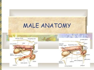
Reproductive System


     VTT 235/245
MALE ANATOMY
Structures
 Testes-
      Male gonad that produces both testosterone
       and germ cells (which become sperm).
      Contained in the scrotum.
 Scrotum- pouch containing the testicles
  and epididymis.
 Seminiferous Tubules-
      Hollow structures where germ cells
       differentiate into spermatozoa.
Structures
 Epididymis-
      Structure adjacent to the testicle.
      3 parts: head, body, and tail.
      Spermatozoa mature in the head and body of
       the epididymis.
 Ductus Deferens (Vas Deferens)-
      The continuation of the epididymal duct at
       the tail of the epididymis.
      It travels up the spermatic cord and through
       the inguinal canal to reach the abdomen.
Structures
 The Spermatic Cord consists of-
   Vas deferens
   Testicular artery, vein, nerve, and
    lymphatics
Accessory Sex Glands
 Prostate
 Seminal vesicles
 Bulbourethral glands
Penis
 The male copulatory organ.
 Provides a passage way for semen
  and urine to the outside of the body.
 Prepuce- the cutaneous sheath
  around the free part of the penis
  when it is not erect.
 Preputial Orifice- the external
  opening of the prepuce to the
  outside environment.
Penis
 Contains the glans penis (head of the
  penis)
 Bulbus Glandis- the caudal part of
  the penis.
   Swells to lock the male into the female
    during copulation.
 +/- Os penis
MALE PHYSIOLOGY
Testosterone
 Produced by the testes.
 Responsible for secondary sex
  characteristics and sex drive.
 An androgen or anabolic steroid.
 Production is stimulated by LH.
Sperm
 Spermatogenesis is stimulated by FSH.
      Head-
        Contains the nucleus and haploid chromosomes.
        Acrosome- a “cap” which contains enzymes to
         permit penetration into the ovum.
      Midpiece-
        “Power plant”
        Numerous mitochondria carry-out metabolism
         that provides ATP for sperm locomotion.
      Tail- consists of flagellum for propulsion.
Seminal Fluid
 Produced by accessory sex organs.
 The medium for survival of the
  sperm.
 Prostatic secretion- alkalinizes the
  vaginal environment to prevent sperm
  death.
MALE PATHOLOGY
Prostatic Disease
   Common in dogs
   Benign Prostatic Hyperplasia
   Prostatic adenocarcinoma
   Bacterial
   All cause enlargement or
    inflammation
Orchitis & Epididymitis
 Acute-
 Caused by trauma, infection, or
  testicular torsion
 Chronic-
 Immune-mediated or neoplastic
 Testicular atrophy and fibrosis
Phimosis
 The inability to extrude the penis
  through an abnormally small preputial
  orifice
 Congenital or it develops due to
  inflammation, neoplasia, edema, or
  fibrosis after trauma, irritation or
  infection
Paraphimosis
 The inability to completely retract
  the penis
 Usually occurs after an erection
 The preputial orifice skin becomes
  inverted and impairs venous drainage
 A medical emergency!!!
Pathologies…
 Inguinal Hernia-
   The protrusion of a loop of organ or
    tissue through the inguinal canal.
 Cryptorchidism-
   Failure of one or both testicles to
    descend into the scrotum.
   The retained testicle can be anywhere
    between the scrotum and the caudal
    pole of the kidney.
FEMALE ANATOMY


    Structures
Structures
 Ovaries
 Oviducts (uterine tubes)
 Uterus- horns and body
 Cervix- a heavy, smooth muscle sphincter
  that is kept tightly closed except during
  estrus and parturition.
 Vagina- glandless mucosa located within
  the pelvic canal.
 Vulva- consists of the vestibule and labia.
Ovaries
 Ovaries- both endocrine (hormone
  producing) and cytogenic (cell
  producing).
   Medulla- vascular center of the ovary.
   Cortex- where follicles can be found,
    both developing and atrophying.
   Functions-
      To produce ova or eggs ready for
       fertilization.
      Acts as an endocrine gland.
Oviducts
 Oviduct- the open end of the uterine
  tube (fallopian tube)
 Functions-
   Collects ova as they are released.
   Conveys ova from the ovaries to the
    uterine horns.
 Infundibulum- funnel-shaped ovarian
  end of the oviduct.
Uterus
 Highly expandable, tubular organ
  where the embryo/fetus develops.
 A hollow structure with 3 parts-
  neck (where the cervix is located),
  body, and horns.
 Function-
    Provides a receptacle for embryos to
     develop.
    Provides nutrients via the PLACENTA.
Uterus
 Uterine Walls- 3 layers
Vagina
 The part of the reproductive tract
  between the cervix and the vulva.
 Along with the vestibule and vulva, it
  is the females copulatory organ and
  birth canal.
 The hymen is the poorly developed,
  vestigial, mucosal folds at the
  junction of the vagina and vestibule.
Other Structures…
 Vulva- the external orifice that
  terminates the genital tract.
 Labia- the Ⓡ and Ⓛ lips of the vulva.
FEMALE PHYSIOLOGY
Types
 Monestrous- usually one cycle per year,
  usually seasonal breeders. (mink)
 Polyestrous- more than one cycle per year,
  continuous. (swine)
 Seasonally Polyestrous- cycles
  continuously in specific seasons.
 Induced Ovulators- requires copulation to
  ovulate.
 Spontaneous Ovulators- ovulation occurs
  naturally, with or without copulation.
Estrous Cycle
 The onset of the estrous cycle begins at
  puberty.
 The purpose is to prepare the uterus to
  receive fertilized ovum.
 Sexual maturity brings about-
      ovarian development, which includes the
       production of ova,
      ovulation,
      and the production of the corpus luteum.
 The estrous cycle is under the control of
  hormones produced by the ovaries and the
  pituitary gland.
 Animals do not undergo menopause.
Estrous Cycle
 At the beginning of each cycle, ova within
  the follicles in the ovaries begin to
  develop.
 One or more follicles (depending on the
  species) continue to develop until they
  reach a ripened follicle
 One or more follicles rupture, (ovulation,
  usually occurs during estrus.)
 Then the ovum is expelled from the ovary
  to the oviduct (uterine tube).
Estrous Cycle
 The ruptured follicle grows larger, filling
  with a yellow, lipoid material and becomes
  the CORPUS LUTEUM (“yellow body”).
 The corpus luteum secretes progesterone.
 If fertilization occurs, the corpus luteum
  continues to secrete progesterone and
  prevents future estrous cycles during
  pregnancy.
Estrous Cycle
 Without fertilization, the corpus
  luteum and its secretions diminish,
  forming a CORPUS ALBICANS
  (“white body”).
 The reduced levels of hormone
  production lead to a new estrous
  cycle.
Stages of the Estrous Cycle
1. Proestrus
 Period of preparation.
 **FSH & LH cause the development
  of the follicle.
 The follicle starts producing
  ESTROGEN.
   Estrogen stimulates the vagina and
    uterus for copulation and pregnancy.
2. Estrus
 Period of female sexual receptivity.
 Uterus and uterine horns are ready
  to receive an embryo.
 Release of LH causes ovulation.
 Dogs may have bloody discharge, cats
  may exhibit behavioral changes.
3. Diestrus & Metestrus
 Post-ovulating phase.
 Each ruptured follicle develops into a
  corpus luteum (CL).
    The CL starts to secrete
     PROGESTERONE which inhibits the
     development of new follicles.
    The CL is also responsible for
     maintaining the uterine lining to
     support the fetus during pregnancy.
3. Diestrus & Metestrus
 If pregnancy does not occur, the CL
  degenerates.
 If pregnancy occurs, the corpus luteum is
  maintained and continues to secrete
  hormones for:
      The entire pregnancy or,
      Until the placenta develops.
        Depends on the species.
4. Anestrus
 Periods of no estrous cycles
     a. Pregnancy
     b. Nursing
     c. Season of year
     d. Poor Nutrition
     e. Pathological Conditions
PREGNANCY
Gestation Periods
   **Dog-               Pig-
       57-63 days              114 days
   **Cat-               Sheep & Goats-
       65 days                 150 days
   Horse-               Mice-
       330 days                19-21 days
   Cow-                 Rats-
       283 days                21-23 days
   Rabbits-             Hamsters-
       30-33 days              15-18 days
   Guinea pigs-         Gerbils-
       59-72 days              23-26 days
Terms
 Gestation- the interval between
  fertilization of the ovum and the birth of
  the offspring.
 Mitosis- cell division, one cell divides into
  2, 2 into 4…
 Zygote- fertilized ovum
 Embryo- stage at which major organs are
  developing.
 Fetus- stage where formation of major
  internal and external structures is
  complete until the time of parturition.
Fertilization & Cell Division
 Ova enter the infundibulum and are
  transported down by muscular
  contractions.
 Sperm travels up the female tract and
  fertilization takes place in the upper part
  of the uterine tube.
 Each ovum is penetrated by one sperm
  which results in a fertilization reaction
  (preventing fertilization by any other
  sperm).
 The fertilized ovum is now a zygote, and
  cell division begins via mitosis.
The Placenta
 A membranous structure that obtains
  nutrients and oxygen from the mother to
  deliver to the fetus.
 Attaches to the endometrial lining of the
  uterus.
      Chorion- outer layer in contact with the
       maternal uterus.
      Amnion- innermost membrane closest to the
       fetus.
      Amnionic Sac- sac in which the fetus is
       located.
Hormones
 Oxytocin-
   **Produced by the Posterior pituitary
   Stimulates milk let-down.
   In the presence of Estrogen, it
    stimulates uterine contractions during
    parturition.
   Stimulates the oviducts to help move
    spermatozoa.
Hormones
 Prolactin-
   **From the Anterior pituitary
   Helps maintain the CL during pregnancy.
   Stimulates the mammary glands to fill
    with milk at parturition.
   Stimulates the replenishment of milk via
    neonatal suckling.
FEMALE PATHOLOGY
Uterine Infection
 Infection of the uterus.
   Endometritis- inflammation of the
    endometrium.
   Metritis- inflammation of all layers.
   Pyometra- accumulation of pus in the
    uterus.
Pyometra
 A hormonally mediated disorder.
 An abnormal uterine endometrium
  combined with a secondary bacterial
  infection.
 Often occurs when progestational
  compounds are administered to delay
  or suppress estrus.
Uterine Prolapse
 The turning inside-out of the uterus
  and vagina causing it to project
  through the vulva.
 Most common in the cow and sow.
 The prolapsed uterus can often be
  pushed back in and sutured in place
  until it heals.
Pseudocyesis
 False pregnancy
 Common in dogs
 Occurs at the end of diestrus,
  characterized by hyperplasia of the
  mammary glands, lactation, and
  behavioral changes
 Falling progesterone and increasing
  prolactin are believed to be the
  cause
LABORATORY ANALYSIS


   VAGINAL CYTOLOGY
     Hendrix p. 327
Anestrus
 Predominantly
  non-cornified
  squamous
  epithelial cells
      Lg nucleus
       and rounded
       edges
Proestrus
 Above- early
  proestrus, below-
  late proestrus
 Cornified
  squamous
  epithelial cells
 Angular with
  jagged borders
 Segs(neutraphils)
  decrease, RBC’s
  increase
Estrus
 All squamous cells
  are cornified
 Segs- absent,
  RBC’s present
Diestrus
 Non-cornified
  squamous and
  abundant cytologic
  debris
 Segs increase,
  RBC’s are absent
 Similar to anestrus
LABORATORY ANALYSIS


    SEMEN ANALYSIS
Semen Collection
Semen Analysis
 Sample Handling-
   Avoid exposure to marked changes in
    temperature
 Supplies-
   Slides, coverslips, pipettes, stains and
    diluents
Semen Analysis

 Evaluation-             Morphology-
      Volume                 Head
      Appearance             Midpiece
      Motility               Tail
      Concentration
      Live:Dead Ratio
Primary Abnormalities
Secondary Abnormalities
Semen Analysis
The End!!
Lp 16a reproductive system

Lp 16a reproductive system

  • 1.
  • 2.
  • 3.
    Structures  Testes-  Male gonad that produces both testosterone and germ cells (which become sperm).  Contained in the scrotum.  Scrotum- pouch containing the testicles and epididymis.  Seminiferous Tubules-  Hollow structures where germ cells differentiate into spermatozoa.
  • 4.
    Structures  Epididymis-  Structure adjacent to the testicle.  3 parts: head, body, and tail.  Spermatozoa mature in the head and body of the epididymis.  Ductus Deferens (Vas Deferens)-  The continuation of the epididymal duct at the tail of the epididymis.  It travels up the spermatic cord and through the inguinal canal to reach the abdomen.
  • 6.
    Structures  The SpermaticCord consists of-  Vas deferens  Testicular artery, vein, nerve, and lymphatics
  • 7.
    Accessory Sex Glands Prostate  Seminal vesicles  Bulbourethral glands
  • 8.
    Penis  The malecopulatory organ.  Provides a passage way for semen and urine to the outside of the body.  Prepuce- the cutaneous sheath around the free part of the penis when it is not erect.  Preputial Orifice- the external opening of the prepuce to the outside environment.
  • 9.
    Penis  Contains theglans penis (head of the penis)  Bulbus Glandis- the caudal part of the penis.  Swells to lock the male into the female during copulation.  +/- Os penis
  • 10.
  • 11.
    Testosterone  Produced bythe testes.  Responsible for secondary sex characteristics and sex drive.  An androgen or anabolic steroid.  Production is stimulated by LH.
  • 12.
    Sperm  Spermatogenesis isstimulated by FSH.  Head-  Contains the nucleus and haploid chromosomes.  Acrosome- a “cap” which contains enzymes to permit penetration into the ovum.  Midpiece-  “Power plant”  Numerous mitochondria carry-out metabolism that provides ATP for sperm locomotion.  Tail- consists of flagellum for propulsion.
  • 13.
    Seminal Fluid  Producedby accessory sex organs.  The medium for survival of the sperm.  Prostatic secretion- alkalinizes the vaginal environment to prevent sperm death.
  • 14.
  • 15.
    Prostatic Disease  Common in dogs  Benign Prostatic Hyperplasia  Prostatic adenocarcinoma  Bacterial  All cause enlargement or inflammation
  • 16.
    Orchitis & Epididymitis Acute-  Caused by trauma, infection, or testicular torsion  Chronic-  Immune-mediated or neoplastic  Testicular atrophy and fibrosis
  • 17.
    Phimosis  The inabilityto extrude the penis through an abnormally small preputial orifice  Congenital or it develops due to inflammation, neoplasia, edema, or fibrosis after trauma, irritation or infection
  • 18.
    Paraphimosis  The inabilityto completely retract the penis  Usually occurs after an erection  The preputial orifice skin becomes inverted and impairs venous drainage  A medical emergency!!!
  • 19.
    Pathologies…  Inguinal Hernia-  The protrusion of a loop of organ or tissue through the inguinal canal.  Cryptorchidism-  Failure of one or both testicles to descend into the scrotum.  The retained testicle can be anywhere between the scrotum and the caudal pole of the kidney.
  • 20.
    FEMALE ANATOMY Structures
  • 21.
    Structures  Ovaries  Oviducts(uterine tubes)  Uterus- horns and body  Cervix- a heavy, smooth muscle sphincter that is kept tightly closed except during estrus and parturition.  Vagina- glandless mucosa located within the pelvic canal.  Vulva- consists of the vestibule and labia.
  • 23.
    Ovaries  Ovaries- bothendocrine (hormone producing) and cytogenic (cell producing).  Medulla- vascular center of the ovary.  Cortex- where follicles can be found, both developing and atrophying.  Functions-  To produce ova or eggs ready for fertilization.  Acts as an endocrine gland.
  • 24.
    Oviducts  Oviduct- theopen end of the uterine tube (fallopian tube)  Functions-  Collects ova as they are released.  Conveys ova from the ovaries to the uterine horns.  Infundibulum- funnel-shaped ovarian end of the oviduct.
  • 26.
    Uterus  Highly expandable,tubular organ where the embryo/fetus develops.  A hollow structure with 3 parts- neck (where the cervix is located), body, and horns.  Function-  Provides a receptacle for embryos to develop.  Provides nutrients via the PLACENTA.
  • 27.
  • 28.
    Vagina  The partof the reproductive tract between the cervix and the vulva.  Along with the vestibule and vulva, it is the females copulatory organ and birth canal.  The hymen is the poorly developed, vestigial, mucosal folds at the junction of the vagina and vestibule.
  • 29.
    Other Structures…  Vulva-the external orifice that terminates the genital tract.  Labia- the Ⓡ and Ⓛ lips of the vulva.
  • 30.
  • 31.
    Types  Monestrous- usuallyone cycle per year, usually seasonal breeders. (mink)  Polyestrous- more than one cycle per year, continuous. (swine)  Seasonally Polyestrous- cycles continuously in specific seasons.  Induced Ovulators- requires copulation to ovulate.  Spontaneous Ovulators- ovulation occurs naturally, with or without copulation.
  • 32.
    Estrous Cycle  Theonset of the estrous cycle begins at puberty.  The purpose is to prepare the uterus to receive fertilized ovum.  Sexual maturity brings about-  ovarian development, which includes the production of ova,  ovulation,  and the production of the corpus luteum.  The estrous cycle is under the control of hormones produced by the ovaries and the pituitary gland.  Animals do not undergo menopause.
  • 33.
    Estrous Cycle  Atthe beginning of each cycle, ova within the follicles in the ovaries begin to develop.  One or more follicles (depending on the species) continue to develop until they reach a ripened follicle  One or more follicles rupture, (ovulation, usually occurs during estrus.)  Then the ovum is expelled from the ovary to the oviduct (uterine tube).
  • 34.
    Estrous Cycle  Theruptured follicle grows larger, filling with a yellow, lipoid material and becomes the CORPUS LUTEUM (“yellow body”).  The corpus luteum secretes progesterone.  If fertilization occurs, the corpus luteum continues to secrete progesterone and prevents future estrous cycles during pregnancy.
  • 35.
    Estrous Cycle  Withoutfertilization, the corpus luteum and its secretions diminish, forming a CORPUS ALBICANS (“white body”).  The reduced levels of hormone production lead to a new estrous cycle.
  • 36.
    Stages of theEstrous Cycle
  • 37.
    1. Proestrus  Periodof preparation.  **FSH & LH cause the development of the follicle.  The follicle starts producing ESTROGEN.  Estrogen stimulates the vagina and uterus for copulation and pregnancy.
  • 38.
    2. Estrus  Periodof female sexual receptivity.  Uterus and uterine horns are ready to receive an embryo.  Release of LH causes ovulation.  Dogs may have bloody discharge, cats may exhibit behavioral changes.
  • 39.
    3. Diestrus &Metestrus  Post-ovulating phase.  Each ruptured follicle develops into a corpus luteum (CL).  The CL starts to secrete PROGESTERONE which inhibits the development of new follicles.  The CL is also responsible for maintaining the uterine lining to support the fetus during pregnancy.
  • 40.
    3. Diestrus &Metestrus  If pregnancy does not occur, the CL degenerates.  If pregnancy occurs, the corpus luteum is maintained and continues to secrete hormones for:  The entire pregnancy or,  Until the placenta develops.  Depends on the species.
  • 41.
    4. Anestrus  Periodsof no estrous cycles  a. Pregnancy  b. Nursing  c. Season of year  d. Poor Nutrition  e. Pathological Conditions
  • 42.
  • 43.
    Gestation Periods  **Dog-  Pig-  57-63 days  114 days  **Cat-  Sheep & Goats-  65 days  150 days  Horse-  Mice-  330 days  19-21 days  Cow-  Rats-  283 days  21-23 days  Rabbits-  Hamsters-  30-33 days  15-18 days  Guinea pigs-  Gerbils-  59-72 days  23-26 days
  • 44.
    Terms  Gestation- theinterval between fertilization of the ovum and the birth of the offspring.  Mitosis- cell division, one cell divides into 2, 2 into 4…  Zygote- fertilized ovum  Embryo- stage at which major organs are developing.  Fetus- stage where formation of major internal and external structures is complete until the time of parturition.
  • 45.
    Fertilization & CellDivision  Ova enter the infundibulum and are transported down by muscular contractions.  Sperm travels up the female tract and fertilization takes place in the upper part of the uterine tube.  Each ovum is penetrated by one sperm which results in a fertilization reaction (preventing fertilization by any other sperm).  The fertilized ovum is now a zygote, and cell division begins via mitosis.
  • 46.
    The Placenta  Amembranous structure that obtains nutrients and oxygen from the mother to deliver to the fetus.  Attaches to the endometrial lining of the uterus.  Chorion- outer layer in contact with the maternal uterus.  Amnion- innermost membrane closest to the fetus.  Amnionic Sac- sac in which the fetus is located.
  • 47.
    Hormones  Oxytocin-  **Produced by the Posterior pituitary  Stimulates milk let-down.  In the presence of Estrogen, it stimulates uterine contractions during parturition.  Stimulates the oviducts to help move spermatozoa.
  • 48.
    Hormones  Prolactin-  **From the Anterior pituitary  Helps maintain the CL during pregnancy.  Stimulates the mammary glands to fill with milk at parturition.  Stimulates the replenishment of milk via neonatal suckling.
  • 49.
  • 50.
    Uterine Infection  Infectionof the uterus.  Endometritis- inflammation of the endometrium.  Metritis- inflammation of all layers.  Pyometra- accumulation of pus in the uterus.
  • 51.
    Pyometra  A hormonallymediated disorder.  An abnormal uterine endometrium combined with a secondary bacterial infection.  Often occurs when progestational compounds are administered to delay or suppress estrus.
  • 52.
    Uterine Prolapse  Theturning inside-out of the uterus and vagina causing it to project through the vulva.  Most common in the cow and sow.  The prolapsed uterus can often be pushed back in and sutured in place until it heals.
  • 53.
    Pseudocyesis  False pregnancy Common in dogs  Occurs at the end of diestrus, characterized by hyperplasia of the mammary glands, lactation, and behavioral changes  Falling progesterone and increasing prolactin are believed to be the cause
  • 54.
    LABORATORY ANALYSIS VAGINAL CYTOLOGY Hendrix p. 327
  • 55.
    Anestrus  Predominantly non-cornified squamous epithelial cells  Lg nucleus and rounded edges
  • 56.
    Proestrus  Above- early proestrus, below- late proestrus  Cornified squamous epithelial cells  Angular with jagged borders  Segs(neutraphils) decrease, RBC’s increase
  • 57.
    Estrus  All squamouscells are cornified  Segs- absent, RBC’s present
  • 58.
    Diestrus  Non-cornified squamous and abundant cytologic debris  Segs increase, RBC’s are absent  Similar to anestrus
  • 59.
    LABORATORY ANALYSIS SEMEN ANALYSIS
  • 60.
  • 61.
    Semen Analysis  SampleHandling-  Avoid exposure to marked changes in temperature  Supplies-  Slides, coverslips, pipettes, stains and diluents
  • 62.
    Semen Analysis  Evaluation-  Morphology-  Volume  Head  Appearance  Midpiece  Motility  Tail  Concentration  Live:Dead Ratio
  • 63.
  • 64.
  • 65.
  • 66.
  • 67.

Editor's Notes

  • #19 Start note cards here!!
  • #42 Know order
  • #55 DON’T NEED TO KNOW PICTURES