Downloaded 273 times

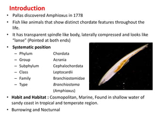


The document provides a detailed overview of the anatomical and physiological features of amphioxus, a marine organism that exhibits chordate characteristics. It covers aspects such as its structure, including the digestive and circulatory systems, sense organs, and excretory mechanisms. The study highlights the organism's unique features, such as its lack of traditional eyes, presence of myotomes, and adaptation to a burrowing lifestyle.


![The animal kingdom.pptxrb[1]](https://cdn.slidesharecdn.com/ss_thumbnails/theanimalkingdom-pptxrb1-121031071444-phpapp02-thumbnail.jpg?width=640&height=640&fit=bounds)


![谷歌留痕技术 [ 𝙩𝙤𝙥 𝟮𝟯𝟯. 𝙘 𝙤𝙢 ]](https://cdn.slidesharecdn.com/ss_thumbnails/top233-260130174328-3833018c-thumbnail.jpg?width=640&height=640&fit=bounds)


