4
The discovery ofGolgi apparatus
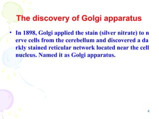
• In 1898, Golgi applied the stain (silver nitrate) to n
erve cells from the cerebellum and discovered a da
rkly stained reticular network located near the cell
nucleus. Named it as Golgi apparatus.
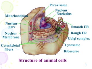
6
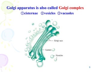
• The Golgiapparatus is usually located near th
e cell nucleus. It consists of a collection of flatt
ened, membrane-bounded sacs (cisternae, sing
ular cisternae), which are piled like stacks of p
lates. Each stack contains three to twenty ciste
rnae.
• The number of Golgi stacks per cell varies gre
atly depending on the cell type: some cells con
tain one large stack, while other contain hund
ereds of very small ones.
I. General organization
8
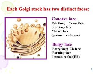
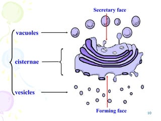
Each Golgi stackhas two distinct faces:
Bulgy face
Entry face; Cis face
Forming face
Immature face(ER)
Concave face
Exit face; Trans face
Secretary face
Mature face
(plasma membrane)
11
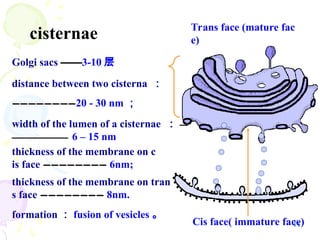
cisternae
Golgi sacs ——
——3-10层
distance between two cisterna :
————————
————————20 - 30 nm ;
Cis face( immature face)
thickness of the membrane on c
is face ————————
———————— 6nm;
formation : fusion of vesicles 。
thickness of the membrane on tran
s face ————————
———————— 8nm.
width of the lumen of a cisternae : —
—
———————
——————— 6 – 15 nm
Trans face (mature fac
e)
12.
12
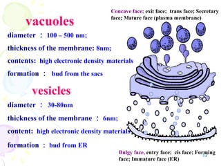
vesicles
diameter : 30-80nm
thicknessof the membrane : 6nm;
content: high electronic density materials
formation : bud from ER
diameter : 100 – 500 nm;
thickness of the membrane: 8nm;
contents: high electronic density materials
formation : bud from the sacs
vacuoles
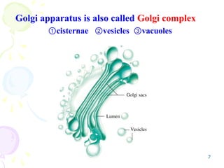
Bulgy face, entry face; cis face; Forming
face; Immature face (ER)
Concave face; exit face; trans face; Secretary
face; Mature face (plasma membrane)
13.
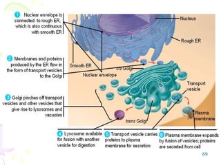
13
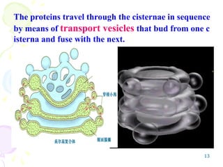
The proteins travelthrough the cisternae in sequence
by means of transport vesicles that bud from one c
isterna and fuse with the next.
14.
14
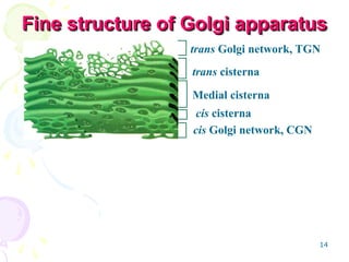
Fine structure ofGolgi apparatus
Fine structure of Golgi apparatus
trans Golgi network, TGN
trans cisterna
Medial cisterna
cis cisterna
cis Golgi network, CGN
15.
15
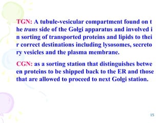
TGN: A tubule-vesicularcompartment found on t
he trans side of the Golgi apparatus and involved i
n sorting of transported proteins and lipids to thei
r correct destinations including lysosomes, secreto
ry vesicles and the plasma membrane.
CGN: as a sorting station that distinguishes betwe
en proteins to be shipped back to the ER and those
that are allowed to proceed to next Golgi station.
16.
16
• Both thecis and trans network are thought to be i
mportant for protein sorting:
• Proteins entering the cis Golgi network can either
move onward through the Golgi stack or, if they c
ontain an ER retetion signal, be returned to the E
R;
• Proteins exiting the trans Golgi network are sorted
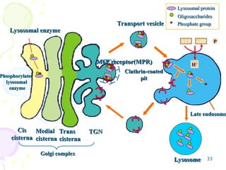
according to whether they are destined for lysoso
mes or for the cell surface.
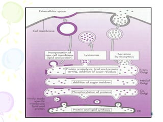
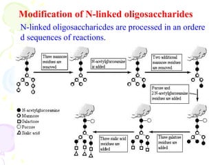
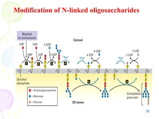
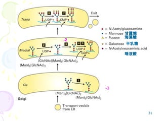
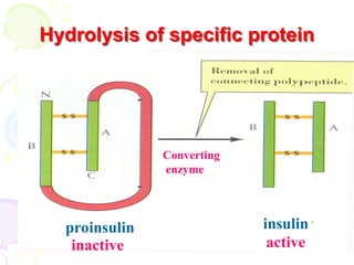
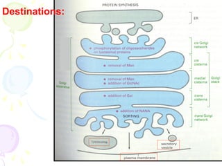
21
• The proteinsprocessed by the Golgi are gly
coproteins (they have N-linked polysacchari
ds attached to them). The oligosaccharids w
ere added in the ER.
• These carbohydrate groups are modified in
the Golgi (removal and addition of carbohy
drate).
*Modification
22.
22
• Because theoligosaccharides are added on t
he luminal side of the ER and Golgi, their di
stribution is asymmetrical.
• As a result,
the oligosaccharides of all membrane-associa
ted glycoproteins and lipoproteins face the
lumen of intracellular membrane
those of the plasma membrane (because of ex
ocytosis) face the outside of the cell.
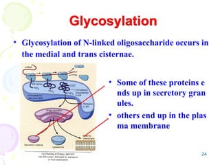
24
• Glycosylation ofN-linked oligosaccharide occurs in
the medial and trans cisternae.
Glycosylation
• Some of these proteins e
nds up in secretory gran
ules.
• others end up in the plas
ma membrane
25.
25
Protein processing withinthe Golgi involves t
he modification and synthesis of the carbohyd
rate portions of glycoproteins.
One of the major aspects of this processing is
the modification of the N-linked oligosacchari
des that were added to proteins in the ER.
Glycosylation
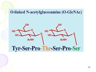
27
• O-linked glycosylation
refersto the connection through the hydrox
yl group of threonine, serine,or hydroxylysi
ne (OH).
• O-linked glycosylation begins in Golgi appa
ratus.
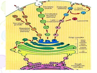
41
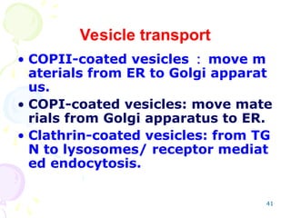
Vesicle transport
• COPII-coatedvesicles : move m
aterials from ER to Golgi apparat
us.
• COPI-coated vesicles: move mate
rials from Golgi apparatus to ER.
• Clathrin-coated vesicles: from TG
N to lysosomes/ receptor mediat
ed endocytosis.
44
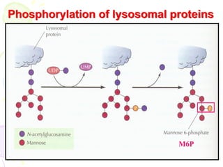
Questions
1. What’s thebasic conformation of G.C.?
2. What’s the main chemical components
in G.C.?
3. What’s the main functions of G.C.?
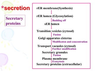
4. Describe briefly the secreting process
of secretory proteins? ( Flow chart )
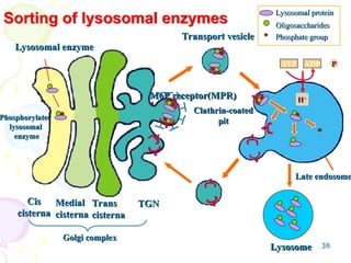
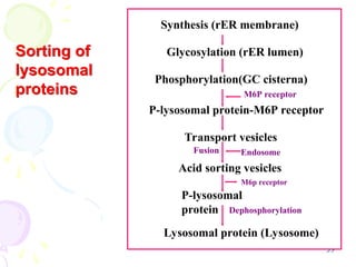
5. Taking lysosomal protein as an example,
explain the sorting function of G.C.
( Flow chart )
57
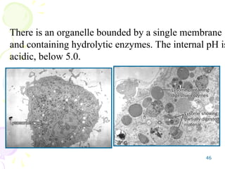
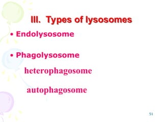
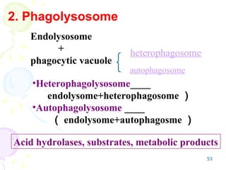
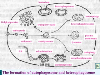
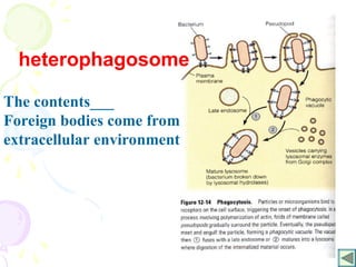
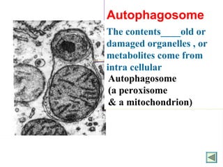
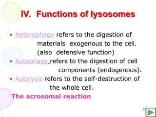
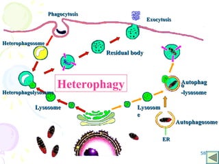
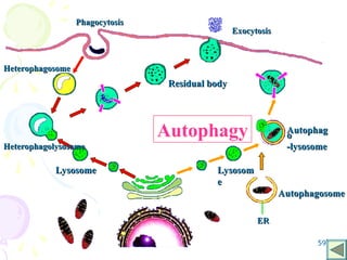
lV. Functions oflysosomes
• Heterophagy refers to the digestion of
materials exogenous to the cell.
(also defensive function)
• Autophagy refers to the digestion of cell
components (endogenous).
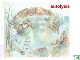
• Autolysis refers to the self-destruction of
the whole cell.
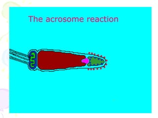
The acrosomal reaction
67
Questions
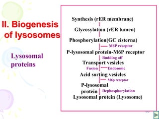
1. Tell thebiogenesis of lysosomes ( flow chart ) .
2. What’s the main type of lysosome? And what’s t
he main difference between these types?
3. What’s the main type of phagolysosome? And w
hat’s the main difference between these types?
4. What’s the main function of lysosome?