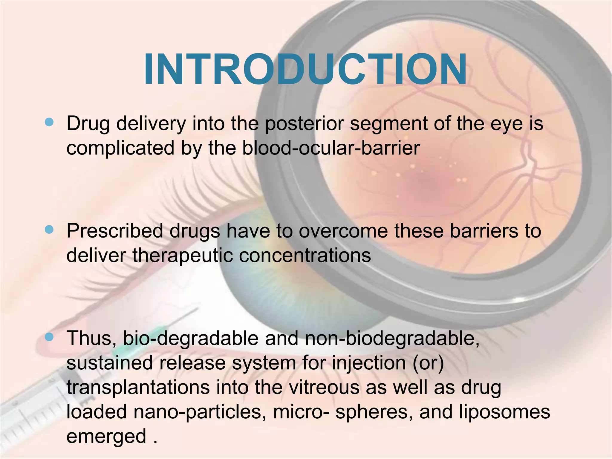
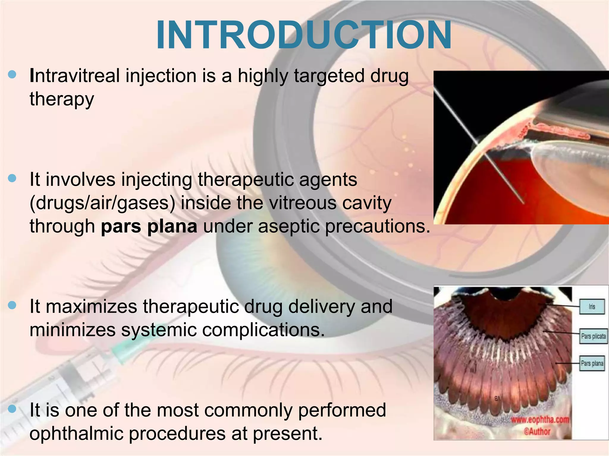
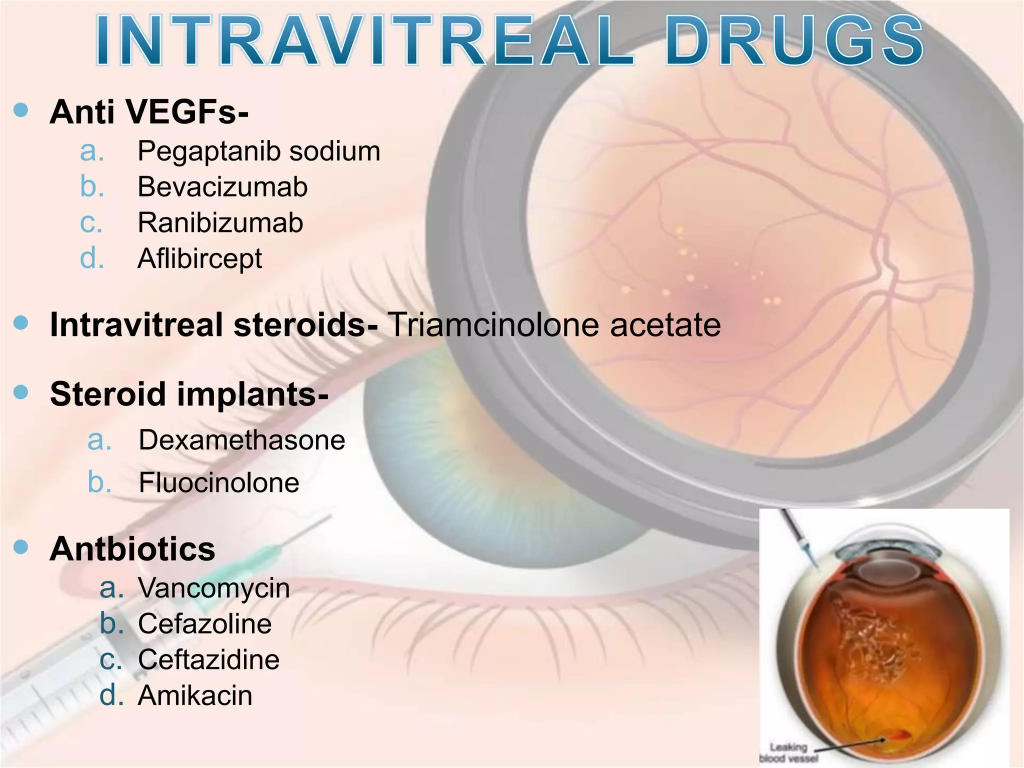
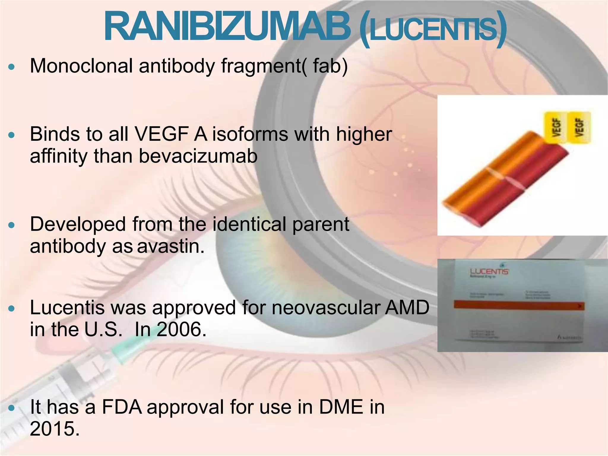
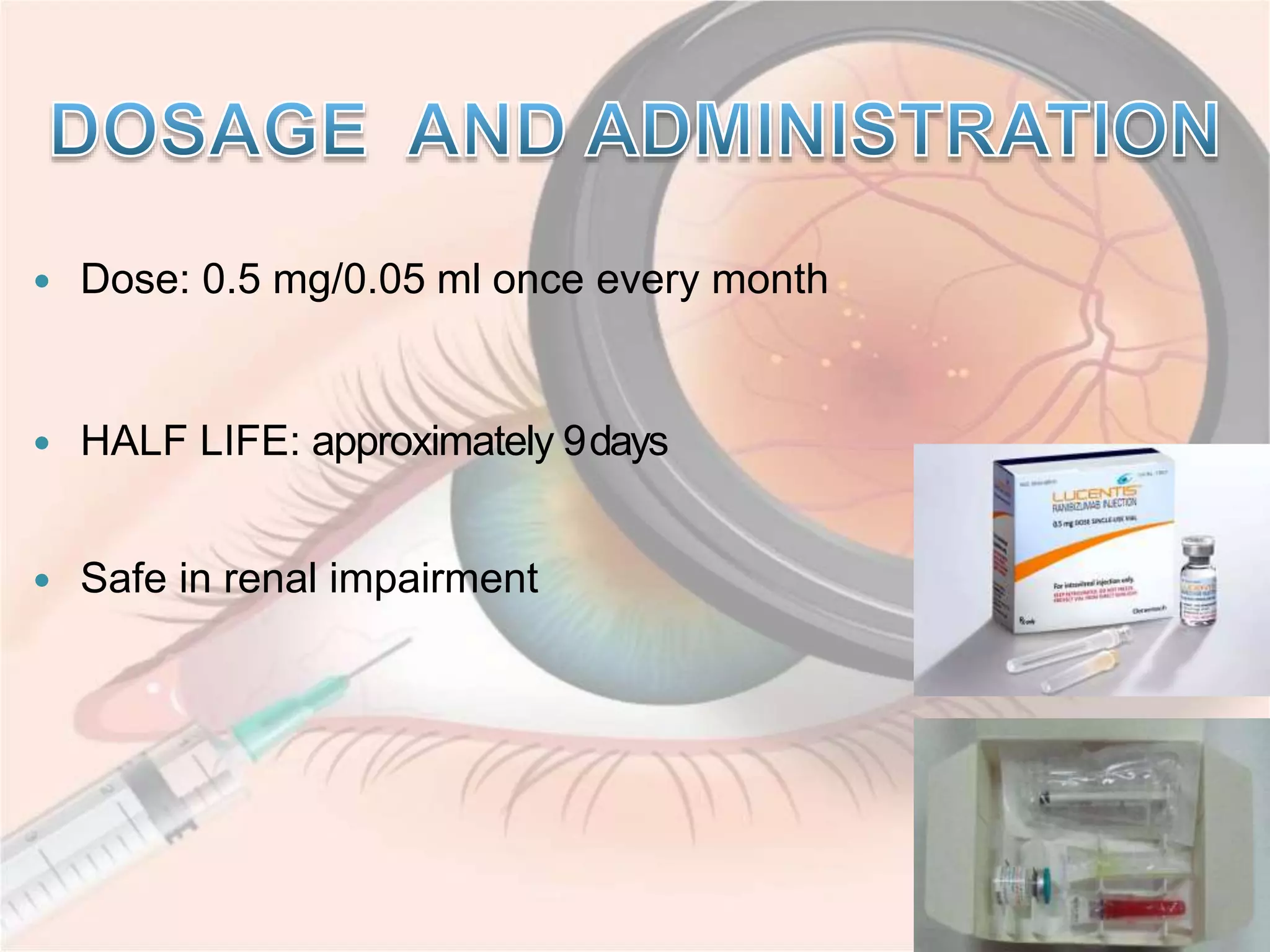
Dr . MAHESHWARI S
INTRODUCTION
 Drug delivery into the posterior segment of the eye is
complicated by the blood-ocular-barrier
 Prescribed drugs have to overcome these barriers to
deliver therapeutic concentrations
 Thus, bio-degradable and non-biodegradable,
sustained release system for injection (or)
transplantations into the vitreous as well as drug
loaded nano-particles, micro- spheres, and liposomes
emerged .
INTRODUCTION
 Intravitreal injection is a highly targeted drug
therapy
 It involves injecting therapeutic agents
(drugs/air/gases) inside the vitreous cavity
through pars plana under aseptic precautions.
 It maximizes therapeutic drug delivery and
minimizes systemic complications.
 It is one of the most commonly performed
ophthalmic procedures at present.
HISTORY
 1911- Intravitreal injection was first described by
Deutschmann and Ohm. First used intravitreal injection was
air bubble to tamponade a retinal detachment.
 Triamcinolone acetonide (Kenalog) became the first
intravitreal drug with widespread application, used as a
treatment for macular edema associated with a variety of
etiologies
 1944- Von Sallmann first demonstrated efficasy of
intravitreal antibiotic injection for the treatment of
endophthalmitis
 1970-1974- PEYMAN et al heralded the use of
intravitreal antibiotics for the successful treatment of
bacterial and fungal endophthalmitis infection and
determined the non-toxic doses of various intravitreal
antibiotics.
 Anti VEGFs-
a. Pegaptanib sodium
b. Bevacizumab
c. Ranibizumab
d. Aflibircept
 Intravitreal steroids- Triamcinolone acetate
 Steroid implants-
a. Dexamethasone
b. Fluocinolone
 Antbiotics
a. Vancomycin
b. Cefazoline
c. Ceftazidine
d. Amikacin
 ANTIVIRALS
a. Ganciclovir
b. Foscarnet
c. Cidofovir
 ANTIFUNGALS
a. Amphotericine B
b. Fluconazole
c. Voriconazole
 AIR AND EXPANSILE GASES
a. Sulfur hexafluoride(SF6)
b. Perfluoropropane (C3F8)
c. Hexafluoroethane(C2F6)
 ANTI METABOLITES
a. Melphalan
b. Topotecan
c. Methotrexate
INDICATIONS
 ANTI VEGF
ARMD
Diabetic Retinopathy
Vascular Occlusion
Retinopathy of Prematurity
Neovascular Glaucoma
IPCV, Coats Disease etc
 ANTIBIOTICS AND ANTI FUNGALS- Endophthalmitis
 ANTI VIRALS – Viral retinitis in immunocompromised
patients
 ANTI METABOLITES- Chemotherapy
 STEROIDS- Macular edema, uveitis
PROCEDURE
INFORMED CONSENT
 Specific mention of the off-label use.
 Anti-VEGF agents- specific mentioning data regarding
thromboembolic events, particularly in high-risk individuals.
 Triamcinolone acetonide - consent should specifically
mention the increased risk of cataract formation, as well as
ocular hypertension or glaucoma.
ANESTHESIA
 Should be given prior to application of a sterile
preparation
 Topical medications such as proparacaine 0.5% drops
or lidocaine jelly to achieve adequate anesthesia
 If the patient has a low threshold for pain,
subconjunctival lidocaine can be administered
adjunctively
 Rarely, peribulbar , retrobulbar or general anesthesia is
required
ASEPSIS
 Several precautions are strongly recommended
to minimize the risk of infection.
 The instruments should be removed from sterile
packaging and dropped onto a sterile field.
 Take a procedural time-out to verify patient,
agent and laterality (2014)
 Once anesthesia has been applied, a 5%
povidone-iodine solution should be applied to the
ocular surface. It is the last agent applied to the
intended injection site before injection. (2014)
Bactericidal with rapid cytotoxicity
 The surgeon should have proper scrubbing as a
part of universal precautions.( 2014)
 Either surgical masks should be used or both the
patient and providers should minimize speaking
during the injection preparation and procedure to
limit aerosolized droplets containing oral
contaminants from the patient and/or provider
(2014)
 Once gloved( sterile/unsterile), the surgeon
applies a fenestrated lid drape similar to those
used in cataract surgery.
 There is no evidence to support the routine use of
a sterile drape.(2014)
 A lid speculum is then applied in
order to retract the eyelid and prevent
the patient from blinking.
 Avoid contamination of the needle
and injection site by the eyelashes or
the eyelid margins.
 Avoid extensive massage of the
eyelids either pre- or post-injection (to
avoid meibomian gland expression).
 Needle size varies according to the substance
injected:
 27-gauge needles used for crystalline
substances such as triamcinolone acetonide
 30-gauge needles commonly used for
the anti-VEGF agents.
 Needle length between ½ - 5/8 inches (12.7 to 15.75 mm) is
recommended
 Separate needles should be used to remove the medication
from the vial and to perform the actual injection. This helps
prevent both contamination and dulling of the needle.
 Studies suggest that smaller, sharper needles require less
force for penetration and result in less drug reflux.
 Longer needles may increase risk of retinal injury if the patient
accidentally moves forward during the procedure
 Any clock hour of the eye can be used
 Injection in the inferotemporal quadrant is
common
AGE INJECTION SITE FROM LIMBUS
1-6 months 1.5mm
6 months – 1 year 2mm
1-2year 2.5mm
2-6years 3mm
>6years 3-3.5mm in pseudophakic/aphakic eyes
3.5 – 4 mm in phakic eyes
NEEDLE INTRODUCTION AND INJECTION
 Sterile ophthalmic calipers used to mark the
injection site and to verify that adequate
anesthesia has been achieved.
 Patient is instructed to look 180° away from
the site in order to better visualize the
quadrant
 The surgeon introduces the needle through
the marked site smoothly and in a single
motion with the needle aimed toward the mid-
vitreous cavity.
 Once the surgeon is assured the needle is in the
vitreous and not within the subretinal space, the
plunger should be pushed in slow, steady
maneuver to prevent a sudden reflux through the
vitreous cavity, as this may disrupt vitreoretinal
adhesions
 The needle is removed, and a sterile cotton swab
is immediately placed over the injection site to
prevent reflux. (2014)
 Routine anterior chamber paracentesis is not
recommended (2014)
 Irrigation of the ocular periocular surface to
remove the povidone- iodine should be performed
INJECTION TECHNIQUE
STEP LIKE ENTRY PATH
- Pulling the conjunctiva
over the injection site with
forceps or a sterile cotton
swab to create a steplike
entry path.
- Decreases drug reflux and
risk of infection
STRAIGHT INJECTION
PATH
 An injection volume of 0.05 mL is most commonly used.
 Maximum safe volume to inject without preinjection paracentesis
is believed to be 0.1 mL to 0.2 mL.
 Larger injection volumes are uncommon, with two exceptions:
• Injection of gas for pneumatic retinopexy
• Injection of multiple intravitreal agents in one session
 Routine anterior chamber paracentesis is not recommended
(2014)
POST-INJECTION ANTIBIOTICS
 Currently, no data clearly demonstrates a reduced risk of endophthalmitis
following intravitreal injection when using post-injection antibiotics.
 Pre-, peri- or post-injection topical antibiotics are unnecessary. (2014)
 Although not required, the use of post-injection antibiotics is left to the discretion
of the treating physician, and typically does not extend beyond 72 hours
 The choice of antibiotic should be determined by community resistance profiles
and the patient’s allergies.
 A broad-spectrum fluoroquinolone is commonly used; however, the clinician may
need to consider other factors including medication allergies.
 Patients should be examined on post injection day 1 and 3.
 Monitoring of post injection IOP (2014)
 Anterior segment examination
 Post-injection dilated examination of the posterior segment (although some
viewed the return of formed vision as sufficient, others routinely dilate the
pupil and examine the posterior segment after injection ) 2014
 Symptoms of infection to be explained
 To share contact information
 To avoid head bath for 1 day and swimming for 3 days
 The use of post-injection antibiotics is left to the discretion of the treating
physician, and typically does not extend beyond 72 hours
 Follow up customised according to the patients status
COMPLICATIONS
ENDOPHTHALMITIS
 Endophthalmitis after injection of triamcinolone acetonide - 0.6%
 All other therapeutics had a risk of 0.1% per injection.
 Staphylococcus and streptococcus is a known cause of endophthalmitis
which has been suggested to be a possible contaminant due to
nasopharyngeal droplets.
 Vitreous wick syndrome is a theory that suggests vitreous incarceration into
a scleral wound which may act as a conduit for bacteria.
Prevention:
 A tunneled injection
 smaller gauge needle
 Placement of a sterile cotton tip applicator over the injection site
HEMORRHAGE
 Subconjunctival hemorrhage may occur due to laceration of
subconjunctival or episcleral vessels
 Vitreous hemorrhage may occur
 If the needle touches the ciliary body or retina
 In patients with abnormal neovascularization such as in
proliferative diabetic retinopathy.
 According to MARINA trial- Patients on warfarin did not have
significant ocular hemorrhagic complications, and
discontinuation of systemic anticoagulation is not
recommended.
INTRAOCULAR PRESSURE CHANGES
 Transient elevations in IOP following intravitreal injections are a
known side effect
 Repeated injections of anti-VEGF have been shown to have elevated
IOP readings over several visits.
 Ocular hypertension is a known side-effect of corticosteroids, but this
mechanism of sustained ocular hypertension is related to the steroid
rather than the injection itself.
 Hypotony has been reported after uncomplicated intravitreal
injections but was believed to be secondary to ciliary body toxicity
from the injected medication.
OTHER COMPLICATIONS
 Retinal vascular occlusion
 Corneal abrasion
 Cataract progression
 Retinal pigment epithelium tear
 Retinal tear or detachment
 Intraocular Inflammation - 1.4-2.9%
VASCULAR ENDOTHELIAL GROWTH
FACTOR (VEGF)
 VEGF was first identified in 1983
 Originally known as vascular permeability factor (VPF)
 It is a signal protein produced by cells that stimulates the
formation of blood vessels.
 It restores the oxygen supply to tissues when blood
circulation is inadequate such as in hypoxic conditions.
.
Characteristics of VEGF induced blood vessels:
 Endothelial cell hyperplasia
 Leaky and friable vessels
 Loss of pericytes
 Increase tortousity
 More propensity for hemorrhage and leakage
ANTI VEGF
Block the VEGF molecules
Decreasing the abnormal and harmful new blood
vessels formation
Decreasing the leakage and swelling of the retina
Stabilization / improvement in vision
INDICATIONS OF
INTRAVITREAL ANTI VEGF
ARMD
Diabetic Retinopathy
Vascular Occlusion
Retinopathy of Prematurity
Neovascular Glaucoma
IPCV,Coats Diseaseetc.
PEGAPTANIB(MACUGEN)
 Pegylated Aptamer
 50 Kda
 MACUGEN ( Pegaptanib sodium)
 Specifically binds to 165 isoform of VEGF A
 Discovered by Gilead Sciences and
licensed in 2000 to EyeTech
Pharmaceuticals.
 Approval was granted by the U.S.
Food and Drug Administration (FDA)
in December 2004.
 Administered in a 0.3 mgdoseonce every
sixweeks by intravitreal injection
 Marketed asapre-filled syringe
 ABSORPTION : Very SLOW systemic absorption
 Plasmat½isabout 10days
 Vitreoust½isabout94hrs
 METABOLISM:ByendoandexonucleasesexcretedbyKIDNEY
 Presentlynotinusebecauseof lower efficacy (doesn’t inhibit VEGF
completely)
BEVACIZUMAB
 Recombinant humanized monoclonal antibody that blocks angiogenesis
by inhibiting VEGFA(allisoforms)
 Received its first approval in 2004, for combination usewith standard
chemotherapy for metastatic coloncancer
 It hassincebeen used for
• Certain lung cancers
• Renalcancers
• Ovarian cancers
• Glioblastoma multiforme of thebrain
 Not yet approved by FDA (Off label use) in Ophthalmology
 Rosenfield et al were the first ones to describe & publish the off label
use of intravitreal Bevacizumab in 2005.
 DOSAGE: 1.25 mg/ 0.05ml in adults, and half that dose in
babies.
 HALF LIFE :Approximately 20 days
 INTERVAL BETWEEN INJECTIONS: Typically administered at
4-6 week intervals, although this varies widely based on
disease and response.
1. CVS- Htn, thromboembolism
2. CNS- headache, dizziness, sensory neuropathy
3. GIT- abdominal pain, vomiting
4. Renal – proteinuria (> 3.5mg dose)
RANIBIZUMAB(LUCENTIS)
 Monoclonal antibody fragment( fab)
 Binds to all VEGF A isoforms with higher
affinity than bevacizumab
 Developed from the identical parent
antibody as avastin.
 Lucentis was approved for neovascular AMD
in the U.S. In 2006.
 It has a FDA approval for use in DME in
2015.
 Dose: 0.5 mg/0.05 ml once every month
 HALF LIFE: approximately 9days
 Safe in renal impairment
DIFFERENCE BETWEEN BEVACIZUMAB AND
RANIBIZUMAB
BEVACIZUMAB (AVASTIN) RANIBIZUMAB
(LUCENTIS)
Full sized antibody Antibody fragment
148 kilodaltons 48 kilodaltons
Half life-20 days Half life- 3 days
Clearance is slow Clearance 100 folds faster
Long action & less dosage 140 times higher affinity
Costs less Costly
AFLIBERCEPT: EYLEA
 Recombinant fusion protein
 Consist of VEGF-bindingportions from the
extracellular domains of human V E GF
receptors 1 and 2, that are fused to the Fc
portion of the human IgG1 immunoglobulin
 Approval was granted by the U.S.
Food and Drug Administration (FDA)
in December 2011.
 DOSAGE: 2 mg/0.05 mL every 4 weeks (monthly) for
the first 12 weeks (3 months), followed by 2 mg/0.05
mL once every 8 weeks (2 months).
 HALF LIFE: 5-6 days
BROLUCIZUMAB
 Humanized single-chain antibody fragment
 Inhibits all isoforms of VEGF-A.
 It is the smallest of the anti-vegf antibodies.
 Brolucizumab has a molecular weight of 26 kda, compared
with 48 kda for ranibizumab, 115 kda for aflibercept and
149 kda for bevacizumab.
 It is possible to concentrate brolucizumab up to 120 mg/ml,
allowing the administration of 6 mg in a single 50-ml ivt
injection.
 On a molar basis, 6 mg of brolucizumab equals
approximately 12 times the 2.0-mg dose of aflibercept and
22 times the 0.5-mg dose of ranibizumab.
 Thus this drug has potential advantages in the treatment of
armd. Assuming comparable half-life, higher molar doses of
drug may be cleared more slowly from the eye, thus
prolonging duration of action.
 Small molecular weight + higher molar doses + high drug
concentration gradient between the vitreous and retina may
support support superior drug distribution into the retina.
Major systemic events in past 3 months
 Stroke
 Cardiacarrest
 Uncontrolledhypertension
 Anticoagulants
 Fibro vascular proliferation threatening macula
 Active ocular or periocular inflammation
 Known hypersensitivity to Anti VEGF agents
ADVERSE OCULAR EVENTS
 Increase in IOP
 Subconjunctival haemorrhage – occurs in 10% of cases,and common
in patients on aspirin
 Intraocular inflammation- 1.4-2.9%
 Cataract
 Rebound macular edema
 CRAO
 Rhegmetogenous RD
 Infectious endophthalmitis- Reported in 0.019 – 1.6% of the cases in
a multicenter trials
SYSTEMIC ADVERSE
EFFECTS
 Significant non ocular haemorrhagic events like
ecchymosis, GI haemorrhages, subdural hematoma etc
 Potential risk of ATE (Arterial thromboembolic event)
 Non fatal stroke
 Non fatal MI
 Vascular deaths
 Intravitreal steroids are used for their anti-inflammatory,
angiostatic, and anti-permeability effects.
 MECHANISM
 Decreases growth factors
 Stabilises endothelial cell tight junctions
 Reduces permeability to water and solutes
INTRAVITREAL
STEROIDS
TRIAMCINOLONE
ACETONIDE
DEXAMETHASONE
FLUCINOLONE
ACETONIDE
TRIAMCENOLONE
ACETONIDE
 Synthetic steroid of the glucocorticoid family with a fluorine in
the ninth position
 It appears as a white- to cream-colored crystalline powder
and it is practically insoluble in water and very soluble in
alcohol
 Therapeutic effects for approximately three months after
4 mg intravitreal TA injection.
 Maximum effect duration - 140 days
 They are considered as off label for intraocular use.
 Kenalog-40 (40 mg/mL, Bristol-Myers
Squibb, NJ) - off-label for intraocular
injections.
 TrivarisTM (80 mg/mL, Allergan Inc.,
Irvine, CA) – FDA approved
 Triesence (40 mg/mL, Alcon Inc., Fort
Worth, TX) - FDA approved
DEXAMETHASONE - OZURDEX
 FDA approved drug
 Five times more potent than TA
 Provides sustained distribution of 700 μg of dexamethasone in the
vitreous cavity
 Formed by a solid biodegradable polymer (Novadurtm, Allergan,
Irvine, CA, USA)
 Degradation produces lactic acid and glycolic acid, which are
subsequently converted to and eliminated as carbon dioxide and
water.
 Implanted using a novel 22-gauge injecting applicator
 Peak effect by 2 months
 Effect lasts for 6 months
FLUOCINOLONE ACETONIDE
 Non bioerodible
 Releases steroid at submicrogram levels for 36 months
 It has high local activity and least systemic effects
Bausch &
Lomb,
Rochester, NY
0.59mg
Anchored to
sclera
Initial release
0f 0.6mic /day
later 0.3-
0.4mic for 30
months
RETISERT
Alimera
Sciences,
Alpharetta,
GA, USA
190 μg
Free floating
low dose of
0.2 μg per
day, with a
delivery
lifespan of
more than 2
years
ILUVIEN
 Intraocular penetration of antibiotics in non vitrectomied
inflamed eyes is unreliable.
 The parameters directing the choice and dosing of intravitreal
antibiotics are
 Ability to achieve and maintain therapeutic levels
 Efficacy and safety of intravitreal antibiotics
 Intraocular distribution
 Clearance from the eye.
 In situations like endophthalmitis intravitreal injections are the
preferred route of drug delivery.
INTRAVITREAL DOSE
 The efficacy of intravitreal antibiotics is based on how long the
intraocular drug level exceeds the minimal inhibitory concentration
(MIC) of a particular drug against the implicated organism.
 The safe and therapeutic intravitreal doses of commonly used
antibiotics have been determined in experimental and clinical
studies.
 Increased retinal toxicity to routinely used doses of intravitreal
antibiotics was demonstrated in eyes filled with silicone oil.
 This was possibly due to reduction of the preretinal space. Using
one quarter of the nontoxic dose could prevent retinal toxicity.
Activity Spectrum and
Choice of Antibiotics
 Prompt clinical, therapeutic and diagnostic decisions have to be
made based on a thorough history and clinical examination
 Bactericidal agents are preferred to bacteriostatic agents as
eye is an immune privileged site.
 The commonly used empirical antibiotic regimen consists of
vancomycin combined with ceftazidime or amikacin.
 THE ENDOPHTHALMITIS VITRECTOMY STUDY
proved the efficacy of the combination of vancomycin to
cover gram-positive bacteria which are the most
common pathogens causing postoperative
endophthalmitis and amikacin to cover gram-negative
bacteria which are rare but cause more fulminant
endophthalmitis
 The choice of antibiotic can be further modified based
on sensitivity spectrum based on the culture reports.
CLEARANCE OF DRUGS
FROM THE EYE
 Clearance of the drug depends on:
 Molecular size
 Solubility coefficient
 Ionic nature
 Surgical status of the eye
 Ocular inflammation
 For the drugs eliminated through posterior route the
drug clearance is retarded and elimination life
prolonged in inflamed eye due to compromise of RPE
pump.
 Drugs eliminated through anterior route have enhanced
removal in an inflamed eye
 Inflammation increases rate of elimination of
vancomycin in aphakic eyes only.
FREQUENCY AND SAFETY OF REPEATED
INTRAVITREAL ANTIBIOTIC INJECTIONS
 Most endophthalmitis cases recover with single intravitreal antibiotic
administration with or without vitrectomy
 Repeat antibiotic injections are required for persistent endophthalmitis and
known to have a worse outcome.
 Decision to repeat intravitreal injections of antibiotics depend on subjective
assessment of the clinical response, microbiological results and toxicity of
the drugs to be given.
 The aim of repeat dosing should be to optimize the duration of drug
exposure to concentrations above the MIC, rather than to aim at higher peak
levels.
 Adequate and safe antibiotic levels can be better
achieved by more frequent rather than higher dosages
with reduced risk of retinal toxicity.
 Retreatment with intravitreal antibiotics with or without
vitrectomy should be considered where stabilization or
improvement is not noted after first 36 to 48 hours or
signs of worsening appear.
 If possible choice of repeat antibiotics should be guided
by culture results of vitreous or aqueous tap.
ANTI FUNGAL INTRAVITREAL
DOSAGE
HALF LIFE IN
VITREOUS (IN
HOURS)
COVERAGE
AMPHOTERICIN B 5-10 mic g 7-15 Candida
Aspergillus
Penicillium
Rhizopus
FLUCANOZOLE 100mic g 3 Candida
Aspergillus
Histoplasma
fusarium
ITRACONOAZOLE 10 mic g As above
VORICONAZOLE 50-100 mic g 2.5 As above
 Intravitreal antiviral medications have been usedfor
treatment of viralretinitis.
 Typically occurs in immunosuppressed patients suffering
from debilitating illnesses
 Canceror AIDSor in patients receiving
systemic corticosteroids
 Immunosuppressive medications for organtransplantattion
ANTI VIRAL INTRAVITREAL
DOSAGE
HALF LIFE IN
VITREOUS (IN
HOURS)
COVERAGE
ACICLOVIR 240mic g HSV, VZV
GANICICLOVIR 2-5MG 7-8 HSV, VZV,CMV
FOSCARNET 1-2.4mg 77 HSV, VZV,CMV
CIDOFOVIR 20-100mic g 24.4 HSV, VZV,CMV
VITRASERT
 Gancicovirimplant.
 Indicated for the treatment of CMV retinitis in
patients with acquired immunodeficiency
syndrome (AIDS)
 4.5 mg drug in 2.5 mm pellet completelycoatedby
drug permeable polyvinylalcoholandincompletely
coatedwith impermeable ethyl vinylacetate.
 Releasesdrug at rate of 1 microgm/hr.
 Mean intravitreal conc. achieved is 4.1
microgm/ml.
 Therapeutic levels upto 8months
 Thefirst IVIof gaswasperformed by Ohmin 1911.
 Ohmpunctured the sclera, drained the fluid, and injected air
into the vitreous cavity to hold the retina in place, but made
aretinal break by administering the injection through the
retina. Hewassuccessfulin two of his fourcases.
COMMONLY USEDGASES
 Sulfur hexafluoride gasexpands2.5 times its volume in 48
hours and maintains aneffective volume for 7daysto 10days
 Perfluoropropane (C3F8)expandsfour times its volume in
96hours and maintains an effective volume for 35days.
PNEUMATIC RETINOPEXY
Usedto manage RDresulting from either
a single break smaller than 1-o’clock hour
and located within the superior 8 hours of
the ocular fundus or by several small
retinal breakswithin1-o’clock hour.
 Thevolume of gasto be injected (0.3– 0.5 mL)isdrawn
through amillipore filter into asyringe.
 Oncethe needle is located in the globe, it is pulled back to
leave only 2 mm in the vitreous, and the volume of the gasis
injected suchthat onlyonebubbleisformed.
 Paracentesis is commonly performed.
METHOTREXATE
 INDICATION:
a. Intraocular lymphoma
b. Noninfectious posterior or panuveitis
 Half life in vitreous – 5 days
 DOSAGE:
400µg/0.1ml Once a month for 3 months
MELPHALAN
 Used for retinoblastoma seeding in vitreous
 DOSAGE:
 20-30micro gram / 0.1 ml
 Half life: 90 min
 PROCEDURE:
With strict asepsis under general anaesthesia
The clock hour of injection was selected based on vitreous
seed activity, with the goal to inject 1 to 2 clock-hours’
distance from the vitreous seeds to avoid direct contact with
the seeds and avoid extraocular extension
Intravitreal injection through pars plana approximately 2.5-
3mm from limbus based on patients age
cryotherapy was applied to the injection site to include the
needle in the ice ball.
The needle was withdrawn through the ice ball during the first
freeze.
Triple-freeze-thaw cryotherapy was completed.
The eyeball was gently moved with forceps back and forth
to cause drug dispersion throughout the vitreous cavity and
preferably to the site of vitreous seeds
TOPOTECAN
 Topoisomerase I inhibitor that can produce lethal
damage in DNA replication of cells
 INDICATION: Used in vitreal seedings of
retinoblastoma adjunctive to melphalan
 DOSAGE: up to 20 µg Intravitreally

INFLIXIMAB
 Acts against Tumour Necrosis
Factor- Alpha (TNF-ά)
 INDICATION:
Chronic non-infectious uveitis
 DOSE: 2mg/0.05ml
SIROLIMUS (RAPAMYCIN)
 Bacteria-derived immunosuppressant
 Inhibits the activation of T cells and B cells and
reduces cytokine production
 INDICATION: Chronic non-infectious uveitis
 DOSAGE:
 Given every 2 months for non infectious Post. Uveitis
 2 different doses: 880µg or 440µg
MARINA STUDY
Minimally Classic/Occult Trial of the Anti-VEGF Antibody
Ranibizumab in the Treatment of Neovascular AMD
 Phase 3, randomized, multicenter, double-masked, sham-
controlled study
 Enrolled 716 patients with minimally classic lesions or occult
with no classic lesions.
 Patients were randomized 1:1:1 to either sham (n=238),
ranibizumab 0.3 mg (n=238), or ranibizumab 0.5 mg (n=240).
 Primary endpoint- Proportion of subjects losing less than 15
ETDRS letters at one year.
 Secondary endpoint- Change in VA from baseline
RESULTS
MAINTAINING VISION
patients losing fewer than 15 letters
95% (452/478) of treated
Vs
62 % (148/238) of control
group (p<.0001)
IMPROVING VISION
patients gaining 15 letters or more
25% (59/238) of 0.3 mg Ranibizumab
vs
34 % (81/240) of 0.5 mg of Ranibizumab
vs
5 % (11/238) of controls
• Ranibizumab therapy was associated with clinically and statistically significant
benefits with respect to visual acuity and angiographic lesions during 2 years of
follow-up in patients with minimally classic or occult lesions with no classic choroidal
neovascularization.
• These efficacy outcomes were achieved with a low rate of serious ocular adverse
events and with no clear difference from the sham-treated group in the rate of
nonocular adverse events.
ANCHOR STUDY
 Phase III, randomised, multi-centre double-masked active treatment-
controlled trial
 Comparing two different doses of Ranibizumab to verteporfin photodynamic
therapy (PDT) in treating subfoveal neovascular macular degeneration.
 423 patients in the United States, Europe and Australia
 Randomised 1:1:1 to either PDT plus sham injection or to placebo PDT
plus Ranibizumab (0.3 mg or 0.5 mg) monthly for 24 months.
 Fluorescein angiography every three months to determine the need for
additional PDT or placebo PDT
 Primary endpoint- Proportion of subjects losing less than 15 ETDRS
letters at one year.
 Secondary endpoint - Change in VA from baseline
RESULTS
MAINTAINING VISION
patients losing fewer than 15 letters at 12
months
94% in Ranibizumab 0.3 mg
Vs
96% in Ranibizumab 0.5 mg
vs.
64% in PDT group (p < 0.0001)
IMPROVING VISION
patients gaining 15 letters or more
36% (Ranibizumab 0.3 mg)
Vs
40% (Ranibizumab 0.5 mg)
Vs
6% (PDT) (p < 0.0001
• The ANCHOR study showed that ranibizumab administered monthly by intravitreal
injection was superior in efficacy to photodynamic therapy with verteporfin in patients
with subfoveal, predominantly classic choroidal neovascularization associated with
age-related macular degeneration.
• The first-year results of our study and the 2-year results of the MARINA study,
considered together, demonstrate that ranibizumab was effective with an acceptable
adverse-event profile in the treatment of all angiographic subtypes of choroidal
neovascularization associated with age-related macular degeneration
CATT STUDY
COMPARISON OF AMD TREAMENT TRIAL
 Multicentre, randomised, double-blind, controlled clinical trial
 Recruitment- 1208 patients
 Primary Outcome- Change in visual acuity
 Secondary outcomes
 Number of treatments
 3-line change in visual acuity (15 letters on ETDRS chart)
 Change in subretinal and intraretinal fluid on OCT
 Change in lesion size on fluorescein angiography
 Incidence of complications of treatment (endophthalmitis, retinal
detachment, cataract, uveitis)
 Cost of treatment
 Dosage: 0.50 mg (in 0.05 ml of solution) for ranibizumab and 1.25 mg (in
0.05 ml of solution) for bevacizumab.
RESULTS
 The CATT trial provided evidence that bevacizumab is an effective
alternative to ranibizumab in the treatment of wet AMD at the 1-year and 2-
year time-points.
 Visual acuity outcomes were not statistically different between the drugs,
when the same dosing regimes were compared.
 The trial confirmed that as required dosing is an effective means of
administration of both anti-VEGF agents, and was not proven to be inferior
to monthly dosing for either drug.
 From a side-effect standpoint, there was no difference in the rates of death
or thrombo-embolic events between the drugs.
 However, somewhat curiously, and reflecting an area of ongoing concern,
the rate of serious adverse events and hospitalisations was higher in
bevacizumab patients, though this difference occurred over conditions not
previously identified as associated with anti-VEGF therapy.
RESTORE study
FOR DME
 Intravitreous injection of ranibizumab (0.5 mg) and
sham laser
 Injection of ranibizumab (0.5 mg) and focal/grid laser
photocoagulation
 sham injection and focal/grid laser photocoagulation
 At 12 months, the mean change in visual acuity in the
group getting ranibizumab alone (+6.1 letters) and in
the group getting ranibizumab plus laser (+5.9 letters)
was significantly better compared with the group
getting laser alone (+0.8 letters; both P <0.0001).
BEAT ROP
NUMBER ENROLLED: 150 infants (67 with zone I, 83 with posterior
zone II) enrolled, 143 survived; 75 randomized to conventional laser, 75
to intravitreal bevacizumab
RATIONALE: Anti-VEGF agents are efficacious in the treatment of stage
3+ ROP in zone I and II
OUTCOME MEASURES: Recurrence of ROP (primary outcme), interval
from treatment to recurrence, need for surgery
RESULTS: Significant treatment effect for zone I ROP (4% recurrence
with intravitreal bevacizumab as compared to 22% recurrence with laser)
but not for zone II disease
REFERENCES
 AAO- Guidelines and technique Of intravitreal injection
 Intravitreal Injection Guidelines - Ekta Rishi and Pramod Bhende
 Endophthalmitis: Current Trends, Drugs and Protocols -Aditya
Verma, Vinata Muralidharan and Eesh Nigam
 Intravitreal injections - Shalabh Sinha
 Intravitreal steroids – Albert J Augustin
 Updated guidelines for intravitreal injection- Colin et al
 Yanoff
Intravitreal injection

Intravitreal injection

  • 1.
  • 3.
    INTRODUCTION  Drug deliveryinto the posterior segment of the eye is complicated by the blood-ocular-barrier  Prescribed drugs have to overcome these barriers to deliver therapeutic concentrations  Thus, bio-degradable and non-biodegradable, sustained release system for injection (or) transplantations into the vitreous as well as drug loaded nano-particles, micro- spheres, and liposomes emerged .
  • 4.
    INTRODUCTION  Intravitreal injectionis a highly targeted drug therapy  It involves injecting therapeutic agents (drugs/air/gases) inside the vitreous cavity through pars plana under aseptic precautions.  It maximizes therapeutic drug delivery and minimizes systemic complications.  It is one of the most commonly performed ophthalmic procedures at present.
  • 5.
    HISTORY  1911- Intravitrealinjection was first described by Deutschmann and Ohm. First used intravitreal injection was air bubble to tamponade a retinal detachment.  Triamcinolone acetonide (Kenalog) became the first intravitreal drug with widespread application, used as a treatment for macular edema associated with a variety of etiologies
  • 6.
     1944- VonSallmann first demonstrated efficasy of intravitreal antibiotic injection for the treatment of endophthalmitis  1970-1974- PEYMAN et al heralded the use of intravitreal antibiotics for the successful treatment of bacterial and fungal endophthalmitis infection and determined the non-toxic doses of various intravitreal antibiotics.
  • 7.
     Anti VEGFs- a.Pegaptanib sodium b. Bevacizumab c. Ranibizumab d. Aflibircept  Intravitreal steroids- Triamcinolone acetate  Steroid implants- a. Dexamethasone b. Fluocinolone  Antbiotics a. Vancomycin b. Cefazoline c. Ceftazidine d. Amikacin
  • 8.
     ANTIVIRALS a. Ganciclovir b.Foscarnet c. Cidofovir  ANTIFUNGALS a. Amphotericine B b. Fluconazole c. Voriconazole  AIR AND EXPANSILE GASES a. Sulfur hexafluoride(SF6) b. Perfluoropropane (C3F8) c. Hexafluoroethane(C2F6)  ANTI METABOLITES a. Melphalan b. Topotecan c. Methotrexate
  • 9.
    INDICATIONS  ANTI VEGF ARMD DiabeticRetinopathy Vascular Occlusion Retinopathy of Prematurity Neovascular Glaucoma IPCV, Coats Disease etc  ANTIBIOTICS AND ANTI FUNGALS- Endophthalmitis  ANTI VIRALS – Viral retinitis in immunocompromised patients  ANTI METABOLITES- Chemotherapy  STEROIDS- Macular edema, uveitis
  • 10.
  • 11.
    INFORMED CONSENT  Specificmention of the off-label use.  Anti-VEGF agents- specific mentioning data regarding thromboembolic events, particularly in high-risk individuals.  Triamcinolone acetonide - consent should specifically mention the increased risk of cataract formation, as well as ocular hypertension or glaucoma.
  • 12.
    ANESTHESIA  Should begiven prior to application of a sterile preparation  Topical medications such as proparacaine 0.5% drops or lidocaine jelly to achieve adequate anesthesia  If the patient has a low threshold for pain, subconjunctival lidocaine can be administered adjunctively  Rarely, peribulbar , retrobulbar or general anesthesia is required
  • 13.
    ASEPSIS  Several precautionsare strongly recommended to minimize the risk of infection.  The instruments should be removed from sterile packaging and dropped onto a sterile field.  Take a procedural time-out to verify patient, agent and laterality (2014)  Once anesthesia has been applied, a 5% povidone-iodine solution should be applied to the ocular surface. It is the last agent applied to the intended injection site before injection. (2014) Bactericidal with rapid cytotoxicity
  • 14.
     The surgeonshould have proper scrubbing as a part of universal precautions.( 2014)  Either surgical masks should be used or both the patient and providers should minimize speaking during the injection preparation and procedure to limit aerosolized droplets containing oral contaminants from the patient and/or provider (2014)  Once gloved( sterile/unsterile), the surgeon applies a fenestrated lid drape similar to those used in cataract surgery.  There is no evidence to support the routine use of a sterile drape.(2014)
  • 15.
     A lidspeculum is then applied in order to retract the eyelid and prevent the patient from blinking.  Avoid contamination of the needle and injection site by the eyelashes or the eyelid margins.  Avoid extensive massage of the eyelids either pre- or post-injection (to avoid meibomian gland expression).
  • 16.
     Needle sizevaries according to the substance injected:  27-gauge needles used for crystalline substances such as triamcinolone acetonide  30-gauge needles commonly used for the anti-VEGF agents.
  • 17.
     Needle lengthbetween ½ - 5/8 inches (12.7 to 15.75 mm) is recommended  Separate needles should be used to remove the medication from the vial and to perform the actual injection. This helps prevent both contamination and dulling of the needle.  Studies suggest that smaller, sharper needles require less force for penetration and result in less drug reflux.  Longer needles may increase risk of retinal injury if the patient accidentally moves forward during the procedure
  • 18.
     Any clockhour of the eye can be used  Injection in the inferotemporal quadrant is common AGE INJECTION SITE FROM LIMBUS 1-6 months 1.5mm 6 months – 1 year 2mm 1-2year 2.5mm 2-6years 3mm >6years 3-3.5mm in pseudophakic/aphakic eyes 3.5 – 4 mm in phakic eyes
  • 19.
    NEEDLE INTRODUCTION ANDINJECTION  Sterile ophthalmic calipers used to mark the injection site and to verify that adequate anesthesia has been achieved.  Patient is instructed to look 180° away from the site in order to better visualize the quadrant  The surgeon introduces the needle through the marked site smoothly and in a single motion with the needle aimed toward the mid- vitreous cavity.
  • 20.
     Once thesurgeon is assured the needle is in the vitreous and not within the subretinal space, the plunger should be pushed in slow, steady maneuver to prevent a sudden reflux through the vitreous cavity, as this may disrupt vitreoretinal adhesions  The needle is removed, and a sterile cotton swab is immediately placed over the injection site to prevent reflux. (2014)  Routine anterior chamber paracentesis is not recommended (2014)  Irrigation of the ocular periocular surface to remove the povidone- iodine should be performed
  • 21.
    INJECTION TECHNIQUE STEP LIKEENTRY PATH - Pulling the conjunctiva over the injection site with forceps or a sterile cotton swab to create a steplike entry path. - Decreases drug reflux and risk of infection STRAIGHT INJECTION PATH
  • 23.
     An injectionvolume of 0.05 mL is most commonly used.  Maximum safe volume to inject without preinjection paracentesis is believed to be 0.1 mL to 0.2 mL.  Larger injection volumes are uncommon, with two exceptions: • Injection of gas for pneumatic retinopexy • Injection of multiple intravitreal agents in one session  Routine anterior chamber paracentesis is not recommended (2014)
  • 24.
    POST-INJECTION ANTIBIOTICS  Currently,no data clearly demonstrates a reduced risk of endophthalmitis following intravitreal injection when using post-injection antibiotics.  Pre-, peri- or post-injection topical antibiotics are unnecessary. (2014)  Although not required, the use of post-injection antibiotics is left to the discretion of the treating physician, and typically does not extend beyond 72 hours  The choice of antibiotic should be determined by community resistance profiles and the patient’s allergies.  A broad-spectrum fluoroquinolone is commonly used; however, the clinician may need to consider other factors including medication allergies.
  • 25.
     Patients shouldbe examined on post injection day 1 and 3.  Monitoring of post injection IOP (2014)  Anterior segment examination  Post-injection dilated examination of the posterior segment (although some viewed the return of formed vision as sufficient, others routinely dilate the pupil and examine the posterior segment after injection ) 2014  Symptoms of infection to be explained  To share contact information  To avoid head bath for 1 day and swimming for 3 days  The use of post-injection antibiotics is left to the discretion of the treating physician, and typically does not extend beyond 72 hours  Follow up customised according to the patients status
  • 26.
  • 27.
    ENDOPHTHALMITIS  Endophthalmitis afterinjection of triamcinolone acetonide - 0.6%  All other therapeutics had a risk of 0.1% per injection.  Staphylococcus and streptococcus is a known cause of endophthalmitis which has been suggested to be a possible contaminant due to nasopharyngeal droplets.  Vitreous wick syndrome is a theory that suggests vitreous incarceration into a scleral wound which may act as a conduit for bacteria. Prevention:  A tunneled injection  smaller gauge needle  Placement of a sterile cotton tip applicator over the injection site
  • 28.
    HEMORRHAGE  Subconjunctival hemorrhagemay occur due to laceration of subconjunctival or episcleral vessels  Vitreous hemorrhage may occur  If the needle touches the ciliary body or retina  In patients with abnormal neovascularization such as in proliferative diabetic retinopathy.  According to MARINA trial- Patients on warfarin did not have significant ocular hemorrhagic complications, and discontinuation of systemic anticoagulation is not recommended.
  • 29.
    INTRAOCULAR PRESSURE CHANGES Transient elevations in IOP following intravitreal injections are a known side effect  Repeated injections of anti-VEGF have been shown to have elevated IOP readings over several visits.  Ocular hypertension is a known side-effect of corticosteroids, but this mechanism of sustained ocular hypertension is related to the steroid rather than the injection itself.  Hypotony has been reported after uncomplicated intravitreal injections but was believed to be secondary to ciliary body toxicity from the injected medication.
  • 30.
    OTHER COMPLICATIONS  Retinalvascular occlusion  Corneal abrasion  Cataract progression  Retinal pigment epithelium tear  Retinal tear or detachment  Intraocular Inflammation - 1.4-2.9%
  • 32.
    VASCULAR ENDOTHELIAL GROWTH FACTOR(VEGF)  VEGF was first identified in 1983  Originally known as vascular permeability factor (VPF)  It is a signal protein produced by cells that stimulates the formation of blood vessels.  It restores the oxygen supply to tissues when blood circulation is inadequate such as in hypoxic conditions.
  • 33.
  • 35.
    Characteristics of VEGFinduced blood vessels:  Endothelial cell hyperplasia  Leaky and friable vessels  Loss of pericytes  Increase tortousity  More propensity for hemorrhage and leakage
  • 36.
    ANTI VEGF Block theVEGF molecules Decreasing the abnormal and harmful new blood vessels formation Decreasing the leakage and swelling of the retina Stabilization / improvement in vision
  • 37.
    INDICATIONS OF INTRAVITREAL ANTIVEGF ARMD Diabetic Retinopathy Vascular Occlusion Retinopathy of Prematurity Neovascular Glaucoma IPCV,Coats Diseaseetc.
  • 39.
    PEGAPTANIB(MACUGEN)  Pegylated Aptamer 50 Kda  MACUGEN ( Pegaptanib sodium)  Specifically binds to 165 isoform of VEGF A  Discovered by Gilead Sciences and licensed in 2000 to EyeTech Pharmaceuticals.  Approval was granted by the U.S. Food and Drug Administration (FDA) in December 2004.
  • 40.
     Administered ina 0.3 mgdoseonce every sixweeks by intravitreal injection  Marketed asapre-filled syringe  ABSORPTION : Very SLOW systemic absorption  Plasmat½isabout 10days  Vitreoust½isabout94hrs  METABOLISM:ByendoandexonucleasesexcretedbyKIDNEY  Presentlynotinusebecauseof lower efficacy (doesn’t inhibit VEGF completely)
  • 41.
    BEVACIZUMAB  Recombinant humanizedmonoclonal antibody that blocks angiogenesis by inhibiting VEGFA(allisoforms)  Received its first approval in 2004, for combination usewith standard chemotherapy for metastatic coloncancer  It hassincebeen used for • Certain lung cancers • Renalcancers • Ovarian cancers • Glioblastoma multiforme of thebrain  Not yet approved by FDA (Off label use) in Ophthalmology  Rosenfield et al were the first ones to describe & publish the off label use of intravitreal Bevacizumab in 2005.
  • 42.
     DOSAGE: 1.25mg/ 0.05ml in adults, and half that dose in babies.  HALF LIFE :Approximately 20 days  INTERVAL BETWEEN INJECTIONS: Typically administered at 4-6 week intervals, although this varies widely based on disease and response.
  • 43.
    1. CVS- Htn,thromboembolism 2. CNS- headache, dizziness, sensory neuropathy 3. GIT- abdominal pain, vomiting 4. Renal – proteinuria (> 3.5mg dose)
  • 44.
    RANIBIZUMAB(LUCENTIS)  Monoclonal antibodyfragment( fab)  Binds to all VEGF A isoforms with higher affinity than bevacizumab  Developed from the identical parent antibody as avastin.  Lucentis was approved for neovascular AMD in the U.S. In 2006.  It has a FDA approval for use in DME in 2015.
  • 45.
     Dose: 0.5mg/0.05 ml once every month  HALF LIFE: approximately 9days  Safe in renal impairment
  • 46.
    DIFFERENCE BETWEEN BEVACIZUMABAND RANIBIZUMAB BEVACIZUMAB (AVASTIN) RANIBIZUMAB (LUCENTIS) Full sized antibody Antibody fragment 148 kilodaltons 48 kilodaltons Half life-20 days Half life- 3 days Clearance is slow Clearance 100 folds faster Long action & less dosage 140 times higher affinity Costs less Costly
  • 47.
    AFLIBERCEPT: EYLEA  Recombinantfusion protein  Consist of VEGF-bindingportions from the extracellular domains of human V E GF receptors 1 and 2, that are fused to the Fc portion of the human IgG1 immunoglobulin  Approval was granted by the U.S. Food and Drug Administration (FDA) in December 2011.
  • 48.
     DOSAGE: 2mg/0.05 mL every 4 weeks (monthly) for the first 12 weeks (3 months), followed by 2 mg/0.05 mL once every 8 weeks (2 months).  HALF LIFE: 5-6 days
  • 49.
    BROLUCIZUMAB  Humanized single-chainantibody fragment  Inhibits all isoforms of VEGF-A.  It is the smallest of the anti-vegf antibodies.  Brolucizumab has a molecular weight of 26 kda, compared with 48 kda for ranibizumab, 115 kda for aflibercept and 149 kda for bevacizumab.  It is possible to concentrate brolucizumab up to 120 mg/ml, allowing the administration of 6 mg in a single 50-ml ivt injection.  On a molar basis, 6 mg of brolucizumab equals approximately 12 times the 2.0-mg dose of aflibercept and 22 times the 0.5-mg dose of ranibizumab.  Thus this drug has potential advantages in the treatment of armd. Assuming comparable half-life, higher molar doses of drug may be cleared more slowly from the eye, thus prolonging duration of action.  Small molecular weight + higher molar doses + high drug concentration gradient between the vitreous and retina may support support superior drug distribution into the retina.
  • 51.
    Major systemic eventsin past 3 months  Stroke  Cardiacarrest  Uncontrolledhypertension  Anticoagulants  Fibro vascular proliferation threatening macula  Active ocular or periocular inflammation  Known hypersensitivity to Anti VEGF agents
  • 52.
    ADVERSE OCULAR EVENTS Increase in IOP  Subconjunctival haemorrhage – occurs in 10% of cases,and common in patients on aspirin  Intraocular inflammation- 1.4-2.9%  Cataract  Rebound macular edema  CRAO  Rhegmetogenous RD  Infectious endophthalmitis- Reported in 0.019 – 1.6% of the cases in a multicenter trials
  • 53.
    SYSTEMIC ADVERSE EFFECTS  Significantnon ocular haemorrhagic events like ecchymosis, GI haemorrhages, subdural hematoma etc  Potential risk of ATE (Arterial thromboembolic event)  Non fatal stroke  Non fatal MI  Vascular deaths
  • 55.
     Intravitreal steroidsare used for their anti-inflammatory, angiostatic, and anti-permeability effects.  MECHANISM  Decreases growth factors  Stabilises endothelial cell tight junctions  Reduces permeability to water and solutes
  • 56.
  • 57.
    TRIAMCENOLONE ACETONIDE  Synthetic steroidof the glucocorticoid family with a fluorine in the ninth position  It appears as a white- to cream-colored crystalline powder and it is practically insoluble in water and very soluble in alcohol  Therapeutic effects for approximately three months after 4 mg intravitreal TA injection.  Maximum effect duration - 140 days  They are considered as off label for intraocular use.
  • 58.
     Kenalog-40 (40 mg/mL,Bristol-Myers Squibb, NJ) - off-label for intraocular injections.  TrivarisTM (80 mg/mL, Allergan Inc., Irvine, CA) – FDA approved  Triesence (40 mg/mL, Alcon Inc., Fort Worth, TX) - FDA approved
  • 59.
    DEXAMETHASONE - OZURDEX FDA approved drug  Five times more potent than TA  Provides sustained distribution of 700 μg of dexamethasone in the vitreous cavity  Formed by a solid biodegradable polymer (Novadurtm, Allergan, Irvine, CA, USA)  Degradation produces lactic acid and glycolic acid, which are subsequently converted to and eliminated as carbon dioxide and water.  Implanted using a novel 22-gauge injecting applicator  Peak effect by 2 months  Effect lasts for 6 months
  • 60.
    FLUOCINOLONE ACETONIDE  Nonbioerodible  Releases steroid at submicrogram levels for 36 months  It has high local activity and least systemic effects Bausch & Lomb, Rochester, NY 0.59mg Anchored to sclera Initial release 0f 0.6mic /day later 0.3- 0.4mic for 30 months RETISERT Alimera Sciences, Alpharetta, GA, USA 190 μg Free floating low dose of 0.2 μg per day, with a delivery lifespan of more than 2 years ILUVIEN
  • 62.
     Intraocular penetrationof antibiotics in non vitrectomied inflamed eyes is unreliable.  The parameters directing the choice and dosing of intravitreal antibiotics are  Ability to achieve and maintain therapeutic levels  Efficacy and safety of intravitreal antibiotics  Intraocular distribution  Clearance from the eye.  In situations like endophthalmitis intravitreal injections are the preferred route of drug delivery.
  • 63.
    INTRAVITREAL DOSE  Theefficacy of intravitreal antibiotics is based on how long the intraocular drug level exceeds the minimal inhibitory concentration (MIC) of a particular drug against the implicated organism.  The safe and therapeutic intravitreal doses of commonly used antibiotics have been determined in experimental and clinical studies.  Increased retinal toxicity to routinely used doses of intravitreal antibiotics was demonstrated in eyes filled with silicone oil.  This was possibly due to reduction of the preretinal space. Using one quarter of the nontoxic dose could prevent retinal toxicity.
  • 67.
    Activity Spectrum and Choiceof Antibiotics  Prompt clinical, therapeutic and diagnostic decisions have to be made based on a thorough history and clinical examination  Bactericidal agents are preferred to bacteriostatic agents as eye is an immune privileged site.  The commonly used empirical antibiotic regimen consists of vancomycin combined with ceftazidime or amikacin.
  • 68.
     THE ENDOPHTHALMITISVITRECTOMY STUDY proved the efficacy of the combination of vancomycin to cover gram-positive bacteria which are the most common pathogens causing postoperative endophthalmitis and amikacin to cover gram-negative bacteria which are rare but cause more fulminant endophthalmitis  The choice of antibiotic can be further modified based on sensitivity spectrum based on the culture reports.
  • 69.
    CLEARANCE OF DRUGS FROMTHE EYE  Clearance of the drug depends on:  Molecular size  Solubility coefficient  Ionic nature  Surgical status of the eye  Ocular inflammation
  • 70.
     For thedrugs eliminated through posterior route the drug clearance is retarded and elimination life prolonged in inflamed eye due to compromise of RPE pump.  Drugs eliminated through anterior route have enhanced removal in an inflamed eye  Inflammation increases rate of elimination of vancomycin in aphakic eyes only.
  • 71.
    FREQUENCY AND SAFETYOF REPEATED INTRAVITREAL ANTIBIOTIC INJECTIONS  Most endophthalmitis cases recover with single intravitreal antibiotic administration with or without vitrectomy  Repeat antibiotic injections are required for persistent endophthalmitis and known to have a worse outcome.  Decision to repeat intravitreal injections of antibiotics depend on subjective assessment of the clinical response, microbiological results and toxicity of the drugs to be given.  The aim of repeat dosing should be to optimize the duration of drug exposure to concentrations above the MIC, rather than to aim at higher peak levels.
  • 72.
     Adequate andsafe antibiotic levels can be better achieved by more frequent rather than higher dosages with reduced risk of retinal toxicity.  Retreatment with intravitreal antibiotics with or without vitrectomy should be considered where stabilization or improvement is not noted after first 36 to 48 hours or signs of worsening appear.  If possible choice of repeat antibiotics should be guided by culture results of vitreous or aqueous tap.
  • 74.
    ANTI FUNGAL INTRAVITREAL DOSAGE HALFLIFE IN VITREOUS (IN HOURS) COVERAGE AMPHOTERICIN B 5-10 mic g 7-15 Candida Aspergillus Penicillium Rhizopus FLUCANOZOLE 100mic g 3 Candida Aspergillus Histoplasma fusarium ITRACONOAZOLE 10 mic g As above VORICONAZOLE 50-100 mic g 2.5 As above
  • 77.
     Intravitreal antiviralmedications have been usedfor treatment of viralretinitis.  Typically occurs in immunosuppressed patients suffering from debilitating illnesses  Canceror AIDSor in patients receiving systemic corticosteroids  Immunosuppressive medications for organtransplantattion
  • 78.
    ANTI VIRAL INTRAVITREAL DOSAGE HALFLIFE IN VITREOUS (IN HOURS) COVERAGE ACICLOVIR 240mic g HSV, VZV GANICICLOVIR 2-5MG 7-8 HSV, VZV,CMV FOSCARNET 1-2.4mg 77 HSV, VZV,CMV CIDOFOVIR 20-100mic g 24.4 HSV, VZV,CMV
  • 79.
    VITRASERT  Gancicovirimplant.  Indicatedfor the treatment of CMV retinitis in patients with acquired immunodeficiency syndrome (AIDS)  4.5 mg drug in 2.5 mm pellet completelycoatedby drug permeable polyvinylalcoholandincompletely coatedwith impermeable ethyl vinylacetate.  Releasesdrug at rate of 1 microgm/hr.  Mean intravitreal conc. achieved is 4.1 microgm/ml.  Therapeutic levels upto 8months
  • 81.
     Thefirst IVIofgaswasperformed by Ohmin 1911.  Ohmpunctured the sclera, drained the fluid, and injected air into the vitreous cavity to hold the retina in place, but made aretinal break by administering the injection through the retina. Hewassuccessfulin two of his fourcases.
  • 82.
    COMMONLY USEDGASES  Sulfurhexafluoride gasexpands2.5 times its volume in 48 hours and maintains aneffective volume for 7daysto 10days  Perfluoropropane (C3F8)expandsfour times its volume in 96hours and maintains an effective volume for 35days.
  • 83.
    PNEUMATIC RETINOPEXY Usedto manageRDresulting from either a single break smaller than 1-o’clock hour and located within the superior 8 hours of the ocular fundus or by several small retinal breakswithin1-o’clock hour.
  • 84.
     Thevolume ofgasto be injected (0.3– 0.5 mL)isdrawn through amillipore filter into asyringe.  Oncethe needle is located in the globe, it is pulled back to leave only 2 mm in the vitreous, and the volume of the gasis injected suchthat onlyonebubbleisformed.  Paracentesis is commonly performed.
  • 86.
    METHOTREXATE  INDICATION: a. Intraocularlymphoma b. Noninfectious posterior or panuveitis  Half life in vitreous – 5 days  DOSAGE: 400µg/0.1ml Once a month for 3 months
  • 87.
    MELPHALAN  Used forretinoblastoma seeding in vitreous  DOSAGE:  20-30micro gram / 0.1 ml  Half life: 90 min  PROCEDURE:
  • 88.
    With strict asepsisunder general anaesthesia The clock hour of injection was selected based on vitreous seed activity, with the goal to inject 1 to 2 clock-hours’ distance from the vitreous seeds to avoid direct contact with the seeds and avoid extraocular extension Intravitreal injection through pars plana approximately 2.5- 3mm from limbus based on patients age cryotherapy was applied to the injection site to include the needle in the ice ball. The needle was withdrawn through the ice ball during the first freeze. Triple-freeze-thaw cryotherapy was completed. The eyeball was gently moved with forceps back and forth to cause drug dispersion throughout the vitreous cavity and preferably to the site of vitreous seeds
  • 89.
    TOPOTECAN  Topoisomerase Iinhibitor that can produce lethal damage in DNA replication of cells  INDICATION: Used in vitreal seedings of retinoblastoma adjunctive to melphalan  DOSAGE: up to 20 µg Intravitreally 
  • 91.
    INFLIXIMAB  Acts againstTumour Necrosis Factor- Alpha (TNF-ά)  INDICATION: Chronic non-infectious uveitis  DOSE: 2mg/0.05ml
  • 92.
    SIROLIMUS (RAPAMYCIN)  Bacteria-derivedimmunosuppressant  Inhibits the activation of T cells and B cells and reduces cytokine production  INDICATION: Chronic non-infectious uveitis  DOSAGE:  Given every 2 months for non infectious Post. Uveitis  2 different doses: 880µg or 440µg
  • 94.
    MARINA STUDY Minimally Classic/OccultTrial of the Anti-VEGF Antibody Ranibizumab in the Treatment of Neovascular AMD  Phase 3, randomized, multicenter, double-masked, sham- controlled study  Enrolled 716 patients with minimally classic lesions or occult with no classic lesions.  Patients were randomized 1:1:1 to either sham (n=238), ranibizumab 0.3 mg (n=238), or ranibizumab 0.5 mg (n=240).  Primary endpoint- Proportion of subjects losing less than 15 ETDRS letters at one year.  Secondary endpoint- Change in VA from baseline
  • 95.
    RESULTS MAINTAINING VISION patients losingfewer than 15 letters 95% (452/478) of treated Vs 62 % (148/238) of control group (p<.0001) IMPROVING VISION patients gaining 15 letters or more 25% (59/238) of 0.3 mg Ranibizumab vs 34 % (81/240) of 0.5 mg of Ranibizumab vs 5 % (11/238) of controls • Ranibizumab therapy was associated with clinically and statistically significant benefits with respect to visual acuity and angiographic lesions during 2 years of follow-up in patients with minimally classic or occult lesions with no classic choroidal neovascularization. • These efficacy outcomes were achieved with a low rate of serious ocular adverse events and with no clear difference from the sham-treated group in the rate of nonocular adverse events.
  • 96.
    ANCHOR STUDY  PhaseIII, randomised, multi-centre double-masked active treatment- controlled trial  Comparing two different doses of Ranibizumab to verteporfin photodynamic therapy (PDT) in treating subfoveal neovascular macular degeneration.  423 patients in the United States, Europe and Australia  Randomised 1:1:1 to either PDT plus sham injection or to placebo PDT plus Ranibizumab (0.3 mg or 0.5 mg) monthly for 24 months.  Fluorescein angiography every three months to determine the need for additional PDT or placebo PDT  Primary endpoint- Proportion of subjects losing less than 15 ETDRS letters at one year.  Secondary endpoint - Change in VA from baseline
  • 97.
    RESULTS MAINTAINING VISION patients losingfewer than 15 letters at 12 months 94% in Ranibizumab 0.3 mg Vs 96% in Ranibizumab 0.5 mg vs. 64% in PDT group (p < 0.0001) IMPROVING VISION patients gaining 15 letters or more 36% (Ranibizumab 0.3 mg) Vs 40% (Ranibizumab 0.5 mg) Vs 6% (PDT) (p < 0.0001 • The ANCHOR study showed that ranibizumab administered monthly by intravitreal injection was superior in efficacy to photodynamic therapy with verteporfin in patients with subfoveal, predominantly classic choroidal neovascularization associated with age-related macular degeneration. • The first-year results of our study and the 2-year results of the MARINA study, considered together, demonstrate that ranibizumab was effective with an acceptable adverse-event profile in the treatment of all angiographic subtypes of choroidal neovascularization associated with age-related macular degeneration
  • 98.
    CATT STUDY COMPARISON OFAMD TREAMENT TRIAL  Multicentre, randomised, double-blind, controlled clinical trial  Recruitment- 1208 patients  Primary Outcome- Change in visual acuity  Secondary outcomes  Number of treatments  3-line change in visual acuity (15 letters on ETDRS chart)  Change in subretinal and intraretinal fluid on OCT  Change in lesion size on fluorescein angiography  Incidence of complications of treatment (endophthalmitis, retinal detachment, cataract, uveitis)  Cost of treatment  Dosage: 0.50 mg (in 0.05 ml of solution) for ranibizumab and 1.25 mg (in 0.05 ml of solution) for bevacizumab.
  • 99.
    RESULTS  The CATTtrial provided evidence that bevacizumab is an effective alternative to ranibizumab in the treatment of wet AMD at the 1-year and 2- year time-points.  Visual acuity outcomes were not statistically different between the drugs, when the same dosing regimes were compared.  The trial confirmed that as required dosing is an effective means of administration of both anti-VEGF agents, and was not proven to be inferior to monthly dosing for either drug.  From a side-effect standpoint, there was no difference in the rates of death or thrombo-embolic events between the drugs.  However, somewhat curiously, and reflecting an area of ongoing concern, the rate of serious adverse events and hospitalisations was higher in bevacizumab patients, though this difference occurred over conditions not previously identified as associated with anti-VEGF therapy.
  • 100.
    RESTORE study FOR DME Intravitreous injection of ranibizumab (0.5 mg) and sham laser  Injection of ranibizumab (0.5 mg) and focal/grid laser photocoagulation  sham injection and focal/grid laser photocoagulation  At 12 months, the mean change in visual acuity in the group getting ranibizumab alone (+6.1 letters) and in the group getting ranibizumab plus laser (+5.9 letters) was significantly better compared with the group getting laser alone (+0.8 letters; both P <0.0001).
  • 101.
    BEAT ROP NUMBER ENROLLED:150 infants (67 with zone I, 83 with posterior zone II) enrolled, 143 survived; 75 randomized to conventional laser, 75 to intravitreal bevacizumab RATIONALE: Anti-VEGF agents are efficacious in the treatment of stage 3+ ROP in zone I and II OUTCOME MEASURES: Recurrence of ROP (primary outcme), interval from treatment to recurrence, need for surgery RESULTS: Significant treatment effect for zone I ROP (4% recurrence with intravitreal bevacizumab as compared to 22% recurrence with laser) but not for zone II disease
  • 102.
    REFERENCES  AAO- Guidelinesand technique Of intravitreal injection  Intravitreal Injection Guidelines - Ekta Rishi and Pramod Bhende  Endophthalmitis: Current Trends, Drugs and Protocols -Aditya Verma, Vinata Muralidharan and Eesh Nigam  Intravitreal injections - Shalabh Sinha  Intravitreal steroids – Albert J Augustin  Updated guidelines for intravitreal injection- Colin et al  Yanoff

Editor's Notes

  • #9 Hyaluronidase / Plasmin/ Chondritinase for PVD induction
  • #12 As with any other procedure, informed consent should be obtained prior to performing the injection. Neither generic triamcinolone acetonide nor bevacizumab has received FDA approval for intraocular use; therefore patients receiving IVT injections of either medication need to be made aware of the off-label use and what that entails.
  • #13 gel may prevent the bactericidal effect of the povidone- iodine have been raised Due to the small needle gauge used, the introduction of the needle into the eye is generally accomplished with minimal discomfort. subconjunctival, following sterile prep and drape, using a 27- or 30-gauge needle in the location where the IVT injection is to be introduced Halting anticoagulant therapy is probably unnecessary as the systemic risk may far outweigh the ocular risks; however, the input of the physician monitoring the anticoagulation profile is valuable.
  • #14 10% povidone solution is keratotoxic. Liberal use of povidone-iodine solution, a sterile lid speculum, and gloves are considered important aseptic techniques. Allergic contact dermatitis and surface irritation are side-effects of povidone- iodine which may result in post-injection discomfort. Chlorhexidine is effective at reducing surgical site infections but is highly toxic to the corneal endothelium and should not be used to prepare the periocular or ocular surface
  • #15 Although a nonfenestrated drape can be used, it requires creating access to the eye, which involves slightly more time and more instrumentation. Aggressive manipulation of the lashes should be avoided to prevent liberation of debris and oils. airborne Streptococcus
  • #17 Needle length should be 1/2 to 5/8 inches to prevent globe trauma. The force required to penetrate the sclera is almost twice as much using a 27-gauge needle compared to 30- or 31-gauge needles
  • #18 ½-5/8 TH INCH
  • #19 These measurements are based on anatomic averages so that the needle can be placed through the pars plana while avoiding the crystalline lens, pars plicata, and retina . It quadrant as it is thought that the medicine will settle faster and limit duration of “floaters”; this may be more critical in patients receiving triamcinolone acetonide. large inferotemporal retinoschisis cavity may benefit from having their injection in another quadrant.
  • #20 or the hub of a sterile tuberculin syringe may be (eg, for an injection site at 7 o’clock of the right eye, the patient is instructed to look superonasally). , akin to a “hot knife through butter,” Some retina specialists use a slight rotary action while advancing the needle forward in order to ease the introduction of the needle through the sclera. A small-toothed forceps may be used to stabilize the eye during needle introduction, although in most cases it is not required (Figure 4)
  • #21 PARAVENTESIS: . In most instances the IOP normalizes rapidly, and performing a paracentesis is not entirely risk-free, as complications such as endophthalmitis can occur.32,33However, in select circumstances, paracentesis may be necessary, and should be done at the treating physician’s discretion  
  • #23 The traditional injection method involves inserting the needle per- pendicular to the ocular scleral surface in a single plane to penetrate the globe. Another tunneling method has been described which involves needle insertion at a 30 degree angle initially with repositioning of the needle to perpendicular and towards the center of the globe. The final needle direction should always be towards the center of the eye to pre- vent damage to other intraocular contents
  • #24  Larger injection volumes are uncommon, with two exceptions: the injection of gas for pneumatic retinopexy and the injection of multiple intravitreal agents in one session.  
  • #25 After ocular circulation is ensured, the lid speculum and drape can be removed.
  • #26 Following application of a post-injection antibiotic, the IOP can be monitored in the office. Usually, the pressure can be checked immediately after the injection and 5 to 10 minutes thereafter. If after 20 minutes, the pressure fails to drop significantly, topical glaucoma medications can be applied. Once the IOP is deemed safe, the patient can be discharged from the office. Although optic nerve and retinal perfusion is of critical importance in the immediate post-injection period, uncontrolled chronically elevated IOP should not be ignored as it can also damage ocular structures.
  • #28 A systematic review of data including references from almost 40years and more than 14000 intravitreal injections reported 0.2% prevalence per injection of endophthalmitis.
  • #29 Discontinuation of antithrombotic or anticoagulation medications may increase the risks of thromboembolic or cerebrovas- cular events and should only be done in coordination with a patient’s primary care physician.
  • #33 They are important signaling proteins involved in both vasculogenesis (thede novo formation of the embryonic circulatory system) and angiogenesis (the growth of blood vessels from pre-existing vasculature
  • #38 Anterior Segment Pterygium Post Keratoplasty Corneal Vascularization Chemical Burns Herpetic Stromal Keratitis Steven Johnson Syndrome As adjuncts with surgical procedures Phacoemulsfication in CSME PPV in PDR and non clearing VH Glaucoma surgery
  • #43 colorless to pale brown, sterile solution with pH 6.2 Estimated half-life is approximately 20 days STORED AT 2-8 DEGREE FOR 45 DAYS
  • #54 In a retrospective study of 1173 patients receiving bevacizumab injections, the reported systemic events included acute blood pressure elevations (0.59%), cerebrovascular accidents (0.5%), myocardial infarctions (0.4%), iliac artery aneurysms (0.17%), and five deaths
  • #56 Due to their ability to inhibit pro-inflammatory cells, pro-angiogenic cytokines such as IL6, and VEGF expression Recalcitrant vision threatening noninfectious uveitis
  • #59 Kenalog-40 (40 mg/mL, Bristol-Myers Squibb, NJ) is the most commonly used intraocular steroid and has been widely utilized as intravitreal injections since 2004 for the treatment of several retinal diseases. This formulation is US Food and Drug Administration (FDA)-approved only for intramuscular and intra-articular use and is currently employed off-label for intraocular injections.
  • #60 short half-life (about 3.5 hours)
  • #61 Depending on its formulation, the insert can deliver a low dose of approximately 0.2 μg per day, with a delivery lifespan of more than 2 years, or a high dose of approximately 0.5 μg per day, with a lifespan of approximately 18 months. Under phase 2 clinical trial
  • #72 In a study by Yoshizumi et al, three repeated intravitreous injections at 48-hour intervals of a combination of vancomycin, ceftazidime, and dexamethasone in rabbit eyes at dosages that approximate drug concentrations recommended for human endophthalmitis were nontoxic. Similar injections at three times higher doses resulted in mild electroretinogram change.22 Another study in aphakic-vitrectomized eyes showed that a single injection of a combination of vancomycin and an aminoglycoside antibiotic is not toxic to aphakic- vitrectomized eyes but repetitive injections may result in increasing toxicity.21
  • #87 advanced proliferative diabetic retinopathy .
  • #88 6 monthly injection
  • #93 PHASE 3 DOUBLE MASKED MULTINATIONAL TRIAL
  • #102 YEAR OF STUDY: 2008-2010 CRITERIA: ≤1,500 g BW ≤ 30 wk 3+ ROP in zone I or II