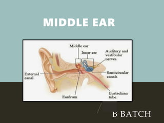
2. MIDDLE EAR
Middle ear cleft- middle ear with eustachian tube
,aditus antrum and mastoid air cells.
Lined by mucous membrane and filled with air.
Divided into:-
Epitympanum- lying above the level of pars tensa
Mesotympanum – lying opp to pars tensa
Hypotympanum- lying below the level of pars
tensa.
3. Roof :- formed by thin plate of bone- tegmen tympani-
separates tympanic cavity from middle cranial fossa
Floor :- thin plate of bone which separates tympanic
cavity from jugular bulb.
Anterior wall:- thin plate of bone which separates the
cavity from internal carotid artery.
Has two openings for eustachian tube and tensor
tympani muscle.
WALLS OF MIDDLE EAR
4. Posterior wall :- lies close to mastoid air cells.
Pyramid - bony projection through which tendon of
stapedius appears.
Aditus – an opening through which middle ear
communicates with antrum.
Facial nerve:-runs behind pyramid
Facial recess:-depression lateral to pyramid
Surgically imp as direct access can be made
through this into middle ear.
5. Medial wall:-
Formed by promontory
Promontory- a bulge due to basal coil of cochlea
Oval window- into which foot plate of stapes is fixed
Round window- covered by secondary tympanic
membrane
Canal for facial nerve
Processus cochleariformis- hook like projection where
the tendon of tensor tympani takes a turn.
Sinus tympani:- deep recess medial to pyramid
6. Lateral wall- formed largely by tympanic membrane, to a
lesser extent by bony wall called scutum
7. MASTOID ANTRUM
Air containing space
Communicates with the middle ear through
additus
Roof- tegmen antri
Marked externally on the surface of mastoid by
suprameatal triangle or Mac Evens triangle
8. ADITUS
Opening through which middle ear
communicates with antrum
MASTOID AND AIR CELL
SYSTEM
Mastoid consists of bony cortex and
honey comb like air cells
3 types
9. 3 types(depending upon development of
air cells)
Cellular – mastoid cells are well
developed
Diploetic – consists of marrow spaces
and few air cells
Sclerotic – no air cells.
10. DEVELOPMENT OF
MASTOID
Develops from squamous and petrous
bones
Petrosquamosal suture may persist as
bony plate-Korner’s septum.
Surgically imp- as it cause difficulty in
locating the antrum.
11. DEPENDING ON THE LOCATION,
MASTOID AIR CELLS ARE DIVIDED
INTO-
Zygomatic cells
Tegmen cells
Perisinus cells
Retrofacial cells
Perilabrinthine cells
Peritubal cells
Tip cells, marginal cells, squamosal cells
12.
13. Malleus :- head, neck, handle, lateral
process, anterior process.
Head and neck lies in the middle ear
Lateral process forms nob like projection
on the outer surface of tympanic
membrane and gives attachment to ant
and post malleal folds.
EAR OSSICLES
Incus:- body, short
process, long process to
which attaches head of
stapes.
Stapes :-head, neck, ant and post crura
and a foot plate(which is held in the oval
window by annular ligament)
Ossicles conduct sound energy from
tympanic membrane to oval window
14. TYMPANIC PLEXUS
Lies on the promontory
Formed by tympanic branch of glossopharynge
N and sympathetic fibres from plexus around
the internal carotid artery
Supplies medial surface of tympanic membrane
tympanic cavity , mastoid air cells ,bony
eustachian tube, secretomotor fibres for the
parotid gland.
.
15. INTRATYMPANIC MUSCLES
1. Tensor tympani- attached to neck of
malleus
Tenses the tympanic membrane Supplied
by mandibular nerve
2. Stapedius – attached to neck of stapes
Helps to dampen loud sounds
supplied by facial nerve
16. CHORDA TYMPANI
Branch of facial nerve
Enters the middle ear through posterior
canaliculus
Runs on the medial surface of tympanic
membrane
Carries taste from ant 2/3rd of tongue
Secretory fibres to sublingual and
submaxillary salivary glands.
17. LINING OF MIDDLE EAR CLEFT
Mucous membrane of nasopharynx
continuous with that of middle ear,
aditus , antrum, mastoid air cells.
It wraps middle ear structures like
peritoneum. Thus middle ear contains
only air, all the structures lie outside
the mucous membrane.
18. Eustachian tube lined by ciliated epithelium –
pseudostratified columnar in cartilaginous part and
columnar in bony part.
Tympanic cavity lined by ciliated columnar epithelium in
ant and inferior part and cuboidal in posterior part
Epitympanum & mastoid air cells-flat non ciliated
epithelium.
BLOOD SUPPLY
2 major arteries
Anterior tympanic branch of maxillary artery-tympanic
membrane
Stylomastoid branch of posterior auricular artery –
middle ear and mastoid air cells.
19. 4 minor vessels
Petrosal branch of middle meningeal artery
Superior tympanic branch of middle meningeal artery
traversing along the canal for tensor tympani muscle
Branch of artery of pterygoid canal runs along the eustaschian
tube
Tympanic branch of internal carotid
Veins drain into pterygoid venous plexus and superior petrosal
sinus
20. LYMPHATIC DRAINAGE
•Middle ear and eustachian tube -
retropharyngeal node & upper jugular nodes
•Pinna - preauricular, postauricular, parotid , infra
auricular, deep jugular & spinal accessory nodes.