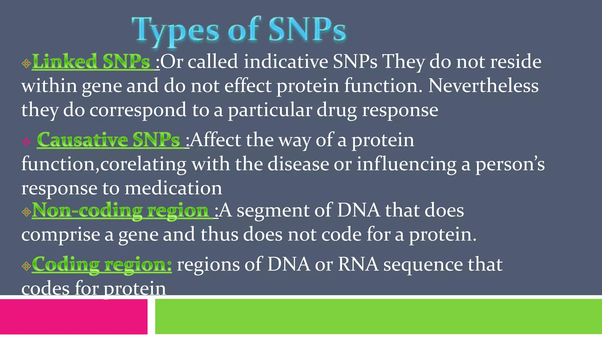
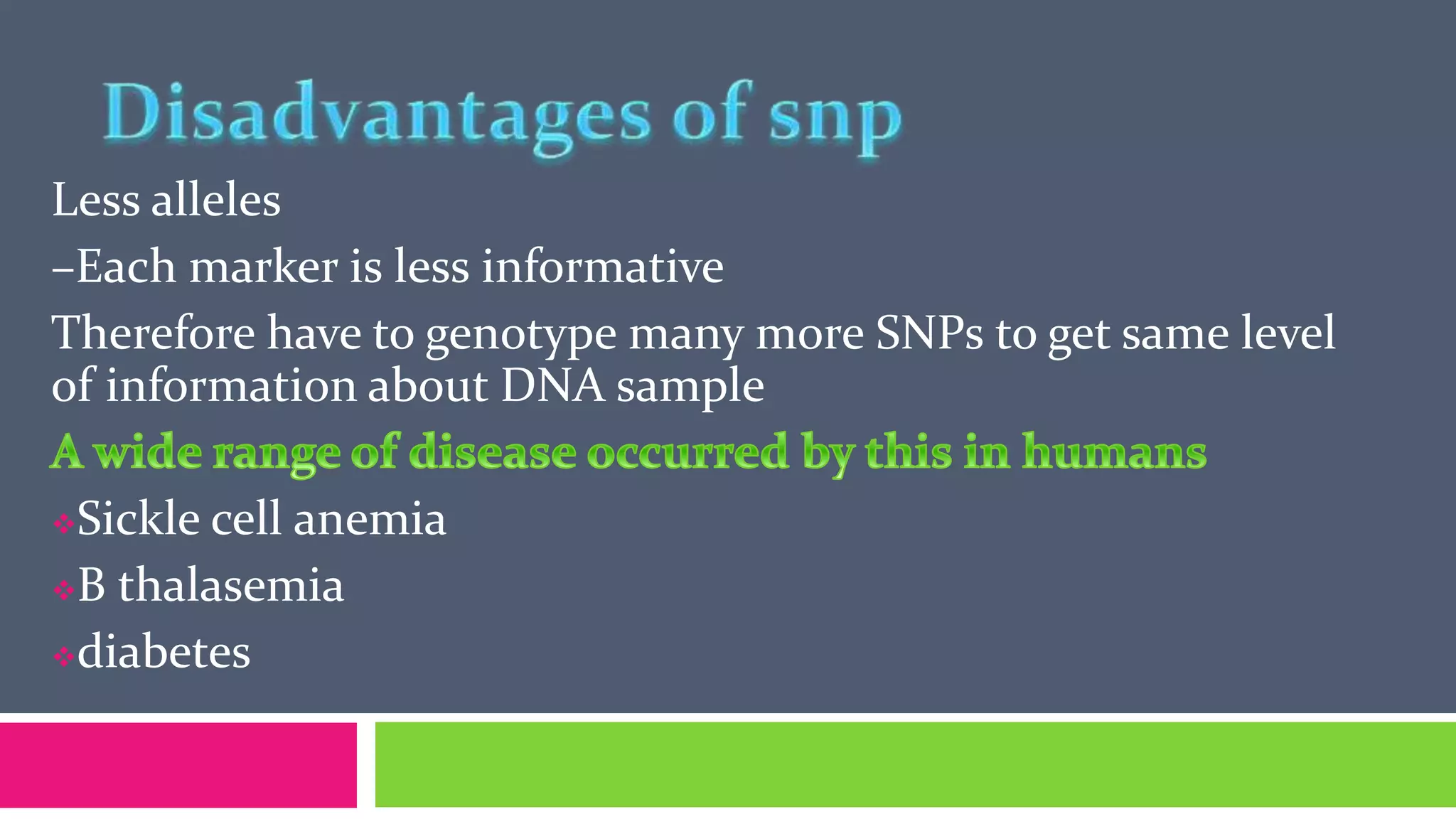
Adil Shehzad
Amna Munir
Asifa Zafar
How can researchers hope to identify and study all
the changes that occur in so many different
diseases?
How can they explain why some people respond to
treatment and not other?
Answer of these question can be given by SNP
poly= many
morphs = shapes
Ability to appear in different shapes
Other terms:
= position on chromosome where sequence or gene
is located
: alternative form of DNA on a locus
A Single Nucleotide Polymorphism, also known as Simple Nucleotide
Polymorphism, is a DNA sequence variation occurring commonly
within a population (e.g. 1%) in which a single nucleotide —
A, T, C or G — in the genome differs between
members of a biological specie .
Pronounced snips
Common type of genetic variation among people
Each SNP represents a difference in a single DNA building block called
as nucleotide
In human 99.9% bases are same Remaining0.1%
bases makes a person unique
in looks, traits and developing diseases
These variations can be
Harmless i.e. phenotype
Harmful i.e. any disease like cancer , diabetes , heart disease
, hemophilia etc
 Latent e.g. susceptibility to lung cancer)
First discovered (early 1980): restriction fragment length
polymorphism
1. SNPs are found in
o coding
o noncoding region
oLocus-specific
2. SNPs in coding regions may alter the protein structure
made by that coding region.
3. Occur with a very high frequency
o about 1 in 1000 bases to 1 in 100 to 300 bases.
4.The abundance of SNPs and the ease with which they
can be measured make these genetic variations significant.
5. SNPs close to particular gene acts as marker for that gene
6.Occur once in every 300 nucleotides on average roughly
there are 10 million SNPs in human genome
Sequence genome of a large no of people
Compare the base sequence to discover SNPs
Generate a single map of human genome that contain
all possible SNPs
Genome of each individual contains
distinct SNP pattern.
People can be grouped based on the SNP
profile.
SNPs Profiles important for identifying
response to Drug Therapy.
Correlations might emerge between
certain SNP profiles and specific responses
to treatment.
Types of SNPs
Linked SNPs Causative SNPs
coding region
Non Coding
region
Synonymous Non synonymous
:Or called indicative SNPs They do not reside
within gene and do not effect protein function. Nevertheless
they do correspond to a particular drug response
 :Affect the way of a protein
function,corelating with the disease or influencing a person’s
response to medication
:A segment of DNA that does
comprise a gene and thus does not code for a protein.
regions of DNA or RNA sequence that
codes for protein
.
Non synonymous SNP may be missense or nonsense.
: Missense SNP leads to a different codon that
codes for other amino acid.
Nonsense mutation in which a codon is changed
to a premature stop codon that results in truncation of the
resulting protein
A set of closely linked genetic markers present on one
chromosome which tend to be inherited together (not easily
separable by recombination)
Phenotype
GATATTCGTACGGA-T
GATGTTCGTACTGAAT
GATATTCGTACGGA-T
GATATTCGTACGGAAT
GATGTTCGTACTGAAT
GATGTTCGTACTGAAT
BLACK EYE
BROWN EYE
BLACK EYE
BROWN EYE
BROWN EYE
123456
Haplotypes
AG- 2/6(BLACK EYE)
GTA 3/6(BROWN EYE)
AGA 1/6 ( )
Haplotypes are not only more informative but also capture
the regional LD information, which is assumed to be powerful
Association of haplotype frequencies with the presence of
desired phenotypic frequencies in the population will help in
utilizing the maximum potential of SNP as a marker.
is a method for separation and analysis of macromolecules and their
fragments Nucleic acid molecules are separated by applying an electric field
to move the negatively charged molecules through a matrix of agarose or
other substances.
Shorter molecules move faster and migrate farther than longer ones.
This phenomenon is called sieving.
Step 1..place DNA into tubes..
Step 2.. The polymerase chain reaction uses a machine called thermocycler to
quickly copy a piece of DNA.
Step 3.. Place restriction
restriction enzyme will cut
Step 4.. Dye DNA and add into gel
..the gel is made of agarose.
restriction enzyme will cut DNA into
different sizes according to its sequnce.
Step 5..
Run electric current
through gel.
Step 6..
DNA is negatively charged and
hence will move towards the positively
charged end of the gel.Smaller pieces
of DNA travel faster than larger pieces
of DNA.
may be used to determine DNA sequences.
mass spectrometry has specifically been investigated as an
alternative method to gel electrophoresis for visualizing DNA
fragments. With this method, DNA fragments are compared
by mass rather than by size. The mass of each nucleotide is
different from the others and this difference is detectable by
mass spectrometry.
Single-nucleotide mutations in a fragment can be more
easily detected with MS than by gel electrophoresis alone
Electrophoretic separation of single-stranded nucleic acids based on subtle differences
in sequence which results in a different secondary structure and a measurable difference
in mobility through a gel. A single nucleotide change in a particular sequence, as
seen in a double-stranded DNA, cannot be distinguished by electrophoresis,
because the physical properties of the double strands are almost identical for both
alleles
SSCP limitations:
• mobilty needs constant temperature.
•under optimal conditions, approximately 80 to 90% of the potential base exchanges are
detectable
• Sensitivity of SSCP is affected by pH.
1) polymerase chain reaction (PCR) amplification of DNA sequence
of interest
(2) denaturation of double-stranded PCR products
(3) cooling of the denatured DNA (single-stranded) to maximize
self-annealing;
(4) detection of mobility difference of the single-stranded DNAs by
electrophoresis under non-denaturing conditions. Several
methods have been developed to visualize the SSCP mobility
shifts. These include the
A
B
A’
B
’
fold into a
three-dimensional
shape according to
their primary
sequence
A
B
A’
B
’
Electrophoresis on
a non denaturing
polyacrylamide gel
Wild type Mutant type
Sample SSCP Gel Result and Interpretation. DNA was
isolated and amplified SCCP analysis of the DNA
shows sets of alleles usually inherited as a unit. Lanes
3 and 4 were identical haplotypes from two
individuals. The difference in band migration in
adjacent lanes is associated with the number of
nucleotide differences (in parentheses): lanes 2-3
(2), lanes 3-4 (0), lanes 4-5 (3), lanes 5-6 (1), lanes 6-7
(3), lanes 7-8 (1), lanes 8-9 (1), and lanes 9-10 (4).
SNPs are the most frequent form of variation
They are disease causing mutations in many genes
They are abundant and slow mutation rates
May work as the nxt generation of genetic markers
IN DISEASE DIAGNOSIS
 IN DRUG DISCOVERY & DEVELOPMENT
 IN DRUG RESPONSE
 INVESTIGATION OF MIGRATION PATTERNS
Less alleles
–Each marker is less informative
Therefore have to genotype many more SNPs to get same level
of information about DNA sample
Sickle cell anemia
B thalasemia
diabetes
 ressecively inherited
chronic hemolytic anemia
nucleotide substition in
b globin gene in
chromosom 11 .
high blood glucose
insulin production is
inadequate
The drug albuterol is Commonly
prescribed to relief symptoms of
asthama
 albuterol effectively releife asthama
but in some people not all. scientist
are studying How people with different
snp Response to treatment
Albuterol acts on b-2-
Adrenergic Receptor
(beta 2 AR protein)
Beta 2 AR encoded by
AdRb2 gene
In analyzing 3000 base
pairs Stretch of ADRB2,
scientist find 13 points
where SNP exist
Scientist have looked up
this region of DNA in different
people and identified 12
halotypes Which are unique
combination of these 13 snps
Single Nucleotide Polymorphism (SNP)

Single Nucleotide Polymorphism (SNP)

  • 3.
  • 4.
    How can researchershope to identify and study all the changes that occur in so many different diseases? How can they explain why some people respond to treatment and not other? Answer of these question can be given by SNP
  • 5.
    poly= many morphs =shapes Ability to appear in different shapes Other terms: = position on chromosome where sequence or gene is located : alternative form of DNA on a locus
  • 6.
    A Single NucleotidePolymorphism, also known as Simple Nucleotide Polymorphism, is a DNA sequence variation occurring commonly within a population (e.g. 1%) in which a single nucleotide — A, T, C or G — in the genome differs between members of a biological specie . Pronounced snips Common type of genetic variation among people Each SNP represents a difference in a single DNA building block called as nucleotide
  • 8.
    In human 99.9%bases are same Remaining0.1% bases makes a person unique in looks, traits and developing diseases These variations can be Harmless i.e. phenotype Harmful i.e. any disease like cancer , diabetes , heart disease , hemophilia etc  Latent e.g. susceptibility to lung cancer) First discovered (early 1980): restriction fragment length polymorphism
  • 9.
    1. SNPs arefound in o coding o noncoding region oLocus-specific 2. SNPs in coding regions may alter the protein structure made by that coding region. 3. Occur with a very high frequency o about 1 in 1000 bases to 1 in 100 to 300 bases. 4.The abundance of SNPs and the ease with which they can be measured make these genetic variations significant. 5. SNPs close to particular gene acts as marker for that gene 6.Occur once in every 300 nucleotides on average roughly there are 10 million SNPs in human genome
  • 10.
    Sequence genome ofa large no of people Compare the base sequence to discover SNPs Generate a single map of human genome that contain all possible SNPs
  • 11.
    Genome of eachindividual contains distinct SNP pattern. People can be grouped based on the SNP profile. SNPs Profiles important for identifying response to Drug Therapy. Correlations might emerge between certain SNP profiles and specific responses to treatment.
  • 12.
    Types of SNPs LinkedSNPs Causative SNPs coding region Non Coding region Synonymous Non synonymous
  • 13.
    :Or called indicativeSNPs They do not reside within gene and do not effect protein function. Nevertheless they do correspond to a particular drug response  :Affect the way of a protein function,corelating with the disease or influencing a person’s response to medication :A segment of DNA that does comprise a gene and thus does not code for a protein. regions of DNA or RNA sequence that codes for protein
  • 14.
  • 15.
    Non synonymous SNPmay be missense or nonsense. : Missense SNP leads to a different codon that codes for other amino acid. Nonsense mutation in which a codon is changed to a premature stop codon that results in truncation of the resulting protein
  • 17.
    A set ofclosely linked genetic markers present on one chromosome which tend to be inherited together (not easily separable by recombination) Phenotype GATATTCGTACGGA-T GATGTTCGTACTGAAT GATATTCGTACGGA-T GATATTCGTACGGAAT GATGTTCGTACTGAAT GATGTTCGTACTGAAT BLACK EYE BROWN EYE BLACK EYE BROWN EYE BROWN EYE 123456 Haplotypes AG- 2/6(BLACK EYE) GTA 3/6(BROWN EYE) AGA 1/6 ( )
  • 18.
    Haplotypes are notonly more informative but also capture the regional LD information, which is assumed to be powerful Association of haplotype frequencies with the presence of desired phenotypic frequencies in the population will help in utilizing the maximum potential of SNP as a marker.
  • 20.
    is a methodfor separation and analysis of macromolecules and their fragments Nucleic acid molecules are separated by applying an electric field to move the negatively charged molecules through a matrix of agarose or other substances. Shorter molecules move faster and migrate farther than longer ones. This phenomenon is called sieving. Step 1..place DNA into tubes.. Step 2.. The polymerase chain reaction uses a machine called thermocycler to quickly copy a piece of DNA.
  • 21.
    Step 3.. Placerestriction restriction enzyme will cut Step 4.. Dye DNA and add into gel ..the gel is made of agarose. restriction enzyme will cut DNA into different sizes according to its sequnce.
  • 22.
    Step 5.. Run electriccurrent through gel. Step 6.. DNA is negatively charged and hence will move towards the positively charged end of the gel.Smaller pieces of DNA travel faster than larger pieces of DNA.
  • 23.
    may be usedto determine DNA sequences. mass spectrometry has specifically been investigated as an alternative method to gel electrophoresis for visualizing DNA fragments. With this method, DNA fragments are compared by mass rather than by size. The mass of each nucleotide is different from the others and this difference is detectable by mass spectrometry. Single-nucleotide mutations in a fragment can be more easily detected with MS than by gel electrophoresis alone
  • 24.
    Electrophoretic separation ofsingle-stranded nucleic acids based on subtle differences in sequence which results in a different secondary structure and a measurable difference in mobility through a gel. A single nucleotide change in a particular sequence, as seen in a double-stranded DNA, cannot be distinguished by electrophoresis, because the physical properties of the double strands are almost identical for both alleles SSCP limitations: • mobilty needs constant temperature. •under optimal conditions, approximately 80 to 90% of the potential base exchanges are detectable • Sensitivity of SSCP is affected by pH.
  • 25.
    1) polymerase chainreaction (PCR) amplification of DNA sequence of interest (2) denaturation of double-stranded PCR products (3) cooling of the denatured DNA (single-stranded) to maximize self-annealing; (4) detection of mobility difference of the single-stranded DNAs by electrophoresis under non-denaturing conditions. Several methods have been developed to visualize the SSCP mobility shifts. These include the
  • 26.
    A B A’ B ’ fold into a three-dimensional shapeaccording to their primary sequence A B A’ B ’ Electrophoresis on a non denaturing polyacrylamide gel
  • 27.
    Wild type Mutanttype Sample SSCP Gel Result and Interpretation. DNA was isolated and amplified SCCP analysis of the DNA shows sets of alleles usually inherited as a unit. Lanes 3 and 4 were identical haplotypes from two individuals. The difference in band migration in adjacent lanes is associated with the number of nucleotide differences (in parentheses): lanes 2-3 (2), lanes 3-4 (0), lanes 4-5 (3), lanes 5-6 (1), lanes 6-7 (3), lanes 7-8 (1), lanes 8-9 (1), and lanes 9-10 (4).
  • 28.
    SNPs are themost frequent form of variation They are disease causing mutations in many genes They are abundant and slow mutation rates May work as the nxt generation of genetic markers IN DISEASE DIAGNOSIS  IN DRUG DISCOVERY & DEVELOPMENT  IN DRUG RESPONSE  INVESTIGATION OF MIGRATION PATTERNS
  • 29.
    Less alleles –Each markeris less informative Therefore have to genotype many more SNPs to get same level of information about DNA sample Sickle cell anemia B thalasemia diabetes
  • 30.
     ressecively inherited chronichemolytic anemia nucleotide substition in b globin gene in chromosom 11 . high blood glucose insulin production is inadequate
  • 31.
    The drug albuterolis Commonly prescribed to relief symptoms of asthama  albuterol effectively releife asthama but in some people not all. scientist are studying How people with different snp Response to treatment
  • 32.
    Albuterol acts onb-2- Adrenergic Receptor (beta 2 AR protein) Beta 2 AR encoded by AdRb2 gene
  • 33.
    In analyzing 3000base pairs Stretch of ADRB2, scientist find 13 points where SNP exist Scientist have looked up this region of DNA in different people and identified 12 halotypes Which are unique combination of these 13 snps

Editor's Notes

  • #25 The mobility of double-stranded DNA in gel electrophoresis is dependent on strand size and length but is relatively independent of the particular nucleotide sequence.  The mobility of single strands, however, is noticeably affected by very small changes in sequence, possibly one changed nucleotide out of several hundred.  Small changes are noticeable because of the relatively unstable nature of single-stranded DNA; in the absence of a complementary strand, the single strand may experience intrastrand base pairing, resulting in loops and folds that give the single strand a unique 3D structure, regardless of its length.  A single nucleotide change could dramatically affect the strand's mobility through a gel by altering the intrastrand base pairing and its resulting 3D conformation (Melcher, 2000).     Single-strand conformation polymorphism analysis takes advantage of this quality of single-stranded DNA.  First announced in 1989 as a new means of detecting DNA polymorphisms, or sequence variations, SSCP analysis offers an inexpensive, convenient, and sensitive method for determining genetic variation (Sunnucks et al., 2000).     Like restriction fragment length polymorphisms (RFLPs),  SSCPs are allelic variants of inherited, genetic traits that can be used as genetic markers.  Unlike RFLP analysis, however, SSCP analysis can detect DNA polymorphisms and mutations at multiple places in DNA fragments (Orita et al., 1989).  As a mutation scanning technique, though, SSCP is more often used to analyze the polymorphisms at single loci, especially when used for medical diagnoses (Sunnucks et al., 2000).
  • #26 SSCP used to be a way to discover new DNA polymorphisms apart from DNA sequencing, but is now being supplanted by sequencing techniques on account of efficiency and accuracy.[
  • #27 1) polymerase chain reaction (PCR) amplification of DNA sequence of interest; (2) denaturation of double-stranded PCR products; (3) cooling of the denatured DNA (single-stranded) to maximize self-annealing; (4) detection of mobility difference of the single-stranded DNAs by electrophoresis under non-denaturing conditions. Several methods have been developed to visualize the SSCP mobility shifts. These include the incorporation of radioisotope labeling, silver staining, fluorescent dye-labeled PCR primers, and more recently, capillary-based electrophoresis. Silver staining is simple, rapid, and cost-effective, and can be routinely performed in clinical laboratories. The use of SSCP analysis to discover and genotype single-nucleotide polymorphisms (SNPs) has been widely applied to the genetics of hypertension, including both monogenic (e.g., Liddle's syndrome) and polygenic disorders (e.g., essential hypertension).
  • #30 Sickle cell anemia: ressecively inherited chronic hemolytic anemia Cause: nucleotide substition in b globin gene in chromosom 11 . GTG GAG substitute valin for glutamate B thalasemia: reduce production of heamoglobin Cause:HBB gene on chromosome 11