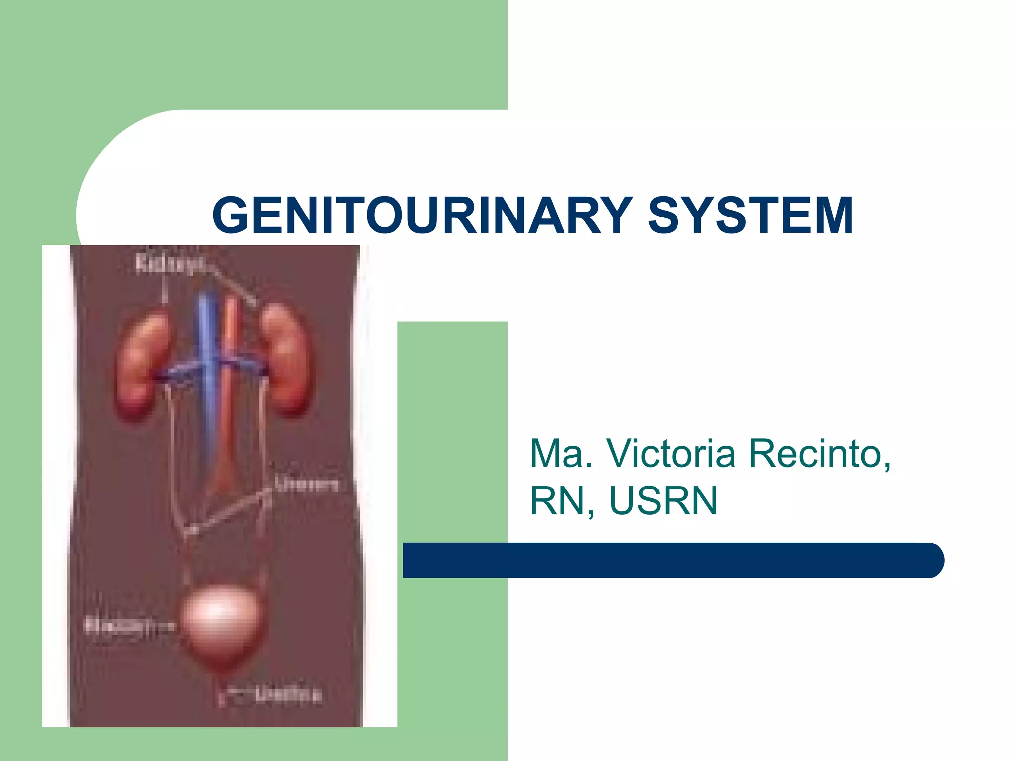
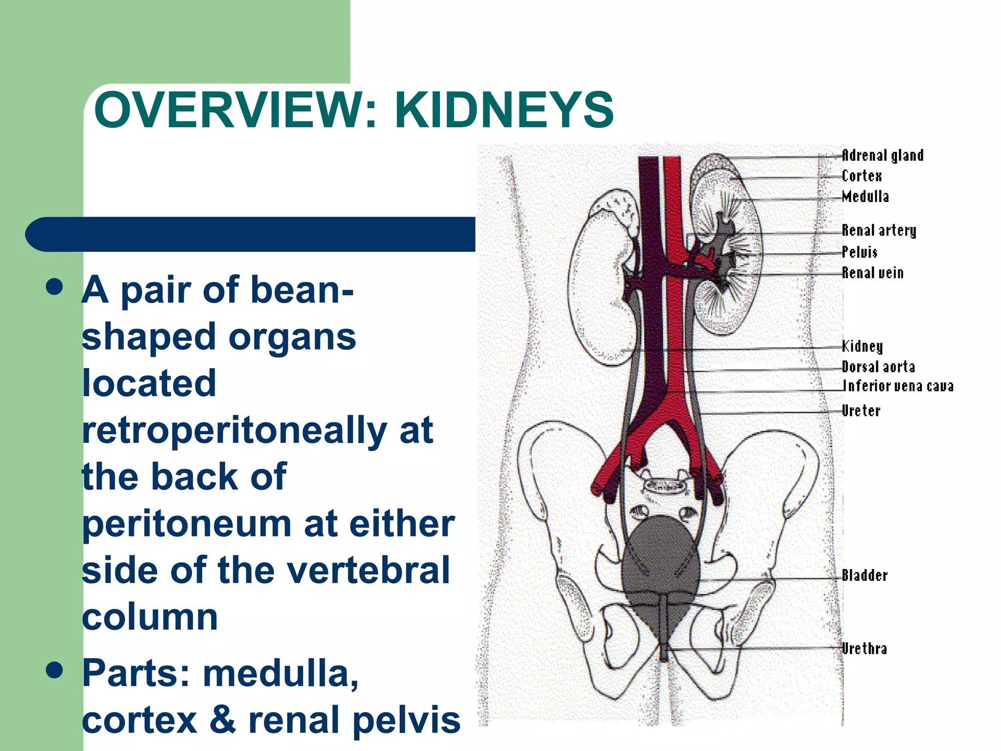
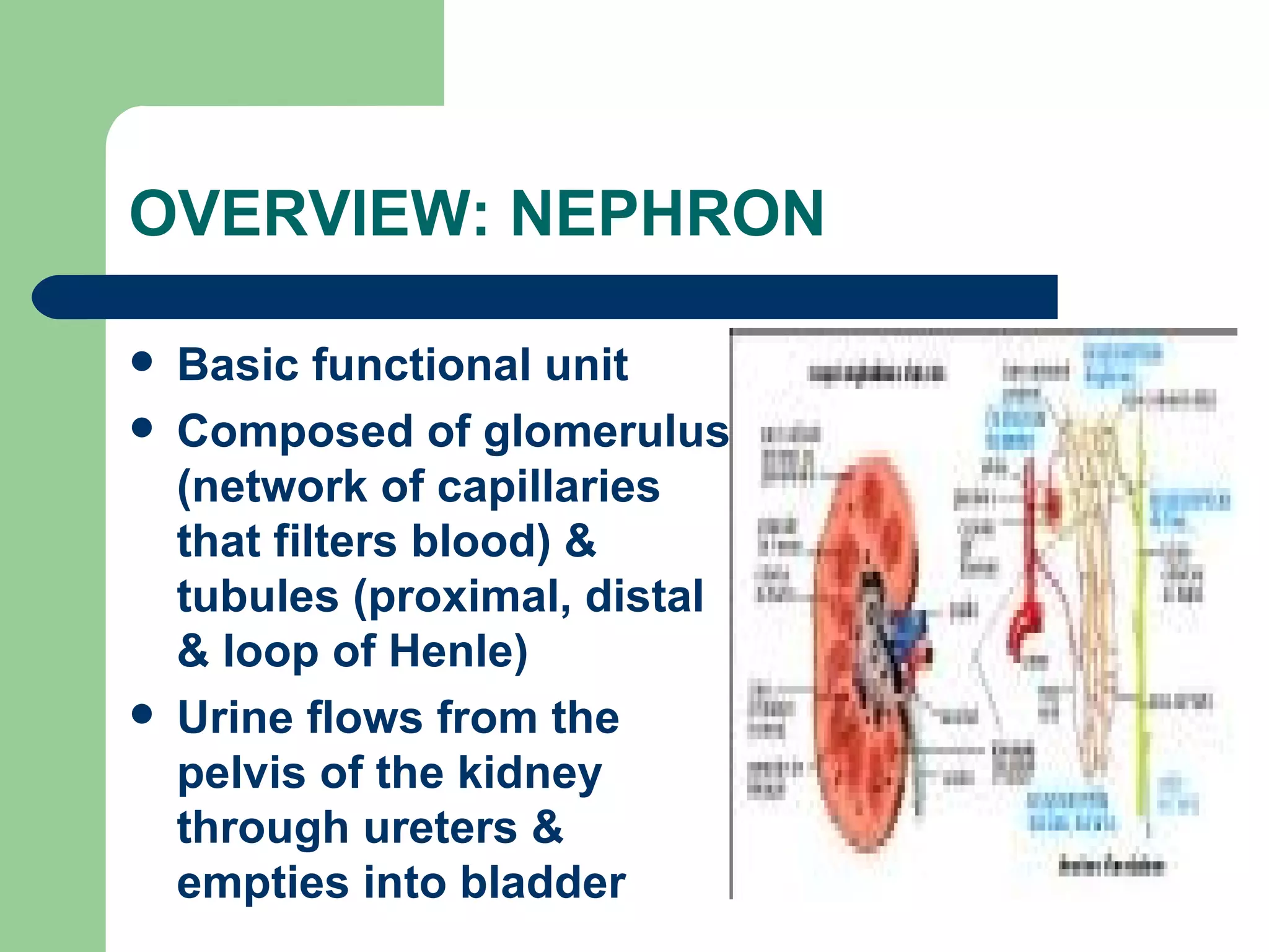
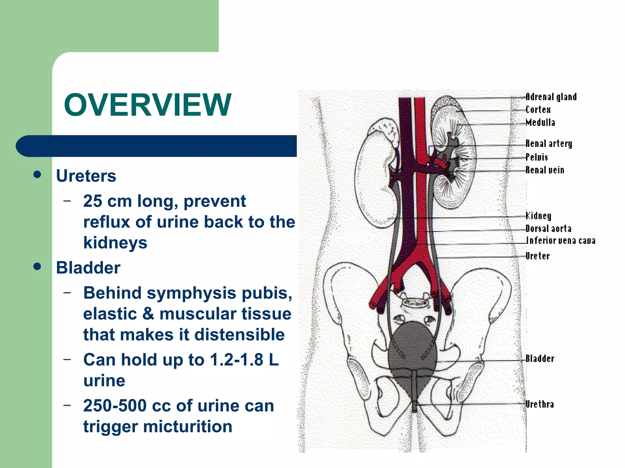
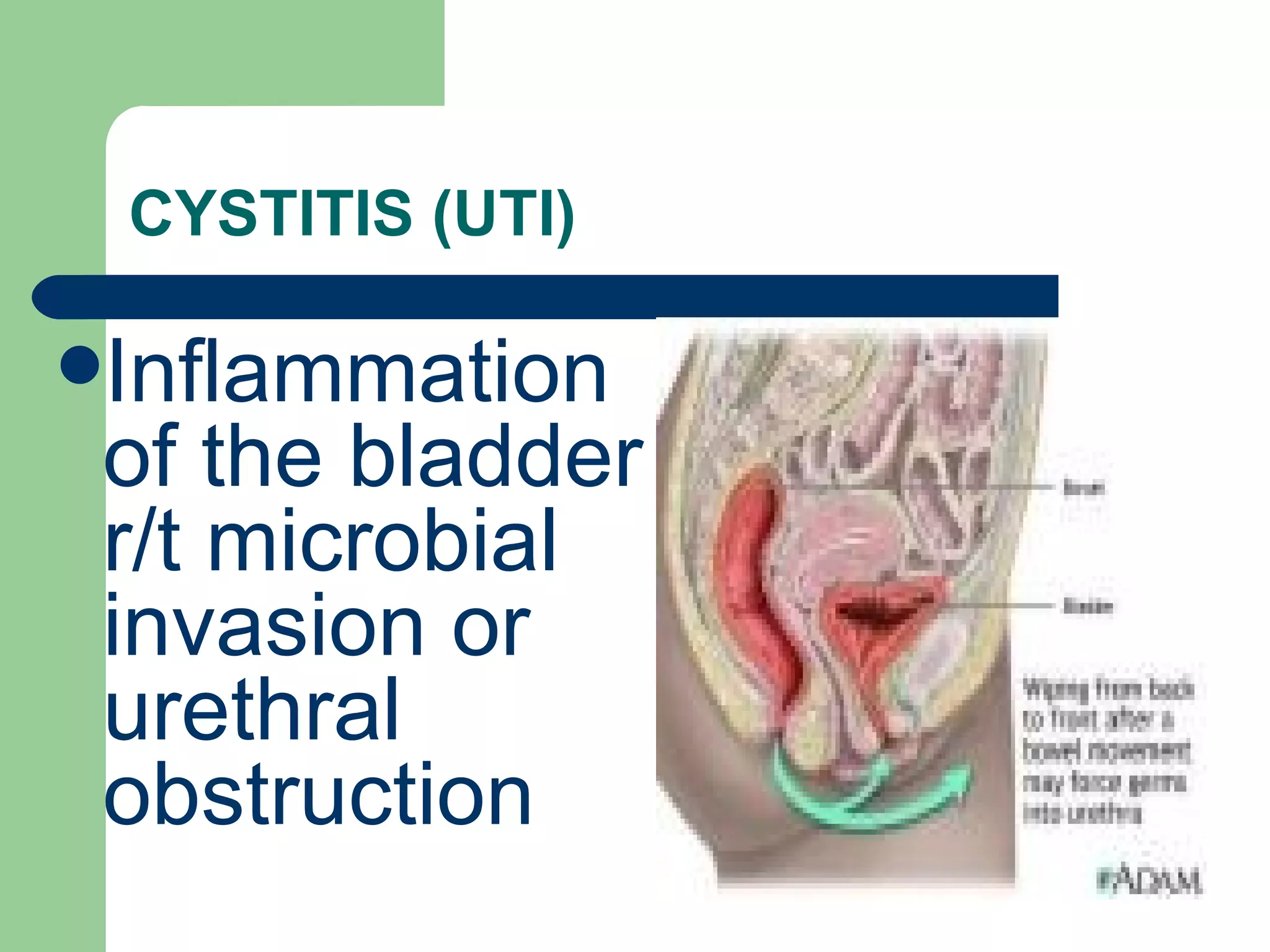
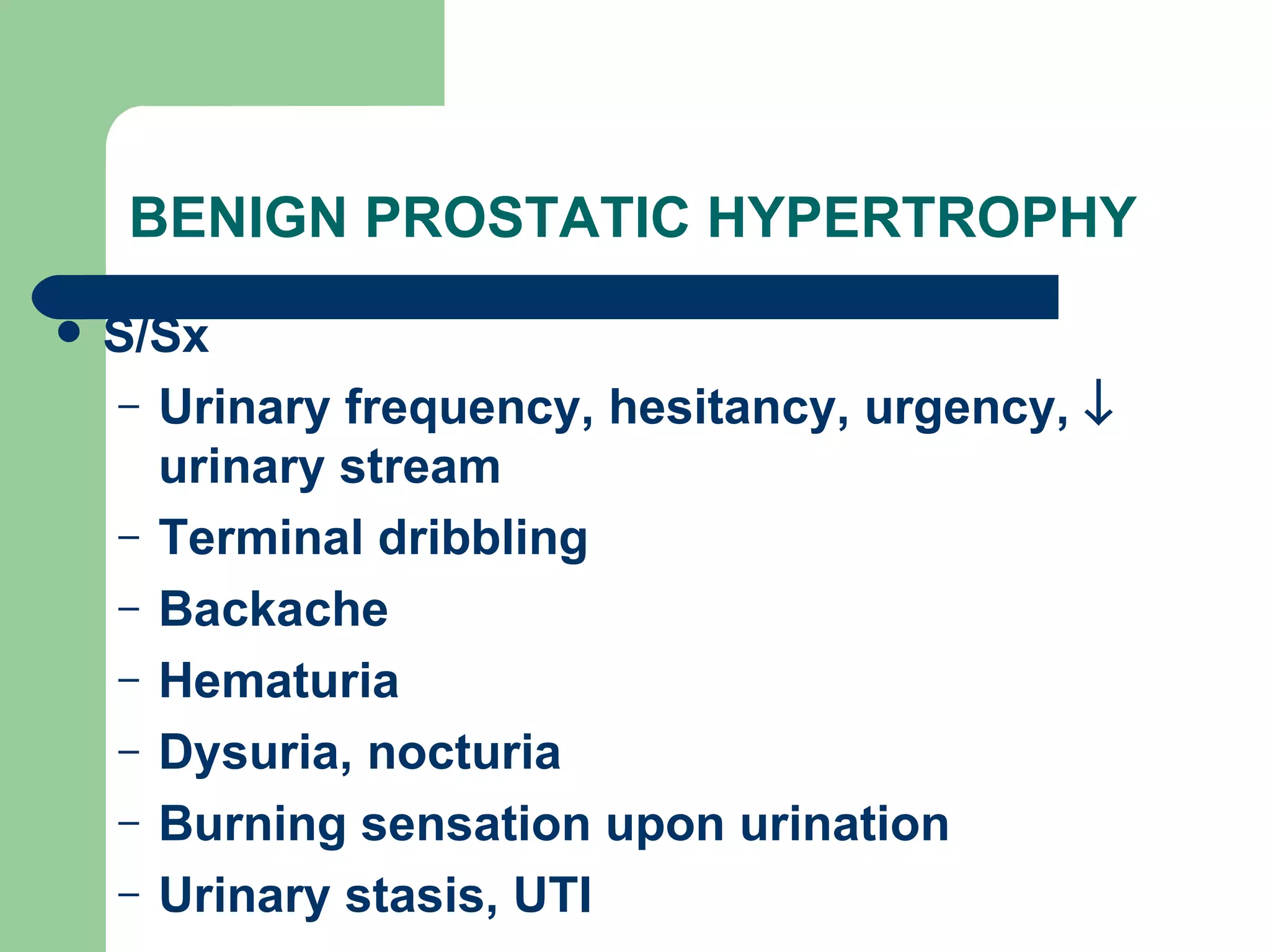
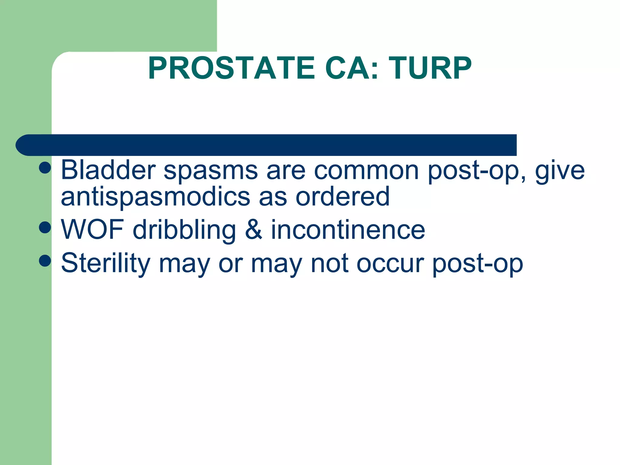
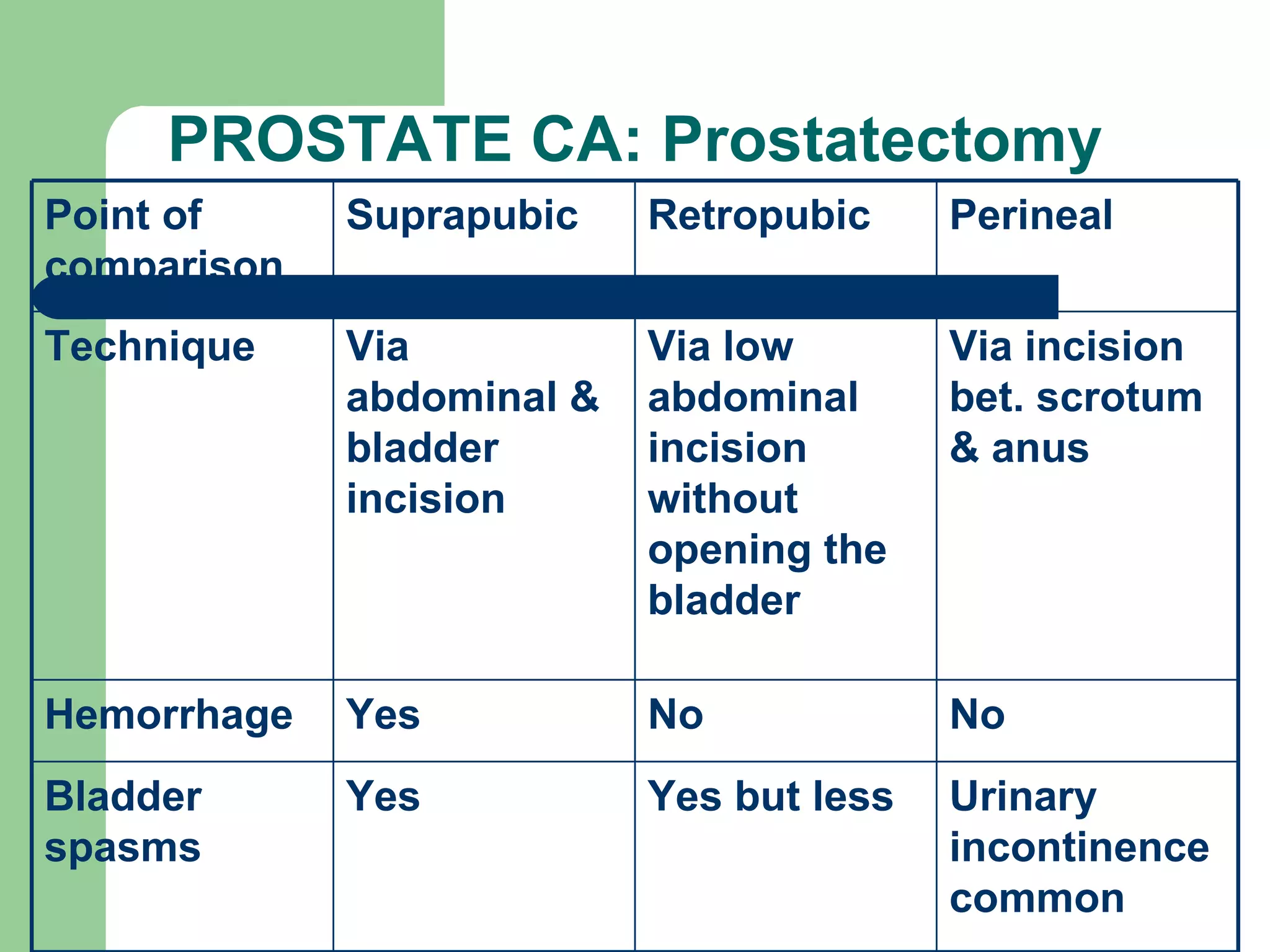
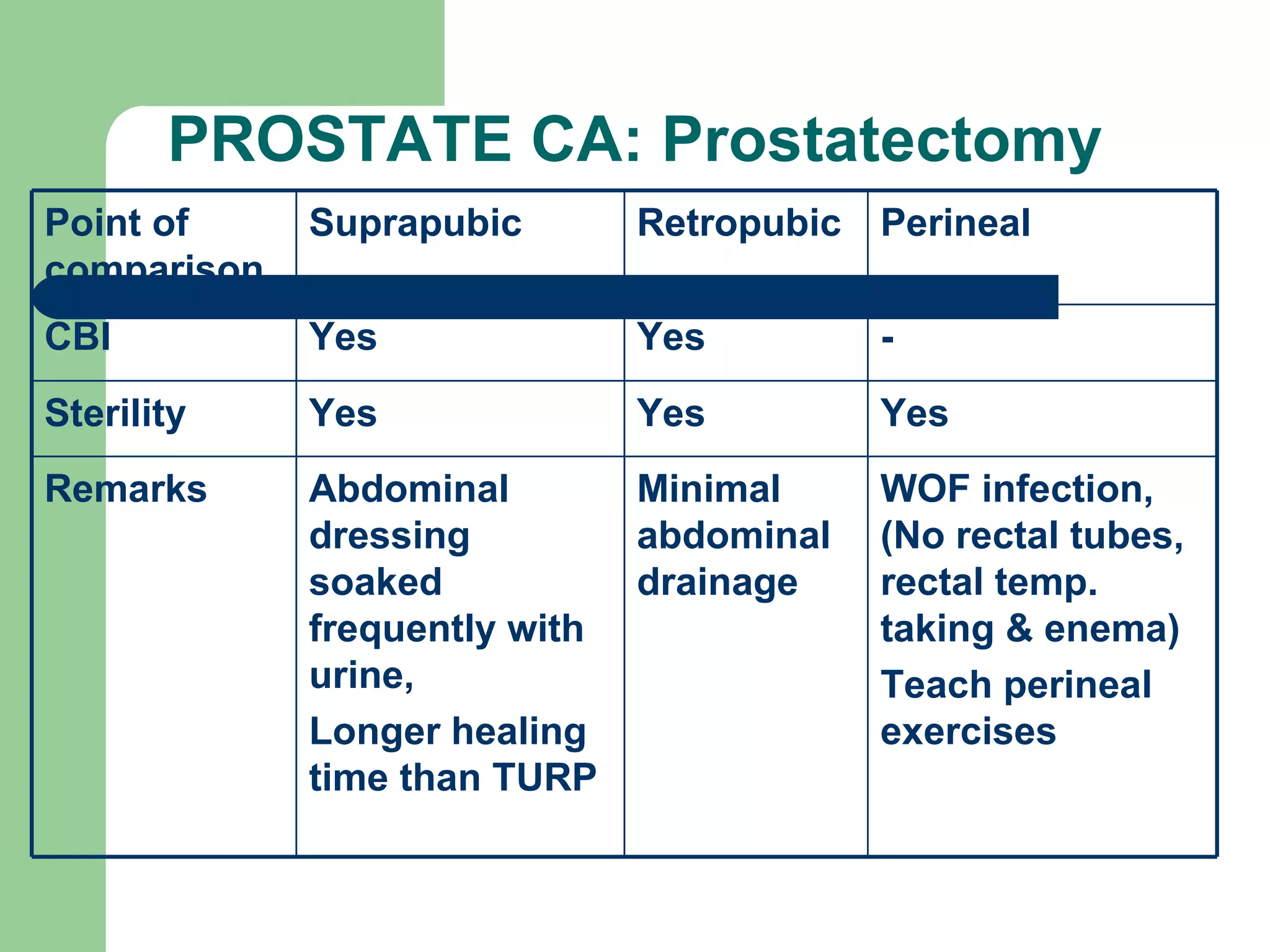
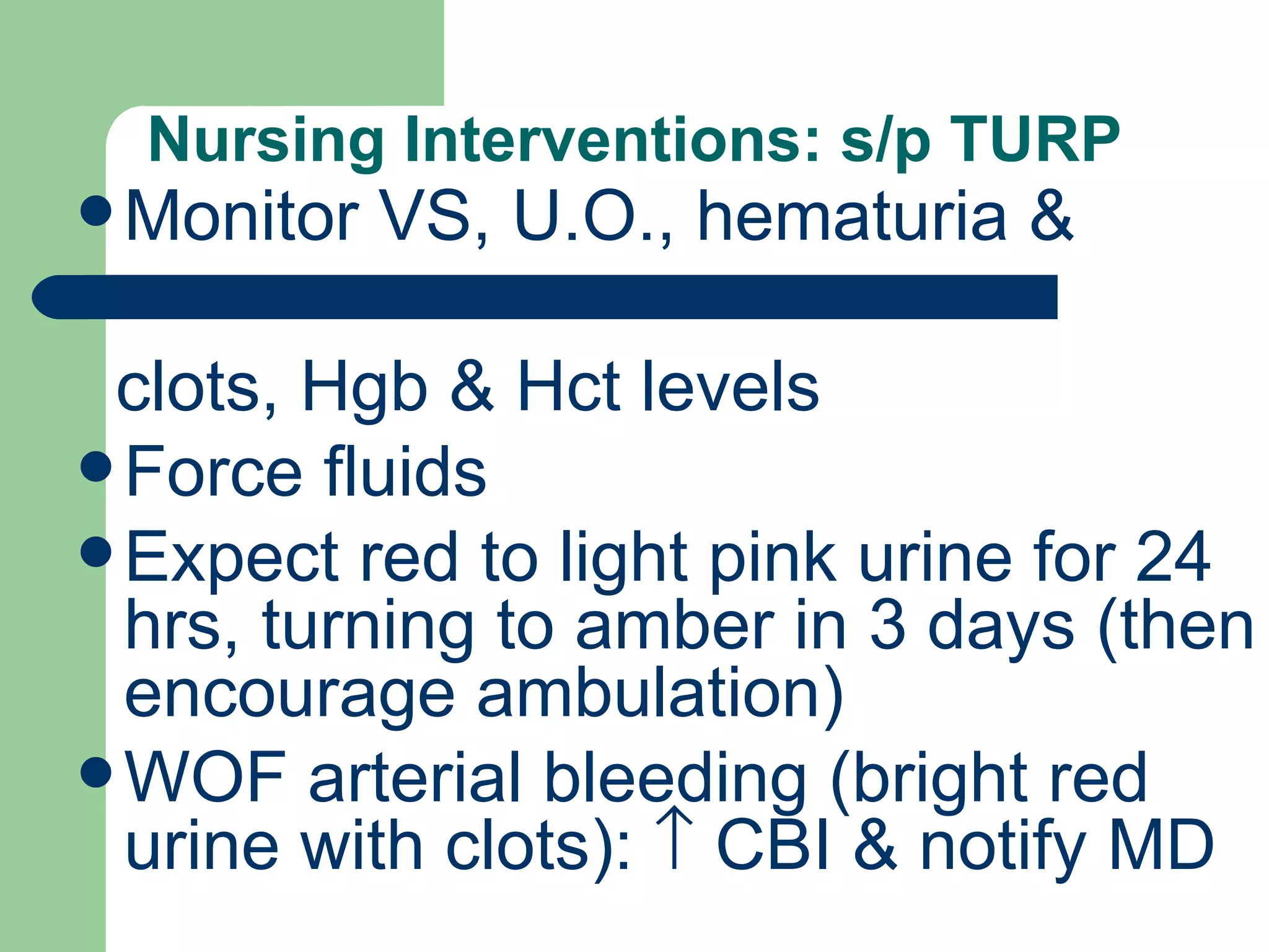
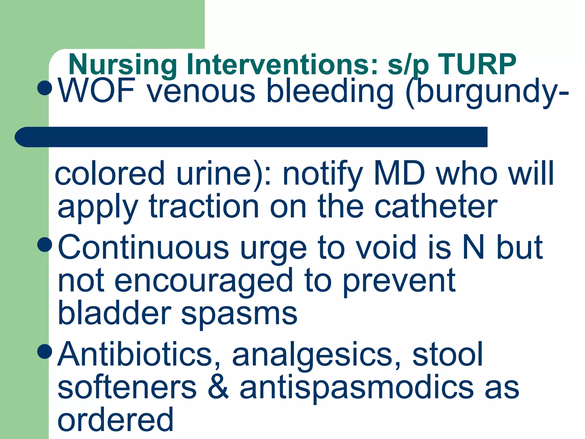
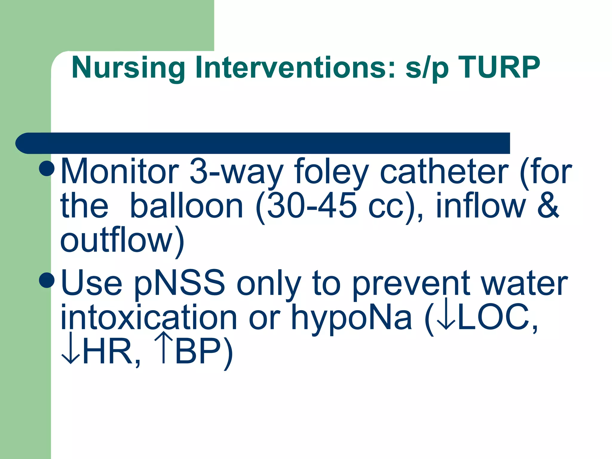
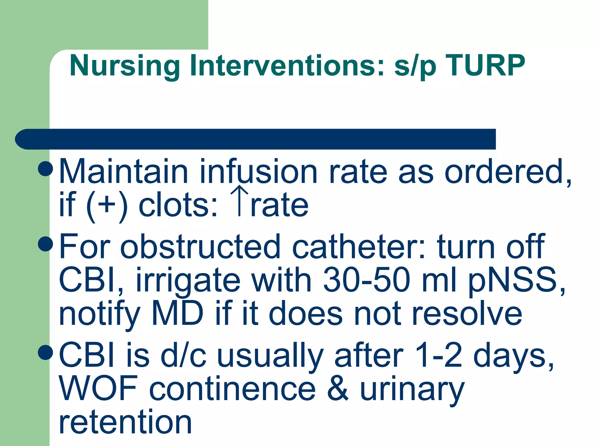
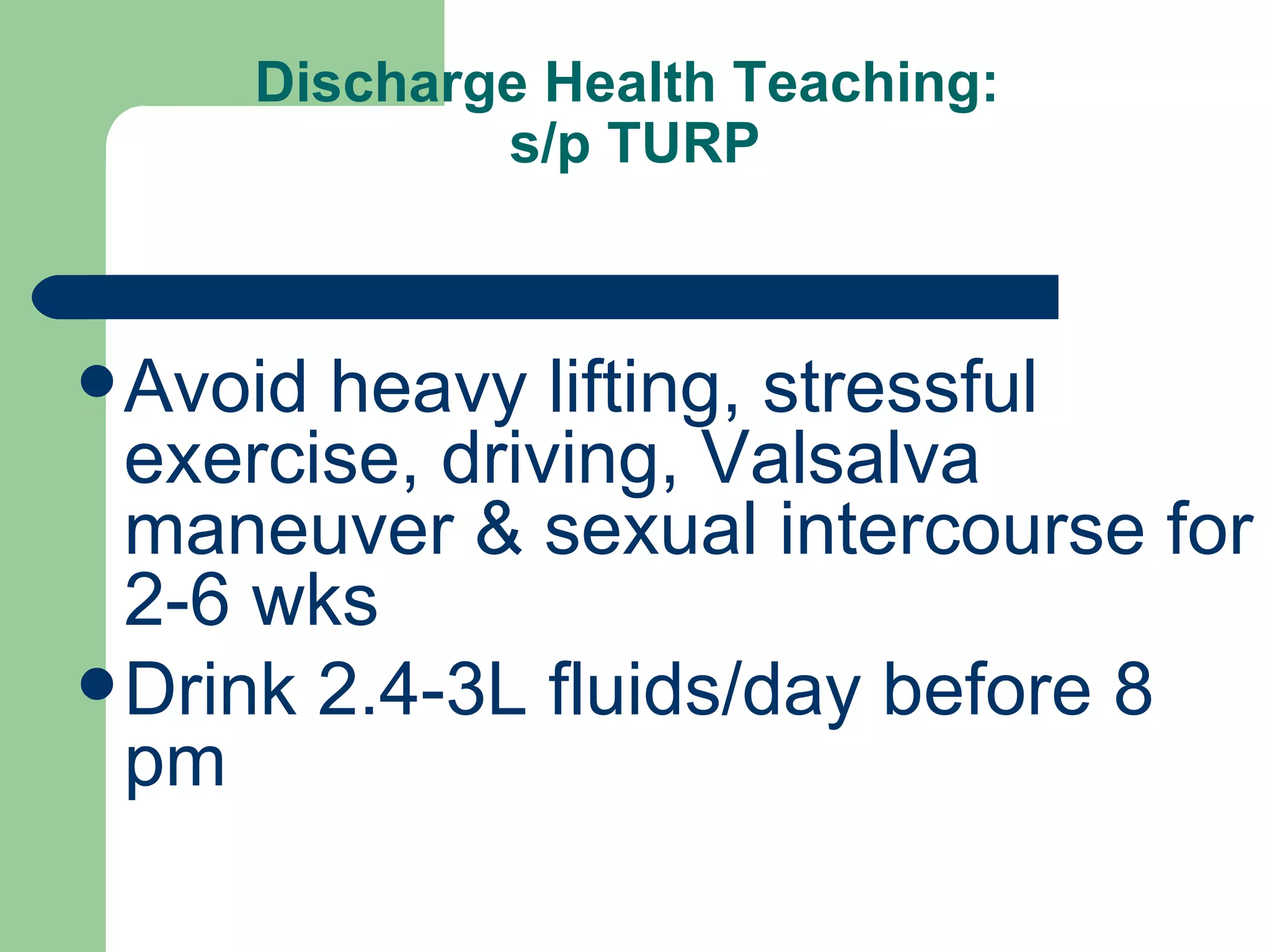
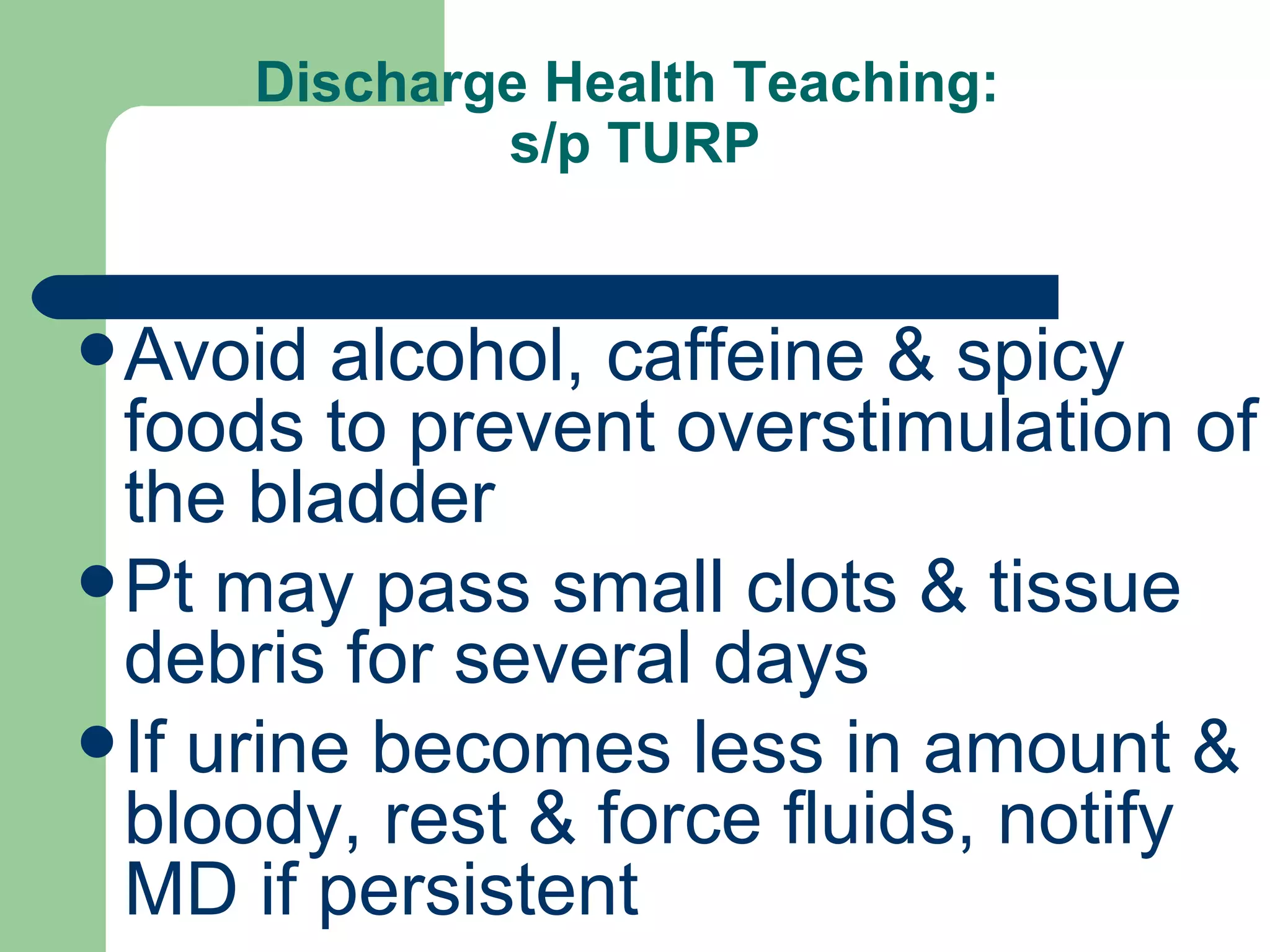
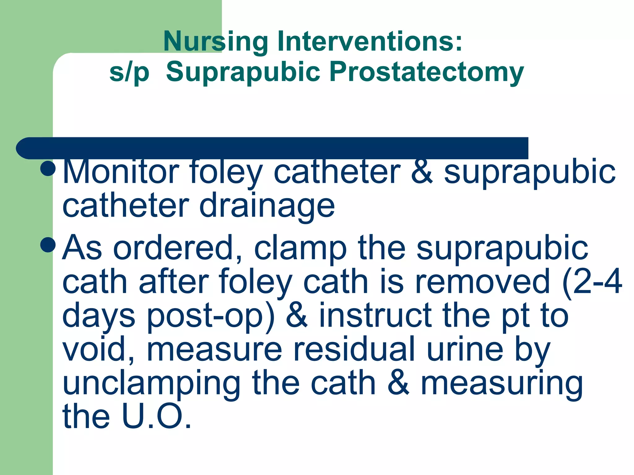
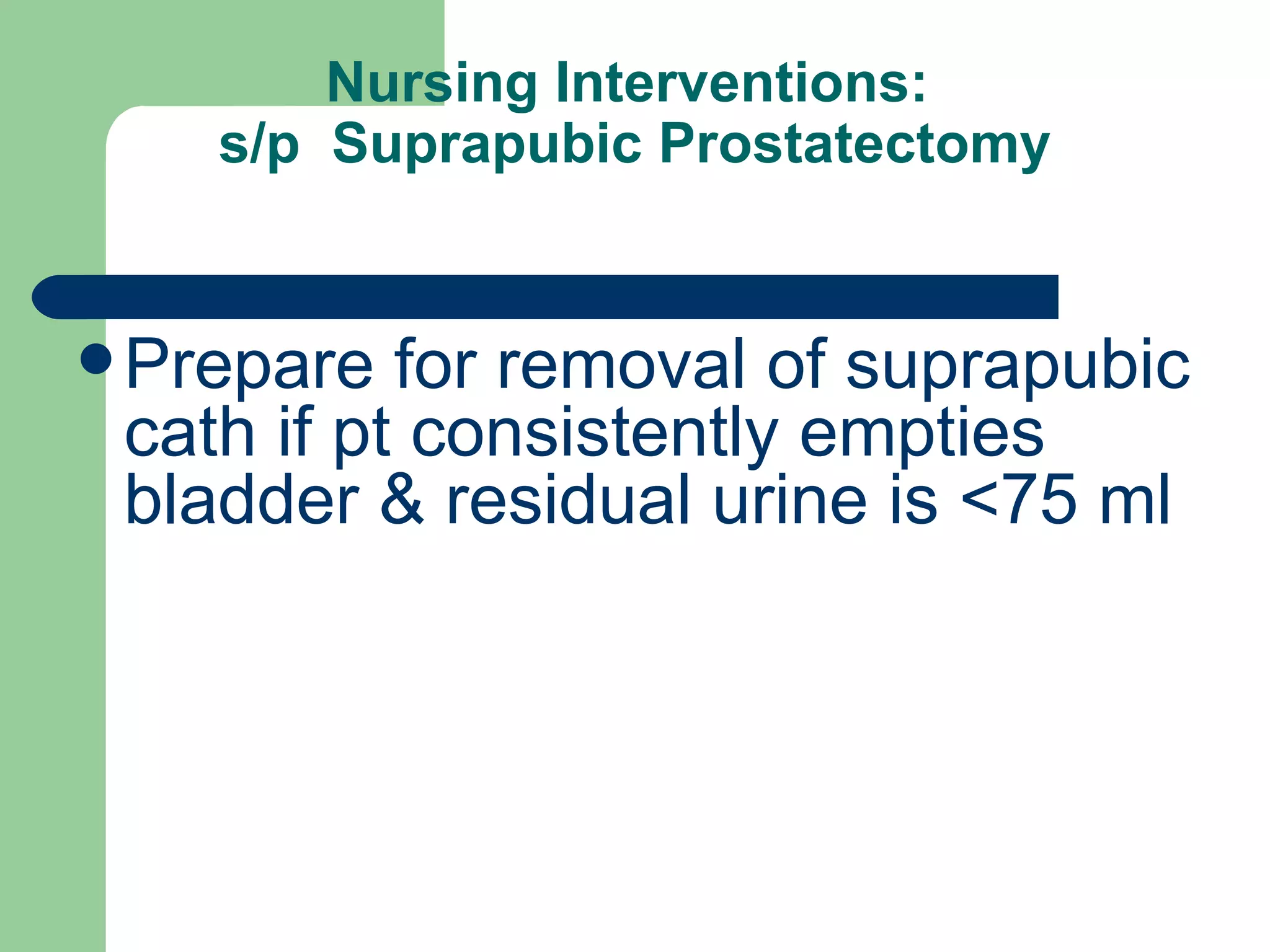
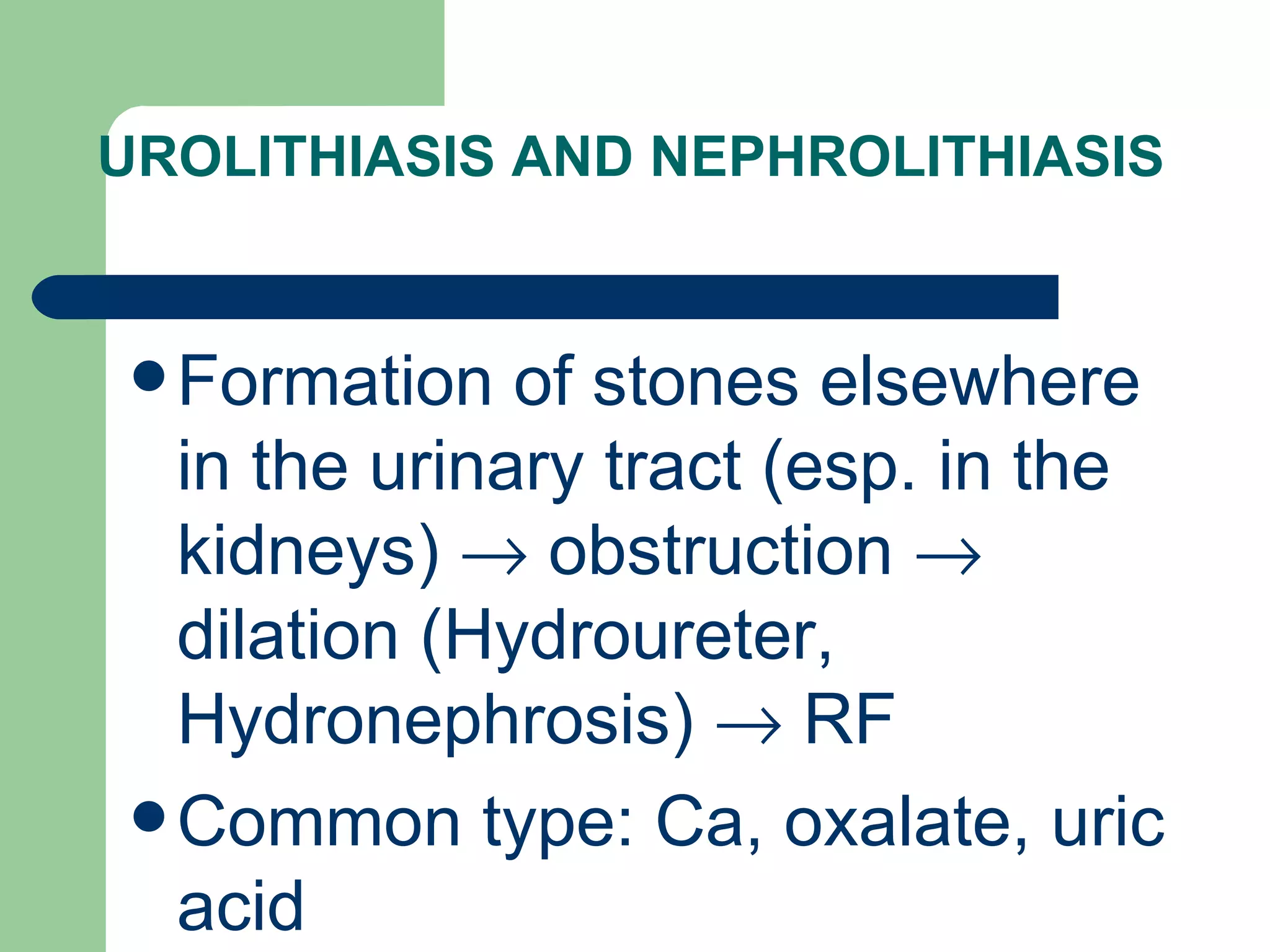
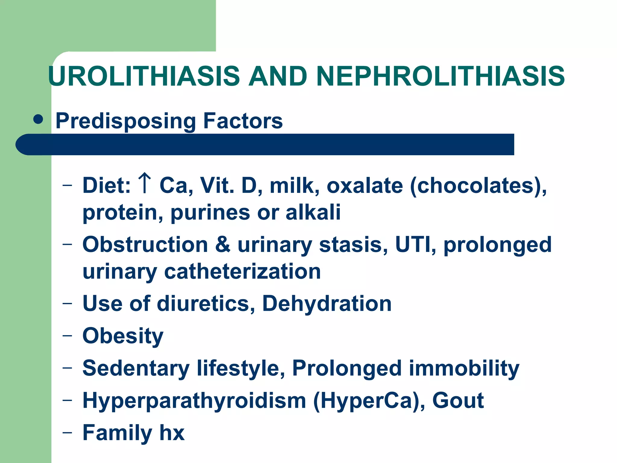
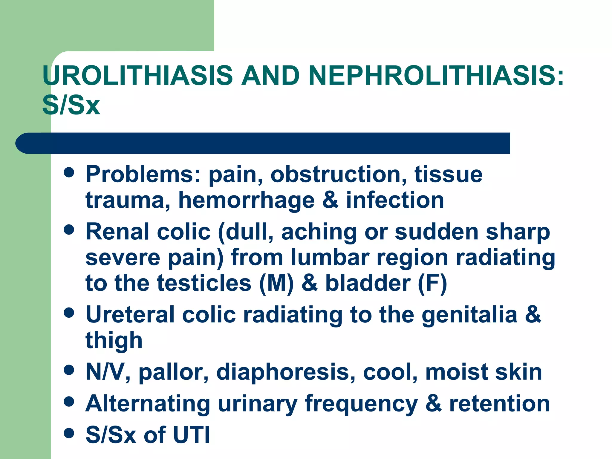
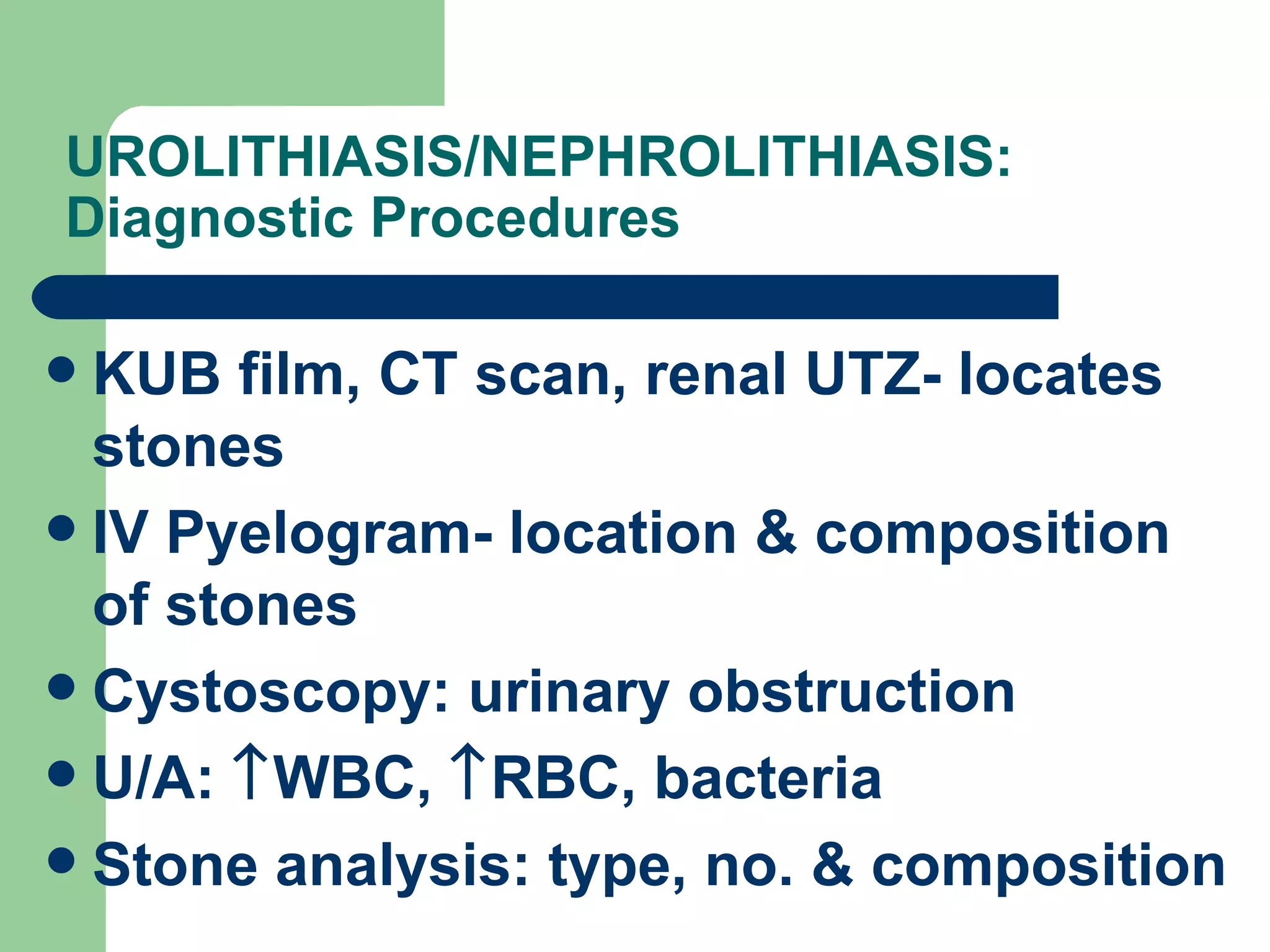
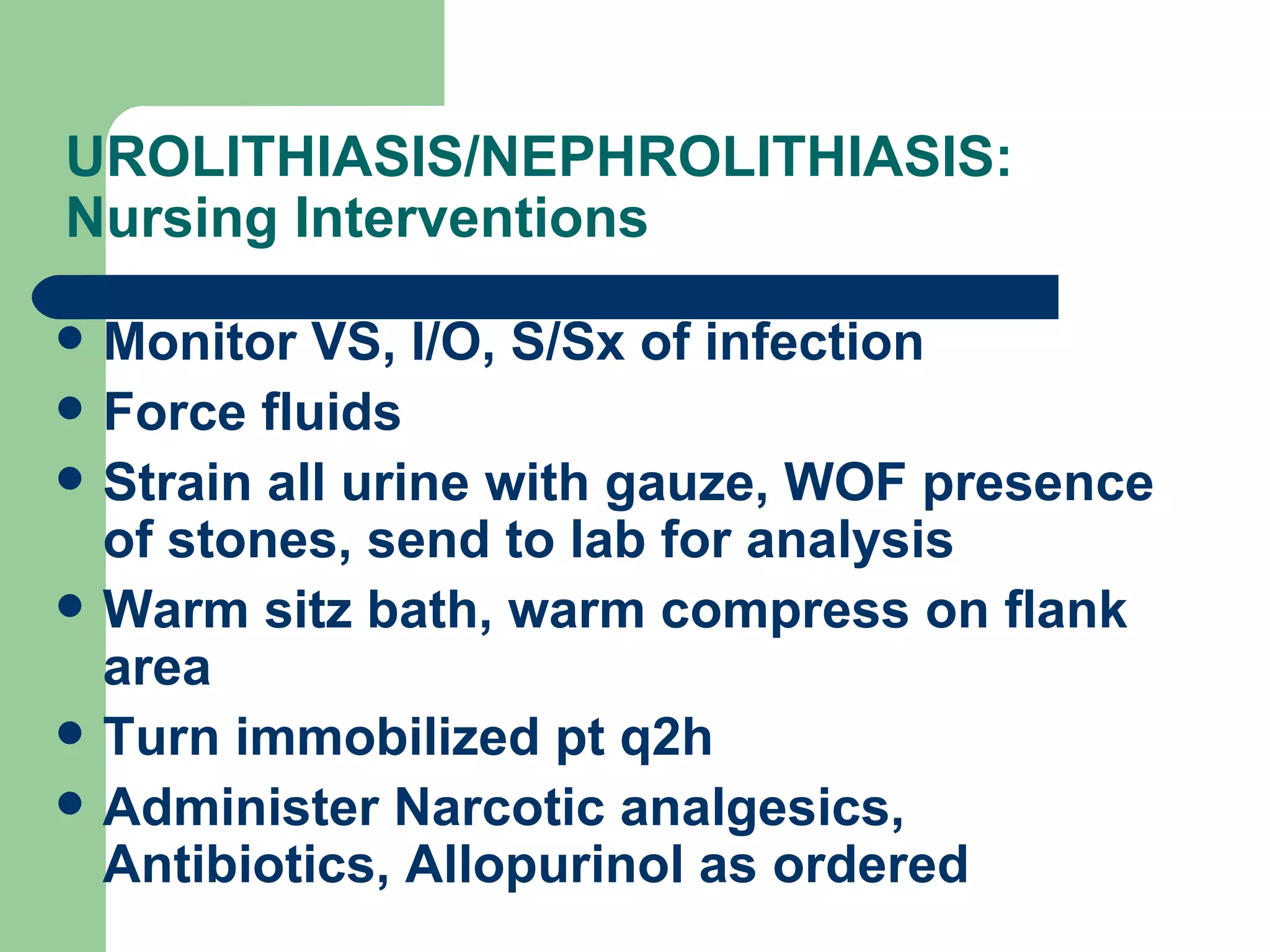
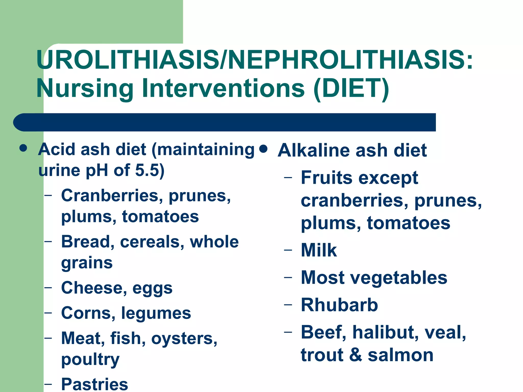
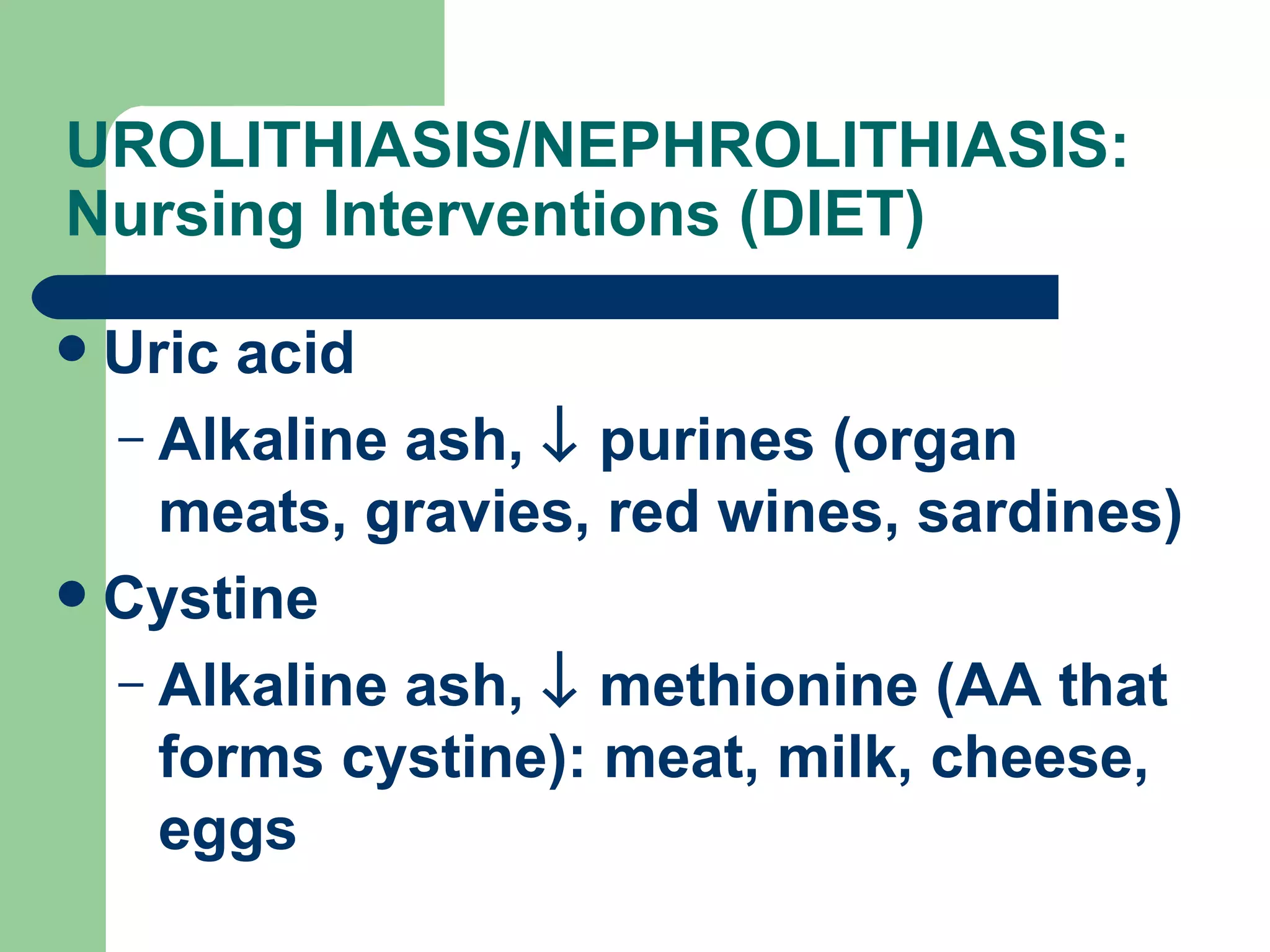
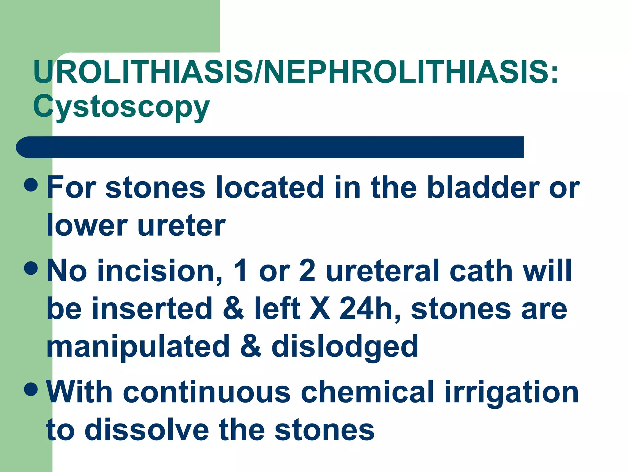
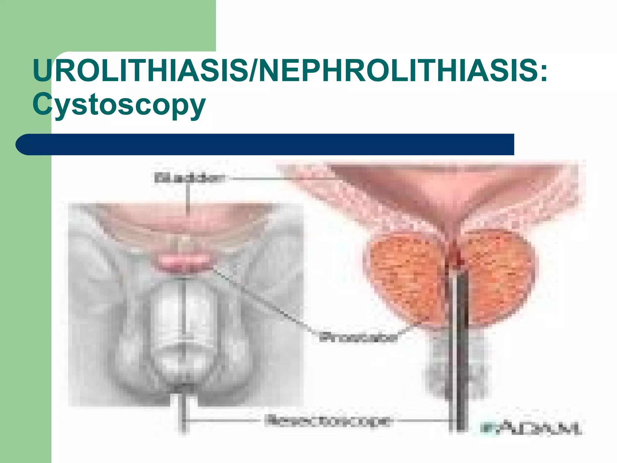
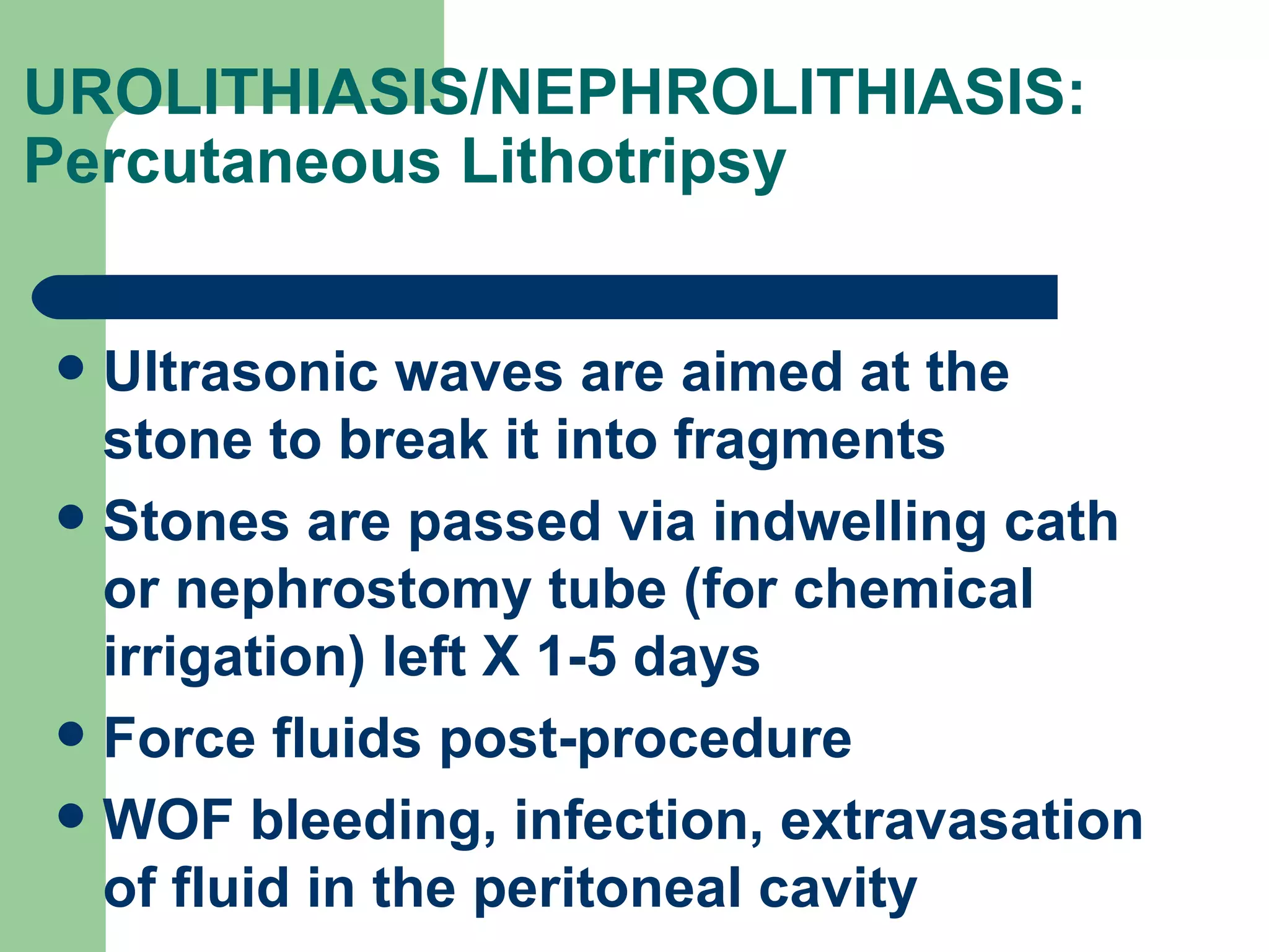
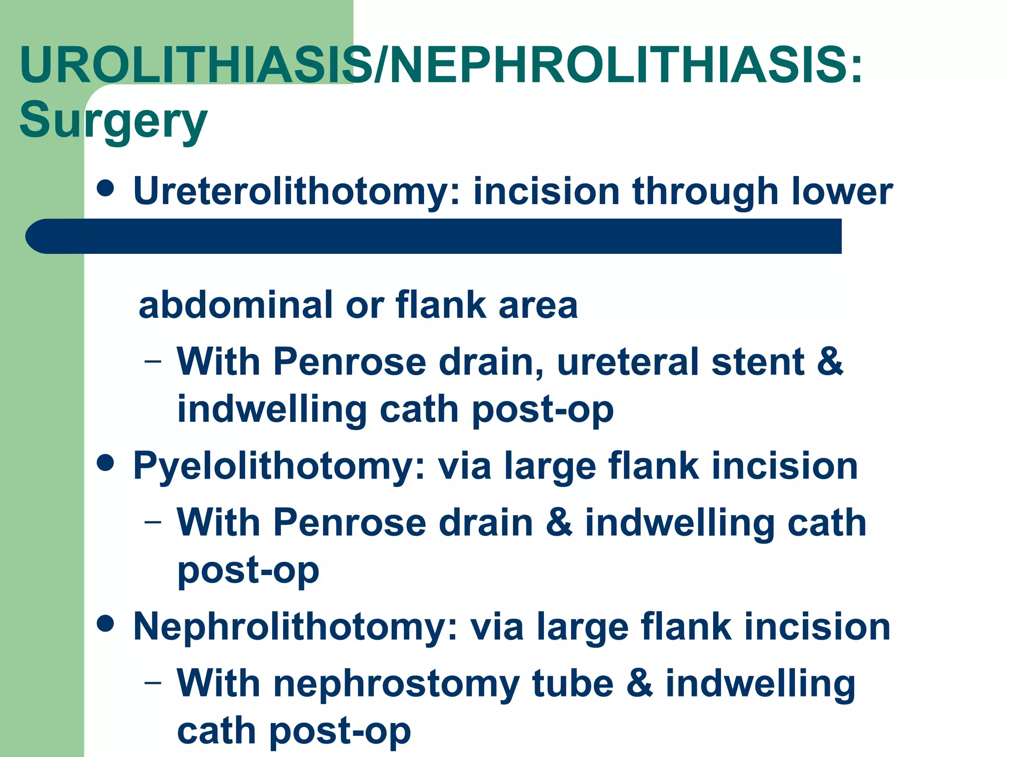
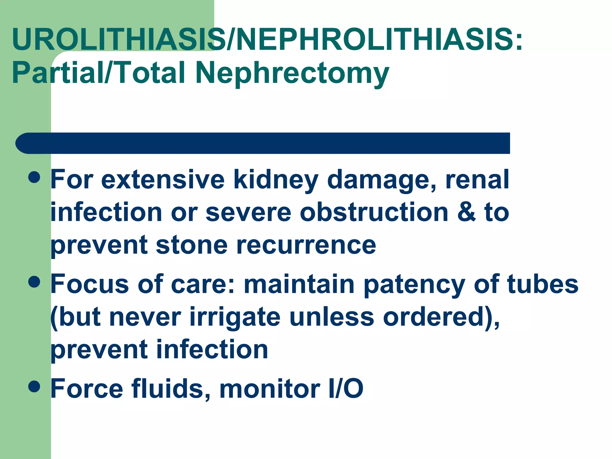
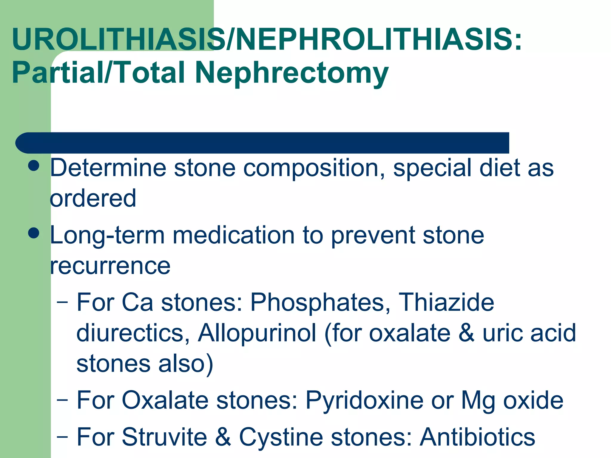
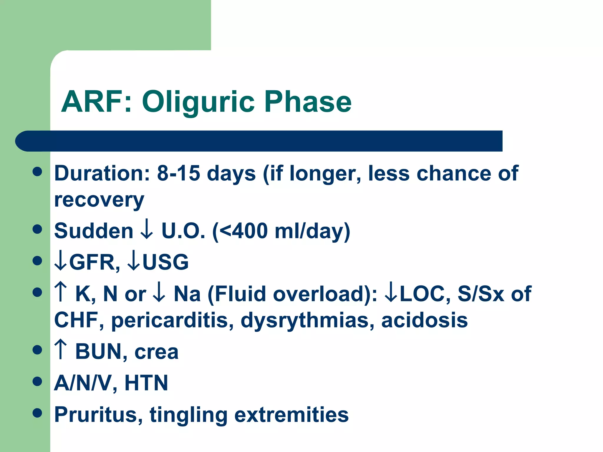
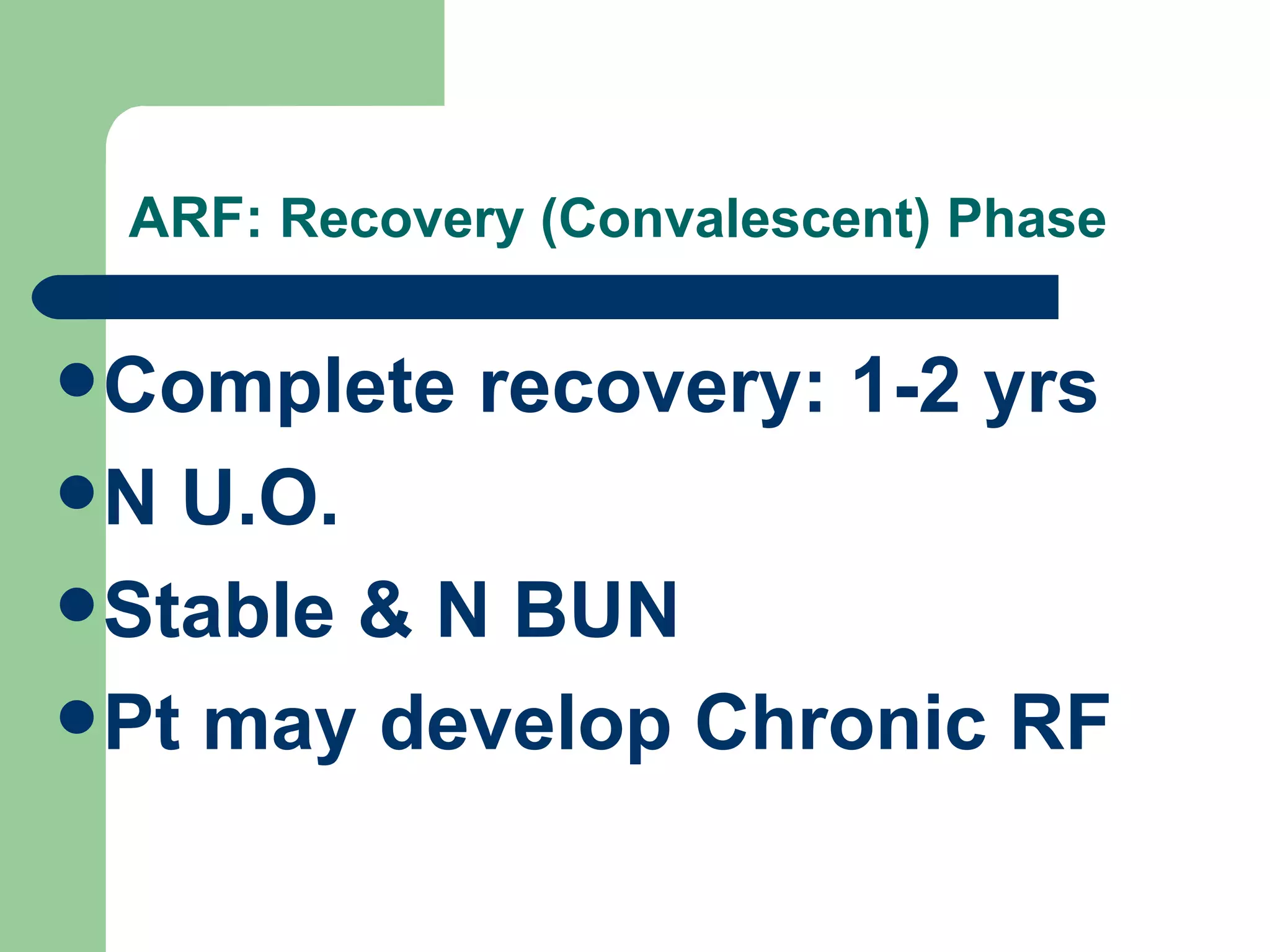
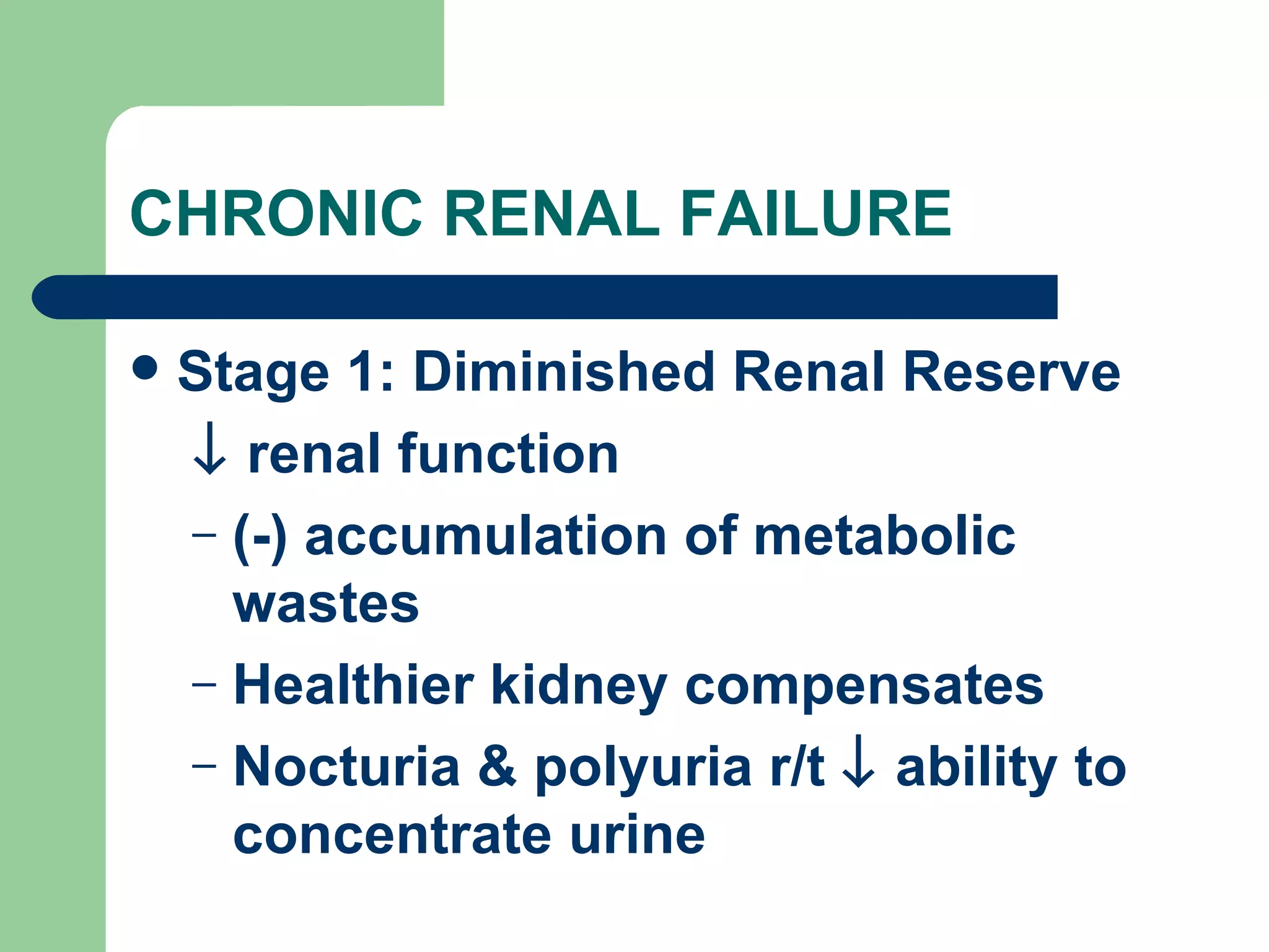
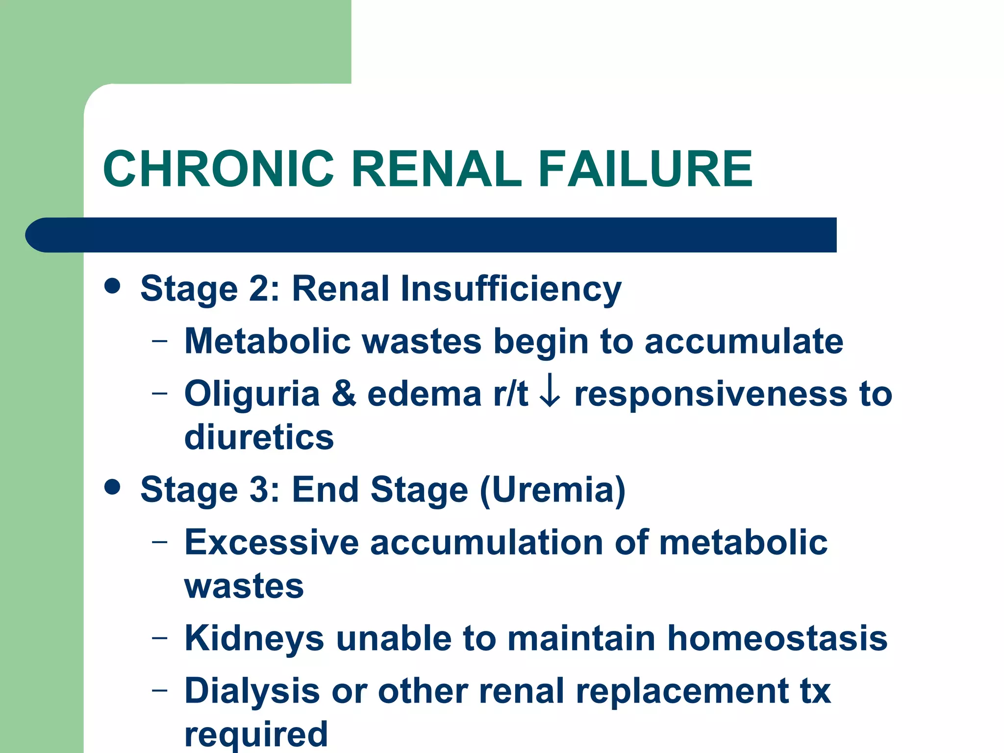
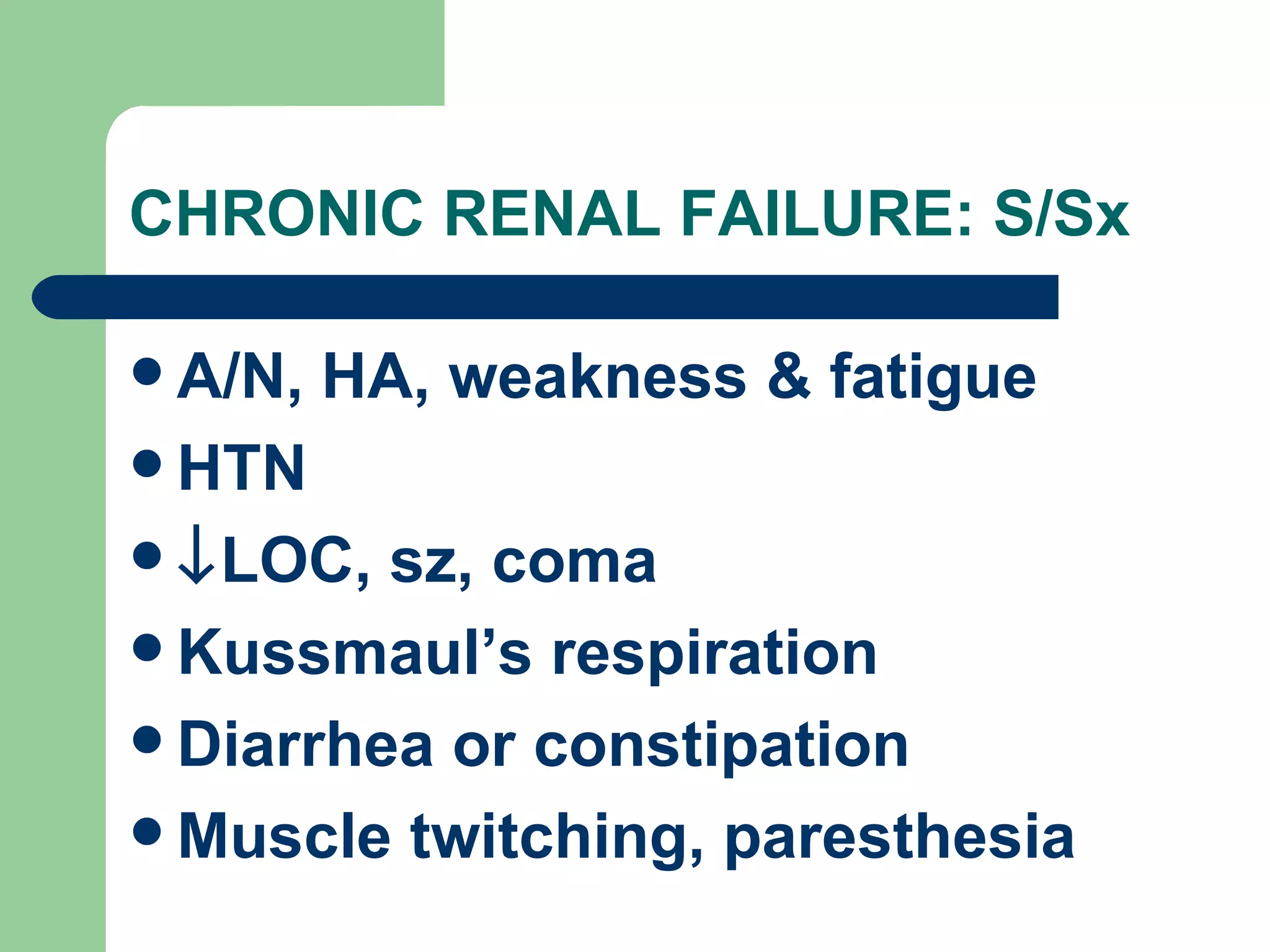
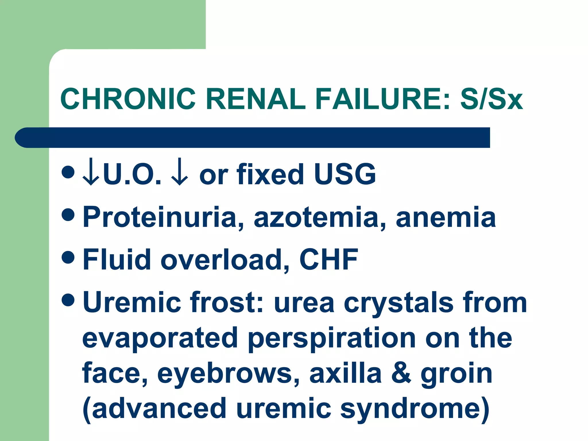
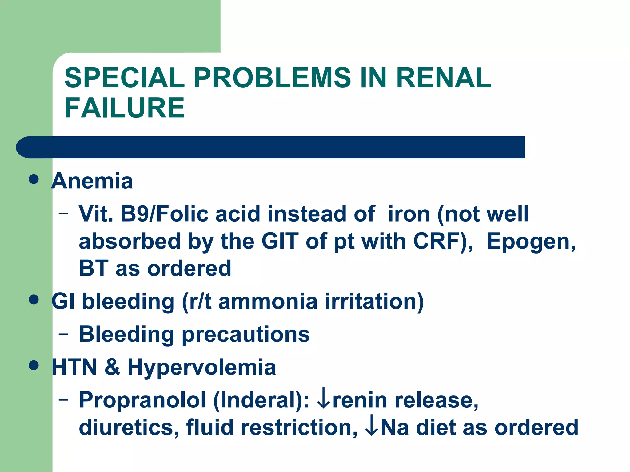
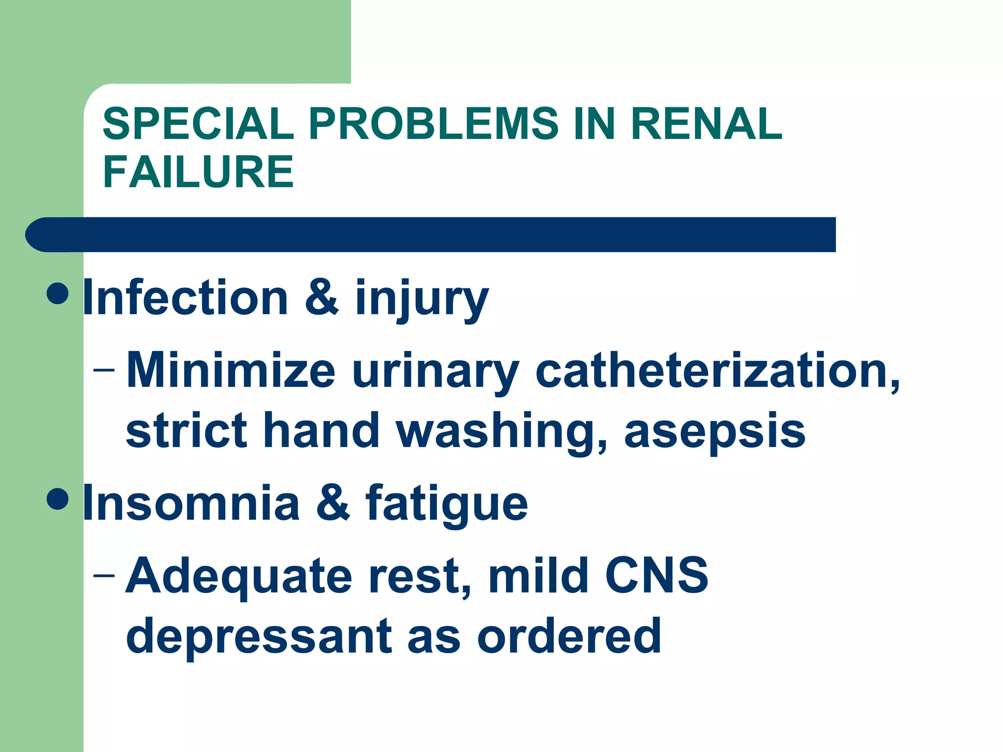
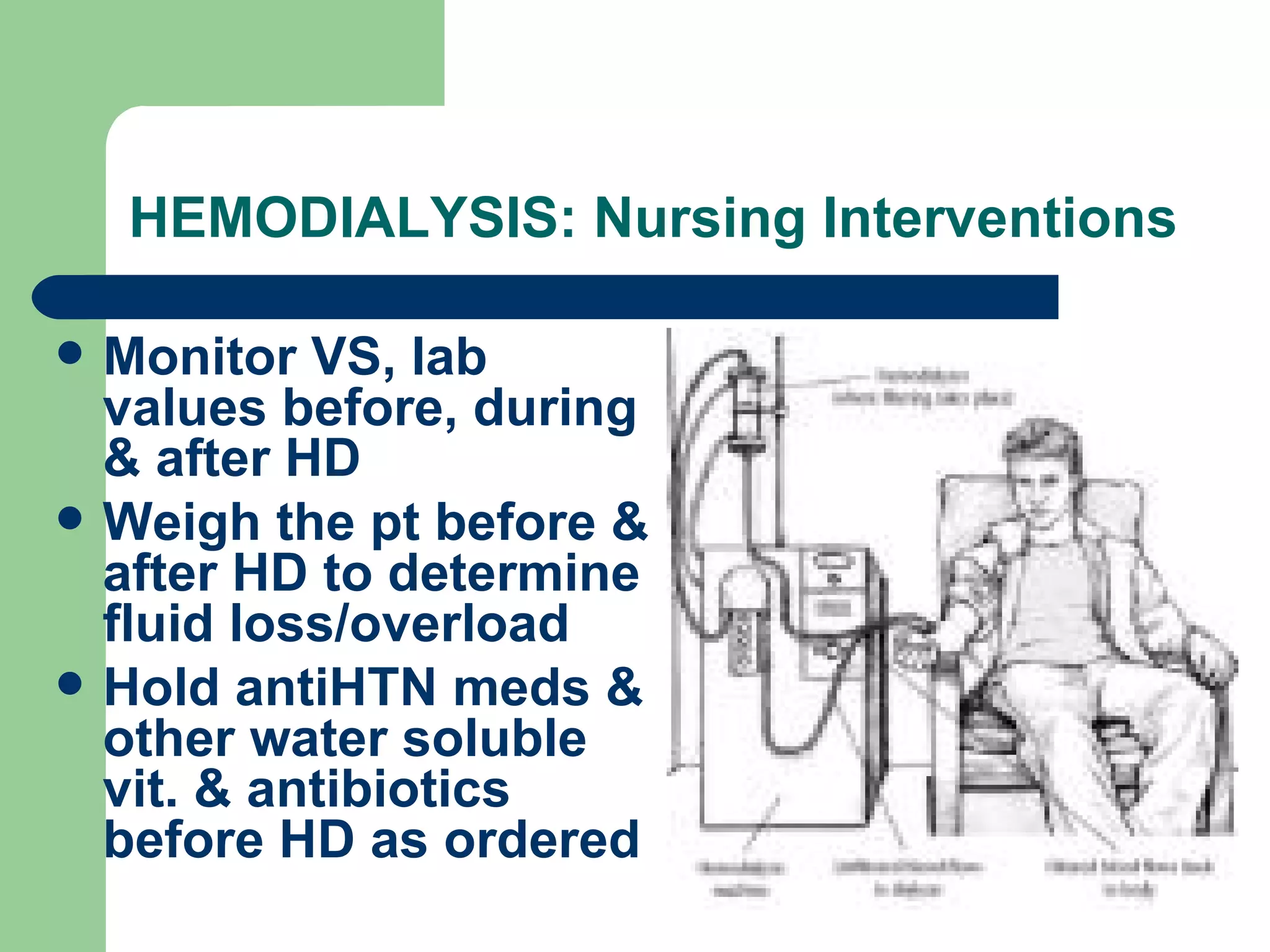
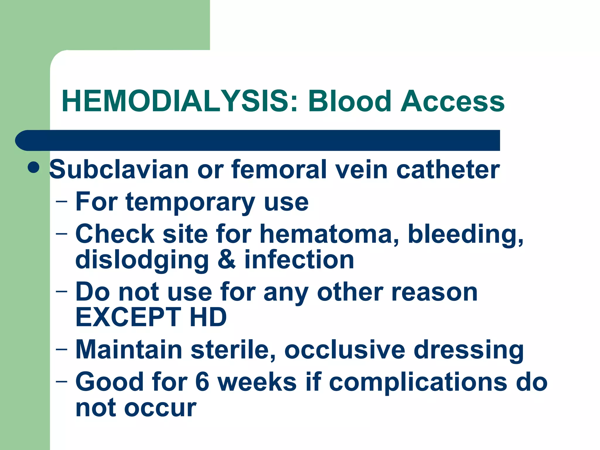
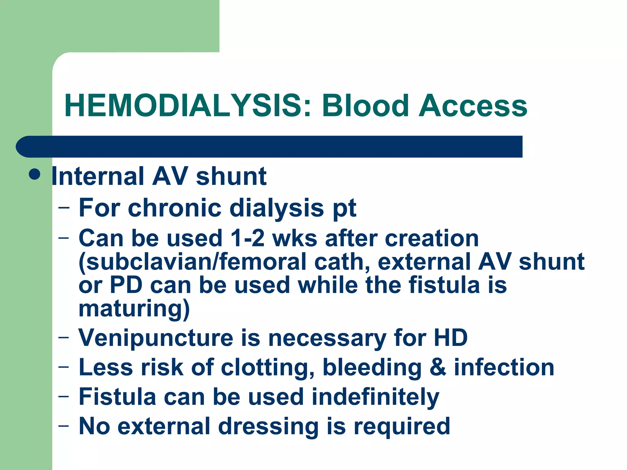
GENITOURINARY SYSTEM Ma. Victoria Recinto, RN, USRN
OVERVIEW Fluid, e+ & acid-base balance Excretion of the nitrogenous waste products, bacterial toxins, water-soluble drugs & drug metabolites Secrete renin & erythropoetin (role in parathyroid hormones & Vit D)
OVERVIEW: KIDNEYS A pair of bean-shaped organs located retroperitoneally at the back of peritoneum at either side of the vertebral column Parts: medulla, cortex & renal pelvis
OVERVIEW: NEPHRON Basic functional unit Composed of glomerulus (network of capillaries that filters blood) & tubules (proximal, distal & loop of Henle) Urine flows from the pelvis of the kidney through ureters & empties into bladder
OVERVIEW Kidney Function Urine formation Stages Filtration: GFR: 125 ml/min Tubular reabsorption: 124 ml reabsorbed Tubular secretion: 1 ml excreted
OVERVIEW Ureters 25 cm long, prevent reflux of urine back to the kidneys Bladder Behind symphysis pubis, elastic & muscular tissue that makes it distensible Can hold up to 1.2-1.8 L urine 250-500 cc of urine can trigger micturition
OVERVIEW Prostate gland Surrounds the male urethra Contains a duct that opens into the prostatic portion of the urethra & secretes the alkaline portion of seminal fluid
OVERVIEW Urethra- extends to the exterior surface of the body F: 2-5 cm/ 1-1.5 in M: 20 cm/ 8 in Catheter: Pedia: 8-10F, Adult F 12-14F, Adult M 14-16 F
CYSTITIS (UTI) Inflammation of the bladder r/t microbial invasion or urethral obstruction
CYSTITIS (UTI) Predisposing Factors Microbial invasion (80%- E. coli, Enterobacter, Pseudomonas & Serratia) Urinary obstruction & stagnation F>M (shorter urethra that is close to the rectum)    estrogen levels (affecting vaginal flora) Sexually active & pregnant woman
CYSTITIS (UTI): Causes Allergens/irritants: soaps, sprays, bubble bath, perfumed sanitary napkins Bladder distention, renal stones Indwelling urethral cath Urinary stasis
CYSTITIS (UTI): Causes Invasive UT procedures Poor-fitting diaphragms, spermicides Sexual intercourse Synthetic underwear & pantyhose Wet bathing suits
CYSTITIS (UTI): S/Sx Flank pain & tenderness Urinary frequency & urgency (incomplete bladder emptying) Dysuria (painful urination), bladder spasms Burning sensation upon urination Cloudy, dark, foul-smelling urine, Hematuria Fever, chills, A/N/V, malaise
CYSTITIS (UTI): Diagnostic Procedure  Urine C/S: determines the causative agent
CYSTITIS (UTI): Nursing Interventions Force fluids Heat on abdomen, Sitz bath as ordered  Monitor for the color, odor, blood in urine Strict asepsis in foley cath. insertion, maintain close system Meticulous perineal care Avoid caffeine & alcohol
CYSTITIS (UTI): Nursing Interventions Administer meds as ordered Systemic Antibiotics (Cephalosporin, Tetracycline, Ampicillin) Sulfonamides (Cotrimoxazole: Bactrim, Gantricin): can cause crystals in concentrated urine Urinary analgesic: Pyridium Antispasmodics
CYSTITIS (UTI): Nursing Interventions Acid ash diet (maintaining urine pH of 5.5) Bread, cereals, whole grains Cheese, eggs Corns, legumes Cranberries, prunes, plums, tomatoes Meat, fish, oysters, poultry Pastries  Prevent Cx: Pyelonephritis
Health Teaching: CYSTITIS (UTI) Prevention Good perineal care (wipe from front to back) Avoid bubble baths, tub baths, vaginal deodorants/sprays Void q 2-3 hrs (esp. for pregnant women) Void & drink a glass of water after intercourse
Health Teaching: CYSTITIS (UTI) Prevention Wear cotton pants, avoid tight clothes or pantyhose with slacks Avoid sitting in a wet bathing suit for prolonged periods of time Use estrogen vaginal creams to restore pH, use water-soluble lubricants for coitus (esp. for menopausal women)
BENIGN PROSTATIC HYPERTROPHY Slow enlargement of the prostate gland   urethral narrowing & obstruction Predisposing factors Male >40-50 y/o r/t hormonal influences
BENIGN PROSTATIC HYPERTROPHY S/Sx Urinary frequency, hesitancy, urgency,    urinary stream Terminal dribbling Backache Hematuria Dysuria, nocturia Burning sensation upon urination Urinary stasis, UTI
BENIGN PROSTATIC HYPERTROPHY Diagnostic Procedures Digital rectal exam: enlarged prostate gland Cystoscopy: urinary obstruction KUB- enlarged prostate gland U/A-   WBC,   RBC
BENIGN PROSTATIC HYPERTROPHY: Nursing Interventions  Force fluids unless contraindicated Bladder drainage via urinary cath as ordered Prostatic massage Administer as ordered Terazosin- relaxes urinary sphincters Finasteride (Proscar)- promotes atrophy of BPH Avoid meds that can cause urinary retention (anticholinergics, antihistamines, decongestants)
BENIGN PROSTATIC HYPERTROPHY: Nursing Interventions  Assist in surgery Prostatectomy (perineal, retropubic & suprapubic) Transurethral Resection of the Prostate (TURP) Cystoclysis: continuous bladder irrigation Irrigate the tube with pNSS to flush the clots WOF bleeding, hemorrhage Strict asepsis
PROSTATE CA: TURP Insertion of a scope into the urethra to excise prostatic tissue Bleeding is common post-op, WOF hemorrhage Continuous bladder irrigation (CBI) post-op to maintain the urine at a pink color
PROSTATE CA: TURP Bladder spasms are common post-op, give antispasmodics as ordered WOF dribbling & incontinence Sterility may or may not occur post-op
PROSTATE CA: Prostatectomy Point of comparison Suprapubic Retropubic Perineal Technique Via abdominal & bladder incision Via low abdominal incision without opening the bladder Via incision bet. scrotum & anus Hemorrhage Yes No  No Bladder spasms Yes Yes but less Urinary incontinence common
PROSTATE CA: Prostatectomy Point of comparison Suprapubic Retropubic Perineal CBI Yes Yes  - Sterility Yes Yes Yes Remarks Abdominal dressing soaked frequently with urine, Longer healing time than TURP Minimal abdominal drainage WOF infection, (No rectal tubes, rectal temp. taking & enema) Teach perineal exercises
Nursing Interventions: s/p TURP Monitor VS, U.O., hematuria &  clots, Hgb & Hct levels Force fluids Expect red to light pink urine for 24 hrs, turning to amber in 3 days (then encourage ambulation) WOF arterial bleeding (bright red urine with clots):    CBI & notify MD
Nursing Interventions: s/p TURP WOF venous bleeding (burgundy- colored urine): notify MD who will apply traction on the catheter Continuous urge to void is N but not encouraged to prevent bladder spasms Antibiotics, analgesics, stool softeners & antispasmodics as ordered
Nursing Interventions: s/p TURP Monitor 3-way foley catheter (for the  balloon (30-45 cc), inflow & outflow) Use pNSS only to prevent water intoxication or hypoNa (  LOC,   HR,   BP)
Nursing Interventions: s/p TURP Maintain infusion rate as ordered, if (+) clots:   rate For obstructed catheter: turn off CBI, irrigate with 30-50 ml pNSS, notify MD if it does not resolve CBI is d/c usually after 1-2 days, WOF continence & urinary retention
Discharge Health Teaching:  s/p TURP Avoid heavy lifting, stressful exercise, driving, Valsalva maneuver & sexual intercourse for 2-6 wks Drink 2.4-3L fluids/day before 8 pm
Discharge Health Teaching:  s/p TURP Avoid alcohol, caffeine & spicy foods to prevent overstimulation of the bladder Pt may pass small clots & tissue debris for several days If urine becomes less in amount & bloody, rest & force fluids, notify MD if persistent
Nursing Interventions:  s/p  Suprapubic Prostatectomy Monitor foley catheter & suprapubic catheter drainage As ordered, clamp the suprapubic cath after foley cath is removed (2-4 days post-op) & instruct the pt to void, measure residual urine by unclamping the cath & measuring the U.O.
Nursing Interventions:  s/p  Suprapubic Prostatectomy Prepare for removal of suprapubic cath if pt consistently empties bladder & residual urine is <75 ml
UROLITHIASIS AND NEPHROLITHIASIS Formation of stones elsewhere in the urinary tract (esp. in the kidneys)    obstruction    dilation (Hydroureter, Hydronephrosis)    RF Common type: Ca, oxalate, uric acid
UROLITHIASIS AND NEPHROLITHIASIS Predisposing Factors Diet:    Ca, Vit. D, milk, oxalate (chocolates), protein, purines or alkali Obstruction & urinary stasis, UTI, prolonged urinary catheterization Use of diuretics, Dehydration Obesity Sedentary lifestyle, Prolonged immobility Hyperparathyroidism (HyperCa), Gout Family hx
UROLITHIASIS AND NEPHROLITHIASIS: S/Sx Problems: pain, obstruction, tissue trauma, hemorrhage & infection Renal colic (dull, aching or sudden sharp severe pain) from lumbar region radiating to the testicles (M) & bladder (F) Ureteral colic radiating to the genitalia & thigh N/V, pallor, diaphoresis, cool, moist skin Alternating urinary frequency & retention S/Sx of UTI
UROLITHIASIS/NEPHROLITHIASIS: Diagnostic Procedures KUB film, CT scan, renal UTZ- locates stones IV Pyelogram- location & composition of stones Cystoscopy: urinary obstruction U/A:   WBC,   RBC, bacteria Stone analysis: type, no. & composition
UROLITHIASIS/NEPHROLITHIASIS: Nursing Interventions  Monitor VS, I/O, S/Sx of infection Force fluids Strain all urine with gauze, WOF presence of stones, send to lab for analysis Warm sitz bath, warm compress on flank area Turn immobilized pt q2h Administer Narcotic analgesics, Antibiotics, Allopurinol as ordered
UROLITHIASIS/NEPHROLITHIASIS: Nursing Interventions (DIET) Acid ash diet (maintaining urine pH of 5.5) Cranberries, prunes, plums, tomatoes Bread, cereals, whole grains Cheese, eggs Corns, legumes Meat, fish, oysters, poultry Pastries Alkaline ash diet  Fruits except cranberries, prunes, plums, tomatoes Milk Most vegetables Rhubarb Beef, halibut, veal, trout & salmon
UROLITHIASIS/NEPHROLITHIASIS: Nursing Interventions (DIET)  Calcium Phosphate Stones Acid ash,    Ca,    Phosphate,    Vit D Calcium Oxalate Acid ash,    Ca,    Oxalate (tea, almonds, cashews, chocolate, cocoa, beans, spinach & rhubarb) Struvite/ Triple Phosphate (Mg & NH3) Caused by urea splitting by bacteria Acid ash,    Phosphate (dairy products, red & organ meats, whole grains)
UROLITHIASIS/NEPHROLITHIASIS: Nursing Interventions (DIET)  Uric acid Alkaline ash,    purines (organ meats, gravies, red wines, sardines) Cystine Alkaline ash,    methionine (AA that forms cystine): meat, milk, cheese, eggs
UROLITHIASIS/NEPHROLITHIASIS: Nursing Interventions  Assist in surgery Nephrectomy: removal of 1 kidney Extracorporeal Shockwave Lithotripsy: if stones are recurrent Prevent Cx: ARF
UROLITHIASIS/NEPHROLITHIASIS: Cystoscopy  For stones located in the bladder or lower ureter No incision, 1 or 2 ureteral cath will be inserted & left X 24h, stones are manipulated & dislodged With continuous chemical irrigation to dissolve the stones
UROLITHIASIS/NEPHROLITHIASIS: Cystoscopy
UROLITHIASIS/NEPHROLITHIASIS: Extracorporeal Shockwave Lithotripsy  Non-invasive mechanical procedure for breaking up stones located in the kidney or upper ureter No incision or drain Fluoroscopy is used to visualize the stones
UROLITHIASIS/NEPHROLITHIASIS: Extracorporeal Shockwave Lithotripsy  Ultrasonic waves are delivered through bath of warm water Stones are passed in the urine within a few days NPO 8 hrs pre-procedure Force fluids & ambulation post-procedure WOF bleeding, pain, S/ of urinary obstruction
UROLITHIASIS/NEPHROLITHIASIS: Percutaneous Lithotripsy  Done via cystoscopy (no incision) or nephroscopy (with small flank incision) to break stones located in the KUB Fluoroscopy is used to visualize the stones
UROLITHIASIS/NEPHROLITHIASIS: Percutaneous Lithotripsy  Ultrasonic waves are aimed at the stone to break it into fragments Stones are passed via indwelling cath or nephrostomy tube (for chemical irrigation) left X 1-5 days Force fluids post-procedure WOF bleeding, infection, extravasation of fluid in the peritoneal cavity
UROLITHIASIS/NEPHROLITHIASIS: Surgery  Ureterolithotomy: incision through lower  abdominal or flank area With Penrose drain, ureteral stent & indwelling cath post-op Pyelolithotomy: via large flank incision  With Penrose drain & indwelling cath post-op Nephrolithotomy: via large flank incision  With nephrostomy tube & indwelling cath post-op
UROLITHIASIS/NEPHROLITHIASIS: Partial/Total Nephrectomy  For extensive kidney damage, renal infection or severe obstruction & to prevent stone recurrence Focus of care: maintain patency of tubes (but never irrigate unless ordered), prevent infection Force fluids, monitor I/O
UROLITHIASIS/NEPHROLITHIASIS: Partial/Total Nephrectomy  Determine stone composition, special diet as ordered Long-term medication to prevent stone recurrence For Ca stones: Phosphates, Thiazide diurectics, Allopurinol (for oxalate & uric acid stones also) For Oxalate stones: Pyridoxine or Mg oxide For Struvite & Cystine stones: Antibiotics
RENAL FAILURE Loss of kidney function S/Sx r/t retention of waste & fluids & inability to regulate e+ Causes Prerenal: DHN, hypovolemic shock,    C.O., vasodilation or vascular obstruction Intrarenal: ATN, nephrotoxicity, altered renal blood flow Postrenal: obstruction of urine flow
ARF: Oliguric Phase Duration: 8-15 days (if longer, less chance of recovery Sudden    U.O. (<400 ml/day)  GFR,   USG    K, N or    Na (Fluid overload):   LOC, S/Sx of CHF, pericarditis, dysrythmias, acidosis    BUN, crea A/N/V, HTN Pruritus, tingling extremities
ARF: Diuretic Phase Slow    U.O. then diuresis (4-5L/day)  GFR  K,   Na Hypovolemia,   BP,   HR, LOC improves Gradual    BUN, crea
ARF:  Recovery (Convalescent) Phase Complete recovery: 1-2 yrs N U.O.  Stable & N BUN Pt may develop Chronic RF
CHRONIC RENAL FAILURE Stage 1: Diminished Renal Reserve    renal function (-) accumulation of metabolic wastes Healthier kidney compensates Nocturia & polyuria r/t    ability to concentrate urine
CHRONIC RENAL FAILURE Stage 2: Renal Insufficiency Metabolic wastes begin to accumulate Oliguria & edema r/t    responsiveness to diuretics Stage 3: End Stage (Uremia) Excessive accumulation of metabolic wastes Kidneys unable to maintain homeostasis Dialysis or other renal replacement tx required
CHRONIC RENAL FAILURE: S/Sx A/N, HA, weakness & fatigue HTN  LOC, sz, coma Kussmaul’s respiration Diarrhea or constipation Muscle twitching, paresthesia
CHRONIC RENAL FAILURE: S/Sx  U.O.    or fixed USG Proteinuria, azotemia, anemia Fluid overload, CHF Uremic frost: urea crystals from evaporated perspiration on the face, eyebrows, axilla & groin (advanced uremic syndrome)
SPECIAL PROBLEMS IN RENAL FAILURE Anemia  Vit. B9/Folic acid instead of  iron (not well absorbed by the GIT of pt with CRF),  Epogen, BT as ordered GI bleeding (r/t ammonia irritation) Bleeding precautions HTN & Hypervolemia Propranolol (Inderal):   renin release, diuretics, fluid restriction,   Na diet as ordered
SPECIAL PROBLEMS IN RENAL FAILURE Infection & injury Minimize urinary catheterization, strict hand washing, asepsis Insomnia & fatigue Adequate rest, mild CNS depressant as ordered
SPECIAL PROBLEMS IN RENAL FAILURE HypoCa, Hyperphosphatemia, HyperK  Diet, dialysis Metabolic acidosis NaHCO3 as ordered (pt with CRF adjusted to low HCO3 levels without getting acutely ill) Muscle cramps e+ replacement, heat & massage as ordered Pruritus (r/t uremic frost)  Skin care, avoid soaps, antipruritics as ordered
SPECIAL PROBLEMS IN RENAL FAILURE Neuro changes Monitor LOC, side rails up, calm & restful env’t, comfort measures & backrubs Occular irritation (r/t Ca deposits in conjunctiva)  Ca & Phosphate binders, Eye drops as ordered Psychosocial problems
HEMODIALYSIS: FUNCTIONS Cleanses the blood with accumulated  waste products (urea, crea & uric acid) Removes excessive fluids Restores buffer system & e+ levels of the body
HEMODIALYSIS: PRINCIPLES Semipermeable membrane: thin, porous cellophane (only water, urea, crea & uric acid can pass through) Proteins, bacteria & some blood cells are too large to pass through Blood flows into the dialyzer & into the dialysate
HEMODIALYSIS: PRINCIPLES Diffusion:  mov’t of particles from greater to lesser concentration Osmosis:  mov’t of fluids across a semipermeable membrane from lesser to greater concentration Ultrafiltration:  mov’t of fluids across a semipermeable membrane due to artificial pressure gradient
HEMODIALYSIS: Nursing Interventions Monitor VS, lab values before, during & after HD Weigh the pt before & after HD to determine fluid loss/overload Hold antiHTN meds & other water soluble vit. & antibiotics before HD as ordered
HEMODIALYSIS: Nursing Interventions Provide adequate nutrition (pt may eat before HD) WOF hypovolemic shock  Assess the patency of blood access device
HEMODIALYSIS: Blood Access Subclavian or femoral vein catheter For temporary use Check site for hematoma, bleeding, dislodging & infection Do not use for any other reason EXCEPT HD Maintain sterile, occlusive dressing Good for 6 weeks if complications do not occur
HEMODIALYSIS: Blood Access External AV shunt Surgical insertion of 2 Silastic cannulas into an artery & a vein (U-shaped) in the forearm or leg to form an external blood path Can be used immediately after creation No venipuncture is necessary for HD Shunt may be disconnected or dislodged WOF hemorrhage, infection, clotting (tingling or discomfort on the extremity) & skin erosion at the cath site
HEMODIALYSIS: Blood Access
HEMODIALYSIS: Blood Access External AV shunt Keep the shunt dressing dry & intact Prepare cannula clamps at bedside No BP taking, blood extraction, injection & venipunctures in the shunt extremity A patent shunt is warm to touch Auscultate for bruit & palpate for thrill WOF circulatory impairment in the shunt extremity
HEMODIALYSIS: Blood Access Internal AV shunt For chronic dialysis pt Can be used 1-2 wks after creation (subclavian/femoral cath, external AV shunt or PD can be used while the fistula is maturing) Venipuncture is necessary for HD Less risk of clotting, bleeding & infection Fistula can be used indefinitely No external dressing is required
HEMODIALYSIS: Blood Access Internal AV shunt Infiltration of needles   hematoma Aneurysm can form in the fistula CHF can occur from    blood flow to the venous system WOF  arterial steal syndrome : compromised arterial perfusion r/t    blood diverted to the vein & refer to MD
HEMODIALYSIS: Blood Access Internal AV graft For chronic dialysis pt who do not have adequate blood vessels for fistula creation Artificial graft (Gore-Tex or bovine carotid artery) is used Can be used 2 wks after creation  Dis/Advantages: same as in internal AV fistula
HEMODIALYSIS: Complications Disequilibrium syndrome Dialysis encephalopathy Electrolyte changes Muscle cramping Blood loss, hypoTN & shock Hepatitis Sepsis
HEMODIALYSIS: Disequilibrium syndrome Solutes are removed from the blood faster than from the CSF & brain   cerebral edema S/Sx: N/V, HA, HTN,   LOC, sz Prepare to dialyze the pt for a shorter pd. at    blood flow rates    env’tal stimuli Refer to MD
HEMODIALYSIS: Dialysis Encephalopathy Al toxicity r/t water source of the dialysate & ingestion of Al-containing antacids (phosphate binders) S/Sx:   LOC, sz, speech disturbance, dementia, bone pain Refer to MD Al-chelating agents as ordered

Genitourinary System

  • 1.
    GENITOURINARY SYSTEM Ma.Victoria Recinto, RN, USRN
  • 2.
    OVERVIEW Fluid, e+& acid-base balance Excretion of the nitrogenous waste products, bacterial toxins, water-soluble drugs & drug metabolites Secrete renin & erythropoetin (role in parathyroid hormones & Vit D)
  • 3.
    OVERVIEW: KIDNEYS Apair of bean-shaped organs located retroperitoneally at the back of peritoneum at either side of the vertebral column Parts: medulla, cortex & renal pelvis
  • 4.
    OVERVIEW: NEPHRON Basicfunctional unit Composed of glomerulus (network of capillaries that filters blood) & tubules (proximal, distal & loop of Henle) Urine flows from the pelvis of the kidney through ureters & empties into bladder
  • 5.
    OVERVIEW Kidney FunctionUrine formation Stages Filtration: GFR: 125 ml/min Tubular reabsorption: 124 ml reabsorbed Tubular secretion: 1 ml excreted
  • 6.
    OVERVIEW Ureters 25cm long, prevent reflux of urine back to the kidneys Bladder Behind symphysis pubis, elastic & muscular tissue that makes it distensible Can hold up to 1.2-1.8 L urine 250-500 cc of urine can trigger micturition
  • 7.
    OVERVIEW Prostate glandSurrounds the male urethra Contains a duct that opens into the prostatic portion of the urethra & secretes the alkaline portion of seminal fluid
  • 8.
    OVERVIEW Urethra- extendsto the exterior surface of the body F: 2-5 cm/ 1-1.5 in M: 20 cm/ 8 in Catheter: Pedia: 8-10F, Adult F 12-14F, Adult M 14-16 F
  • 9.
    CYSTITIS (UTI) Inflammationof the bladder r/t microbial invasion or urethral obstruction
  • 10.
    CYSTITIS (UTI) PredisposingFactors Microbial invasion (80%- E. coli, Enterobacter, Pseudomonas & Serratia) Urinary obstruction & stagnation F>M (shorter urethra that is close to the rectum)  estrogen levels (affecting vaginal flora) Sexually active & pregnant woman
  • 11.
    CYSTITIS (UTI): CausesAllergens/irritants: soaps, sprays, bubble bath, perfumed sanitary napkins Bladder distention, renal stones Indwelling urethral cath Urinary stasis
  • 12.
    CYSTITIS (UTI): CausesInvasive UT procedures Poor-fitting diaphragms, spermicides Sexual intercourse Synthetic underwear & pantyhose Wet bathing suits
  • 13.
    CYSTITIS (UTI): S/SxFlank pain & tenderness Urinary frequency & urgency (incomplete bladder emptying) Dysuria (painful urination), bladder spasms Burning sensation upon urination Cloudy, dark, foul-smelling urine, Hematuria Fever, chills, A/N/V, malaise
  • 14.
    CYSTITIS (UTI): DiagnosticProcedure Urine C/S: determines the causative agent
  • 15.
    CYSTITIS (UTI): NursingInterventions Force fluids Heat on abdomen, Sitz bath as ordered Monitor for the color, odor, blood in urine Strict asepsis in foley cath. insertion, maintain close system Meticulous perineal care Avoid caffeine & alcohol
  • 16.
    CYSTITIS (UTI): NursingInterventions Administer meds as ordered Systemic Antibiotics (Cephalosporin, Tetracycline, Ampicillin) Sulfonamides (Cotrimoxazole: Bactrim, Gantricin): can cause crystals in concentrated urine Urinary analgesic: Pyridium Antispasmodics
  • 17.
    CYSTITIS (UTI): NursingInterventions Acid ash diet (maintaining urine pH of 5.5) Bread, cereals, whole grains Cheese, eggs Corns, legumes Cranberries, prunes, plums, tomatoes Meat, fish, oysters, poultry Pastries Prevent Cx: Pyelonephritis
  • 18.
    Health Teaching: CYSTITIS(UTI) Prevention Good perineal care (wipe from front to back) Avoid bubble baths, tub baths, vaginal deodorants/sprays Void q 2-3 hrs (esp. for pregnant women) Void & drink a glass of water after intercourse
  • 19.
    Health Teaching: CYSTITIS(UTI) Prevention Wear cotton pants, avoid tight clothes or pantyhose with slacks Avoid sitting in a wet bathing suit for prolonged periods of time Use estrogen vaginal creams to restore pH, use water-soluble lubricants for coitus (esp. for menopausal women)
  • 20.
    BENIGN PROSTATIC HYPERTROPHYSlow enlargement of the prostate gland  urethral narrowing & obstruction Predisposing factors Male >40-50 y/o r/t hormonal influences
  • 21.
    BENIGN PROSTATIC HYPERTROPHYS/Sx Urinary frequency, hesitancy, urgency,  urinary stream Terminal dribbling Backache Hematuria Dysuria, nocturia Burning sensation upon urination Urinary stasis, UTI
  • 22.
    BENIGN PROSTATIC HYPERTROPHYDiagnostic Procedures Digital rectal exam: enlarged prostate gland Cystoscopy: urinary obstruction KUB- enlarged prostate gland U/A-  WBC,  RBC
  • 23.
    BENIGN PROSTATIC HYPERTROPHY:Nursing Interventions Force fluids unless contraindicated Bladder drainage via urinary cath as ordered Prostatic massage Administer as ordered Terazosin- relaxes urinary sphincters Finasteride (Proscar)- promotes atrophy of BPH Avoid meds that can cause urinary retention (anticholinergics, antihistamines, decongestants)
  • 24.
    BENIGN PROSTATIC HYPERTROPHY:Nursing Interventions Assist in surgery Prostatectomy (perineal, retropubic & suprapubic) Transurethral Resection of the Prostate (TURP) Cystoclysis: continuous bladder irrigation Irrigate the tube with pNSS to flush the clots WOF bleeding, hemorrhage Strict asepsis
  • 25.
    PROSTATE CA: TURPInsertion of a scope into the urethra to excise prostatic tissue Bleeding is common post-op, WOF hemorrhage Continuous bladder irrigation (CBI) post-op to maintain the urine at a pink color
  • 26.
    PROSTATE CA: TURPBladder spasms are common post-op, give antispasmodics as ordered WOF dribbling & incontinence Sterility may or may not occur post-op
  • 27.
    PROSTATE CA: ProstatectomyPoint of comparison Suprapubic Retropubic Perineal Technique Via abdominal & bladder incision Via low abdominal incision without opening the bladder Via incision bet. scrotum & anus Hemorrhage Yes No No Bladder spasms Yes Yes but less Urinary incontinence common
  • 28.
    PROSTATE CA: ProstatectomyPoint of comparison Suprapubic Retropubic Perineal CBI Yes Yes - Sterility Yes Yes Yes Remarks Abdominal dressing soaked frequently with urine, Longer healing time than TURP Minimal abdominal drainage WOF infection, (No rectal tubes, rectal temp. taking & enema) Teach perineal exercises
  • 29.
    Nursing Interventions: s/pTURP Monitor VS, U.O., hematuria & clots, Hgb & Hct levels Force fluids Expect red to light pink urine for 24 hrs, turning to amber in 3 days (then encourage ambulation) WOF arterial bleeding (bright red urine with clots):  CBI & notify MD
  • 30.
    Nursing Interventions: s/pTURP WOF venous bleeding (burgundy- colored urine): notify MD who will apply traction on the catheter Continuous urge to void is N but not encouraged to prevent bladder spasms Antibiotics, analgesics, stool softeners & antispasmodics as ordered
  • 31.
    Nursing Interventions: s/pTURP Monitor 3-way foley catheter (for the balloon (30-45 cc), inflow & outflow) Use pNSS only to prevent water intoxication or hypoNa (  LOC,  HR,  BP)
  • 32.
    Nursing Interventions: s/pTURP Maintain infusion rate as ordered, if (+) clots:  rate For obstructed catheter: turn off CBI, irrigate with 30-50 ml pNSS, notify MD if it does not resolve CBI is d/c usually after 1-2 days, WOF continence & urinary retention
  • 33.
    Discharge Health Teaching: s/p TURP Avoid heavy lifting, stressful exercise, driving, Valsalva maneuver & sexual intercourse for 2-6 wks Drink 2.4-3L fluids/day before 8 pm
  • 34.
    Discharge Health Teaching: s/p TURP Avoid alcohol, caffeine & spicy foods to prevent overstimulation of the bladder Pt may pass small clots & tissue debris for several days If urine becomes less in amount & bloody, rest & force fluids, notify MD if persistent
  • 35.
    Nursing Interventions: s/p Suprapubic Prostatectomy Monitor foley catheter & suprapubic catheter drainage As ordered, clamp the suprapubic cath after foley cath is removed (2-4 days post-op) & instruct the pt to void, measure residual urine by unclamping the cath & measuring the U.O.
  • 36.
    Nursing Interventions: s/p Suprapubic Prostatectomy Prepare for removal of suprapubic cath if pt consistently empties bladder & residual urine is <75 ml
  • 37.
    UROLITHIASIS AND NEPHROLITHIASISFormation of stones elsewhere in the urinary tract (esp. in the kidneys)  obstruction  dilation (Hydroureter, Hydronephrosis)  RF Common type: Ca, oxalate, uric acid
  • 38.
    UROLITHIASIS AND NEPHROLITHIASISPredisposing Factors Diet:  Ca, Vit. D, milk, oxalate (chocolates), protein, purines or alkali Obstruction & urinary stasis, UTI, prolonged urinary catheterization Use of diuretics, Dehydration Obesity Sedentary lifestyle, Prolonged immobility Hyperparathyroidism (HyperCa), Gout Family hx
  • 39.
    UROLITHIASIS AND NEPHROLITHIASIS:S/Sx Problems: pain, obstruction, tissue trauma, hemorrhage & infection Renal colic (dull, aching or sudden sharp severe pain) from lumbar region radiating to the testicles (M) & bladder (F) Ureteral colic radiating to the genitalia & thigh N/V, pallor, diaphoresis, cool, moist skin Alternating urinary frequency & retention S/Sx of UTI
  • 40.
    UROLITHIASIS/NEPHROLITHIASIS: Diagnostic ProceduresKUB film, CT scan, renal UTZ- locates stones IV Pyelogram- location & composition of stones Cystoscopy: urinary obstruction U/A:  WBC,  RBC, bacteria Stone analysis: type, no. & composition
  • 41.
    UROLITHIASIS/NEPHROLITHIASIS: Nursing Interventions Monitor VS, I/O, S/Sx of infection Force fluids Strain all urine with gauze, WOF presence of stones, send to lab for analysis Warm sitz bath, warm compress on flank area Turn immobilized pt q2h Administer Narcotic analgesics, Antibiotics, Allopurinol as ordered
  • 42.
    UROLITHIASIS/NEPHROLITHIASIS: Nursing Interventions(DIET) Acid ash diet (maintaining urine pH of 5.5) Cranberries, prunes, plums, tomatoes Bread, cereals, whole grains Cheese, eggs Corns, legumes Meat, fish, oysters, poultry Pastries Alkaline ash diet Fruits except cranberries, prunes, plums, tomatoes Milk Most vegetables Rhubarb Beef, halibut, veal, trout & salmon
  • 43.
    UROLITHIASIS/NEPHROLITHIASIS: Nursing Interventions(DIET) Calcium Phosphate Stones Acid ash,  Ca,  Phosphate,  Vit D Calcium Oxalate Acid ash,  Ca,  Oxalate (tea, almonds, cashews, chocolate, cocoa, beans, spinach & rhubarb) Struvite/ Triple Phosphate (Mg & NH3) Caused by urea splitting by bacteria Acid ash,  Phosphate (dairy products, red & organ meats, whole grains)
  • 44.
    UROLITHIASIS/NEPHROLITHIASIS: Nursing Interventions(DIET) Uric acid Alkaline ash,  purines (organ meats, gravies, red wines, sardines) Cystine Alkaline ash,  methionine (AA that forms cystine): meat, milk, cheese, eggs
  • 45.
    UROLITHIASIS/NEPHROLITHIASIS: Nursing Interventions Assist in surgery Nephrectomy: removal of 1 kidney Extracorporeal Shockwave Lithotripsy: if stones are recurrent Prevent Cx: ARF
  • 46.
    UROLITHIASIS/NEPHROLITHIASIS: Cystoscopy For stones located in the bladder or lower ureter No incision, 1 or 2 ureteral cath will be inserted & left X 24h, stones are manipulated & dislodged With continuous chemical irrigation to dissolve the stones
  • 47.
  • 48.
    UROLITHIASIS/NEPHROLITHIASIS: Extracorporeal ShockwaveLithotripsy Non-invasive mechanical procedure for breaking up stones located in the kidney or upper ureter No incision or drain Fluoroscopy is used to visualize the stones
  • 49.
    UROLITHIASIS/NEPHROLITHIASIS: Extracorporeal ShockwaveLithotripsy Ultrasonic waves are delivered through bath of warm water Stones are passed in the urine within a few days NPO 8 hrs pre-procedure Force fluids & ambulation post-procedure WOF bleeding, pain, S/ of urinary obstruction
  • 50.
    UROLITHIASIS/NEPHROLITHIASIS: Percutaneous Lithotripsy Done via cystoscopy (no incision) or nephroscopy (with small flank incision) to break stones located in the KUB Fluoroscopy is used to visualize the stones
  • 51.
    UROLITHIASIS/NEPHROLITHIASIS: Percutaneous Lithotripsy Ultrasonic waves are aimed at the stone to break it into fragments Stones are passed via indwelling cath or nephrostomy tube (for chemical irrigation) left X 1-5 days Force fluids post-procedure WOF bleeding, infection, extravasation of fluid in the peritoneal cavity
  • 52.
    UROLITHIASIS/NEPHROLITHIASIS: Surgery Ureterolithotomy: incision through lower abdominal or flank area With Penrose drain, ureteral stent & indwelling cath post-op Pyelolithotomy: via large flank incision With Penrose drain & indwelling cath post-op Nephrolithotomy: via large flank incision With nephrostomy tube & indwelling cath post-op
  • 53.
    UROLITHIASIS/NEPHROLITHIASIS: Partial/Total Nephrectomy For extensive kidney damage, renal infection or severe obstruction & to prevent stone recurrence Focus of care: maintain patency of tubes (but never irrigate unless ordered), prevent infection Force fluids, monitor I/O
  • 54.
    UROLITHIASIS/NEPHROLITHIASIS: Partial/Total Nephrectomy Determine stone composition, special diet as ordered Long-term medication to prevent stone recurrence For Ca stones: Phosphates, Thiazide diurectics, Allopurinol (for oxalate & uric acid stones also) For Oxalate stones: Pyridoxine or Mg oxide For Struvite & Cystine stones: Antibiotics
  • 55.
    RENAL FAILURE Lossof kidney function S/Sx r/t retention of waste & fluids & inability to regulate e+ Causes Prerenal: DHN, hypovolemic shock,  C.O., vasodilation or vascular obstruction Intrarenal: ATN, nephrotoxicity, altered renal blood flow Postrenal: obstruction of urine flow
  • 56.
    ARF: Oliguric PhaseDuration: 8-15 days (if longer, less chance of recovery Sudden  U.O. (<400 ml/day)  GFR,  USG  K, N or  Na (Fluid overload):  LOC, S/Sx of CHF, pericarditis, dysrythmias, acidosis  BUN, crea A/N/V, HTN Pruritus, tingling extremities
  • 57.
    ARF: Diuretic PhaseSlow  U.O. then diuresis (4-5L/day)  GFR  K,  Na Hypovolemia,  BP,  HR, LOC improves Gradual  BUN, crea
  • 58.
    ARF: Recovery(Convalescent) Phase Complete recovery: 1-2 yrs N U.O. Stable & N BUN Pt may develop Chronic RF
  • 59.
    CHRONIC RENAL FAILUREStage 1: Diminished Renal Reserve  renal function (-) accumulation of metabolic wastes Healthier kidney compensates Nocturia & polyuria r/t  ability to concentrate urine
  • 60.
    CHRONIC RENAL FAILUREStage 2: Renal Insufficiency Metabolic wastes begin to accumulate Oliguria & edema r/t  responsiveness to diuretics Stage 3: End Stage (Uremia) Excessive accumulation of metabolic wastes Kidneys unable to maintain homeostasis Dialysis or other renal replacement tx required
  • 61.
    CHRONIC RENAL FAILURE:S/Sx A/N, HA, weakness & fatigue HTN  LOC, sz, coma Kussmaul’s respiration Diarrhea or constipation Muscle twitching, paresthesia
  • 62.
    CHRONIC RENAL FAILURE:S/Sx  U.O.  or fixed USG Proteinuria, azotemia, anemia Fluid overload, CHF Uremic frost: urea crystals from evaporated perspiration on the face, eyebrows, axilla & groin (advanced uremic syndrome)
  • 63.
    SPECIAL PROBLEMS INRENAL FAILURE Anemia Vit. B9/Folic acid instead of iron (not well absorbed by the GIT of pt with CRF), Epogen, BT as ordered GI bleeding (r/t ammonia irritation) Bleeding precautions HTN & Hypervolemia Propranolol (Inderal):  renin release, diuretics, fluid restriction,  Na diet as ordered
  • 64.
    SPECIAL PROBLEMS INRENAL FAILURE Infection & injury Minimize urinary catheterization, strict hand washing, asepsis Insomnia & fatigue Adequate rest, mild CNS depressant as ordered
  • 65.
    SPECIAL PROBLEMS INRENAL FAILURE HypoCa, Hyperphosphatemia, HyperK Diet, dialysis Metabolic acidosis NaHCO3 as ordered (pt with CRF adjusted to low HCO3 levels without getting acutely ill) Muscle cramps e+ replacement, heat & massage as ordered Pruritus (r/t uremic frost) Skin care, avoid soaps, antipruritics as ordered
  • 66.
    SPECIAL PROBLEMS INRENAL FAILURE Neuro changes Monitor LOC, side rails up, calm & restful env’t, comfort measures & backrubs Occular irritation (r/t Ca deposits in conjunctiva) Ca & Phosphate binders, Eye drops as ordered Psychosocial problems
  • 67.
    HEMODIALYSIS: FUNCTIONS Cleansesthe blood with accumulated waste products (urea, crea & uric acid) Removes excessive fluids Restores buffer system & e+ levels of the body
  • 68.
    HEMODIALYSIS: PRINCIPLES Semipermeablemembrane: thin, porous cellophane (only water, urea, crea & uric acid can pass through) Proteins, bacteria & some blood cells are too large to pass through Blood flows into the dialyzer & into the dialysate
  • 69.
    HEMODIALYSIS: PRINCIPLES Diffusion: mov’t of particles from greater to lesser concentration Osmosis: mov’t of fluids across a semipermeable membrane from lesser to greater concentration Ultrafiltration: mov’t of fluids across a semipermeable membrane due to artificial pressure gradient
  • 70.
    HEMODIALYSIS: Nursing InterventionsMonitor VS, lab values before, during & after HD Weigh the pt before & after HD to determine fluid loss/overload Hold antiHTN meds & other water soluble vit. & antibiotics before HD as ordered
  • 71.
    HEMODIALYSIS: Nursing InterventionsProvide adequate nutrition (pt may eat before HD) WOF hypovolemic shock Assess the patency of blood access device
  • 72.
    HEMODIALYSIS: Blood AccessSubclavian or femoral vein catheter For temporary use Check site for hematoma, bleeding, dislodging & infection Do not use for any other reason EXCEPT HD Maintain sterile, occlusive dressing Good for 6 weeks if complications do not occur
  • 73.
    HEMODIALYSIS: Blood AccessExternal AV shunt Surgical insertion of 2 Silastic cannulas into an artery & a vein (U-shaped) in the forearm or leg to form an external blood path Can be used immediately after creation No venipuncture is necessary for HD Shunt may be disconnected or dislodged WOF hemorrhage, infection, clotting (tingling or discomfort on the extremity) & skin erosion at the cath site
  • 74.
  • 75.
    HEMODIALYSIS: Blood AccessExternal AV shunt Keep the shunt dressing dry & intact Prepare cannula clamps at bedside No BP taking, blood extraction, injection & venipunctures in the shunt extremity A patent shunt is warm to touch Auscultate for bruit & palpate for thrill WOF circulatory impairment in the shunt extremity
  • 76.
    HEMODIALYSIS: Blood AccessInternal AV shunt For chronic dialysis pt Can be used 1-2 wks after creation (subclavian/femoral cath, external AV shunt or PD can be used while the fistula is maturing) Venipuncture is necessary for HD Less risk of clotting, bleeding & infection Fistula can be used indefinitely No external dressing is required
  • 77.
    HEMODIALYSIS: Blood AccessInternal AV shunt Infiltration of needles  hematoma Aneurysm can form in the fistula CHF can occur from  blood flow to the venous system WOF arterial steal syndrome : compromised arterial perfusion r/t  blood diverted to the vein & refer to MD
  • 78.
    HEMODIALYSIS: Blood AccessInternal AV graft For chronic dialysis pt who do not have adequate blood vessels for fistula creation Artificial graft (Gore-Tex or bovine carotid artery) is used Can be used 2 wks after creation Dis/Advantages: same as in internal AV fistula
  • 79.
    HEMODIALYSIS: Complications Disequilibriumsyndrome Dialysis encephalopathy Electrolyte changes Muscle cramping Blood loss, hypoTN & shock Hepatitis Sepsis
  • 80.
    HEMODIALYSIS: Disequilibrium syndromeSolutes are removed from the blood faster than from the CSF & brain  cerebral edema S/Sx: N/V, HA, HTN,  LOC, sz Prepare to dialyze the pt for a shorter pd. at  blood flow rates  env’tal stimuli Refer to MD
  • 81.
    HEMODIALYSIS: Dialysis EncephalopathyAl toxicity r/t water source of the dialysate & ingestion of Al-containing antacids (phosphate binders) S/Sx:  LOC, sz, speech disturbance, dementia, bone pain Refer to MD Al-chelating agents as ordered