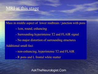
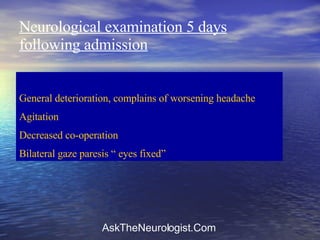
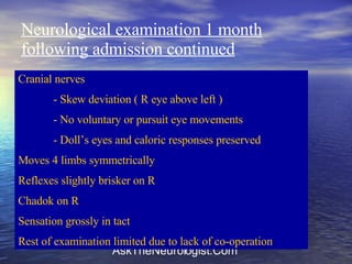
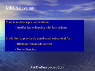
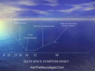
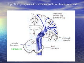
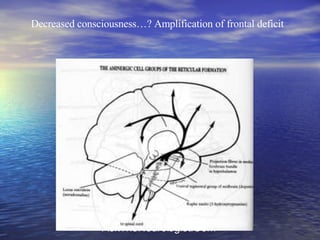
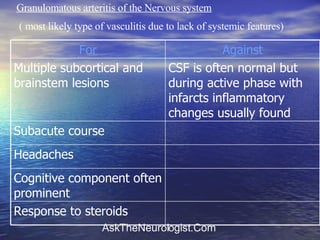
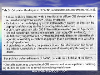
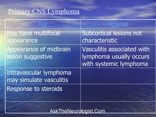
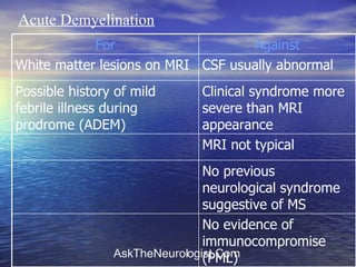
Vip Call Girls Anna Salai Chennai đ 8250192130 âŁď¸đŻ Top Class Girls Available
Â
Frontal syndrome and mid-brain lesion
1. 56 year old, lady, widowed + 3, Syrian descent presented to ER with speech difficulty, dizziness and general weakness. 3 weeks prior to admission, around the time of family bereavement (mother dying of lymphoma aged 87) began to complain of occipital headache. At the same time multiple complaints - âblurred visionâ unable to define accurately - general weakness, unclear history of mild febrile illness - articulation difficulty - peri-oral paraesthesia - left hand paraesthesia - bilateral arm weakness - left leg weakness
2. Examined in neurology clinic 4 days previously - examination at that time normal - sent for CT ambulatory 2 days prior to admission presented to ER due to worsening of above symptoms - examination normal - treatment commenced with aspirin and patient advised to continue investigation on ambulatory basis CT performed on day prior to admission normal On day of admission worsening of above symptoms as well as a brief episode of diplopia and difficulty swallowing.
3. Past History Mild hypothyroidism S/P R lobectomy for hyperthyroidism Hypercholesterolemia S/P head radiation for tinea capitis On admission receiving aspirin 100mg only
4. Examination on admission General examination unremarkable Fully conscious and orientated No language deficit Speech mildly dysarthric Cranial nerves in tact Power preserved, reflexes symmetrical, no pyramidal signs, Sensory examination in tact No cerebellar signs Gait in tact
5. Neurological examination on day following admission Fully conscious and orientated Speech: - mild cerebellar dysarthria with nasal quality Upgaze nystagmus. Cranial nerves otherwise in tact Tone, power, reflexes symmetrical and in tact No pyramidal signs Sensory examination in tact Mild cerebellar signs bilaterally Gait mildly ataxic
6. Neurological examination 2 days following admission Fully conscious and orientated PEARLA Skew deviation with no clear cranial nerve lesion Rest of examination as above Psychiatric examination revealed â normal grief reactionâ Treatment with thiamine commenced
7. Investigations performed which were normal Blood count, biochemistry ESR ( 17 ) LP: - Pressure 17, no cells, TP = 475, OCB âve Tumour markers AchR Abâs B1 EBV, CMV â no sign of acute infection Whole body CT
8. Other Investigations performed Thyroid USS multinodular goitre left lobe, R lobe absent Thyroid FNAC consistent with benign multinodular goitre Thyroid function VDRL ANA Anti-cardiolipin
9. MRI at this stage Mass in middle aspect of lower midbrain / junction with pons - 1cm, round, enhancing - Surrounding hyperintense T2 and FLAIR signal - No major distortion of surrounding structures Additional small foci - non-enhancing, hyperintense T2 and FLAIR - R pons and L frontal white matter
10. Neurological examination 5 days following admission General deterioration, complains of worsening headache Agitation Decreased co-operation Bilateral gaze paresis â eyes fixedâ
11. Neurological examination 9 days following admission Deterioration in level of consciousness - Unresponsive to painful stimuli - No eye opening - No vocalisation - Corneal reflexes and Dollâs eyes preserved - not moving arms - spontaneous symmetrical leg movements HEAD CT â revealed hypodense lesion in midbrain Whole body PET: no evidence of lesions outside of brain Treatment with dexamethasone initiated
12. Neurological examination 12 days following admission Improvement in conscious level Orientated in time and place Moves all 4 limbs symmetrical No pyramidal signs Over following 2 weeks ( until now ) progressive improvement in conscious state and orientation In parallel with above improvement, development of frontal syndromeâŚâŚâŚ.
13. Neurological examination 1 month following admission ( up to date ) Fully conscious and orientated Minimal co-operation with examination - disinhibited speech - Echolalia - perseveration - hyper-orality - utilisation behaviour
15. Neurological examination 1 month following admission continued Cranial nerves - Skew deviation ( R eye above left ) - No voluntary or pursuit eye movements - Dollâs eyes and caloric responses preserved Moves 4 limbs symmetrically Reflexes slightly brisker on R Chadok on R Sensation grossly in tact Rest of examination limited due to lack of co-operation
16. MRI 6 days ago Mass in middle aspect of midbrain - smaller less enhancing with less oedema In addition to previously noted small subcortical foci: - Bilateral frontal sub-cortical - Non-enhancing
17. 0 21 23 26 30 32 50 Vague symptoms until admission Development of brainstem signs Coma: steroids initiated Recovery of conscious state Stable but with severe frontal syndrome DAYS SINCE SYMPTOM ONSET
18. Summary 56 year old lady Progressive brainstem syndrome with subsequent development of a severe frontal syndrome following improvement of conscious state under steroid treatment
20. Can MRI findings explain clinical picture? Brainstem syndrome including coma readily explained by lesion in lower midbrain Can frontal syndrome be explained on basis of lesions?
23. â Disorders of higher mental function due to single infarctions in the thalamus and in the area of the thalamofrontal tractsâ Kalashnikova et al Neuroscience and Behavioural Physiology 1999 9 patients with small infarcts in the thalamus or thalamo-frontal tracts described in which the result was an acute cognitive syndrome. The complex of changes most closely approximated a â frontal syndromeâ ; either apathetic, akinetic, or disinhibited type. Lesions were all in positions corresponding to structures traversed by pathways connecting the reticular formation to the frontal lobes Suggest that âfrontal syndromesâ may be caused by â functional inactivation of frontal cortexâ
27. Granulomatous arteritis of the Nervous system ( most likely type of vasculitis due to lack of systemic features) Response to steroids Cognitive component often prominent Headaches Subacute course CSF is often normal but during active phase with infarcts inflammatory changes usually found Multiple subcortical and brainstem lesions Against For
32. Primary CNS Lymphoma Response to steroids Intravascular lymphoma may simulate vasculitis Vasculitis associated with lymphoma usually occurs with systemic lymphoma Appearance of midbrain lesion suggestive Subcortical lesions not characteristic May have multifocal appearance Against For
33. Localized isolated angiitis of the central nervous system associated with primary intracerebral lymphoma . Borenstein et al Cancer 1988 When GANS is associated with malignancy, it is usually due to lymphoma outside of the CNS Present case of 52 year-old man presenting with a focal neurological syndrome attributable to the midbrain and headaches. GANS was confirmed via angiography and brain biopsy Good response to treatment initially achieved with cyclophosphamide and steroids Patient subsequently admitted with features of a left occipital lobe infarct Patient died due to complications. Autopsy revealed primary CNS lymphoma in midbrain adjacent to severe area of vasculitis Believe primary disease was lymphoma; but cannot exclude possibility that lymphoma was secondary to cytotoxic treatment
34. Acute Demyelination No previous neurological syndrome suggestive of MS No evidence of immunocompromise (PML) MRI not typical Clinical syndrome more severe than MRI appearance Possible history of mild febrile illness during prodrome (ADEM) CSF usually abnormal White matter lesions on MRI Against For
36. Whippleâs disease Although oculomasticatory and oculo-facial-skeletal myorhythmia is said to be pathognomonic, it is only present in 20% of cases Focal enhancing lesions may be present or absent Frontal syndrome never documented Most common neurological manifestation is cognitive / behavioural / psychiatric alone Very rarely occurs without GIT involvement Global supranuclear opthalmoplegia Against For
37. HIV CSF may be normal.May be combined with other related conditions such as lymphoma, endocarditis , syphilis or opportunistic infections leading to focal clinical features. No other clinical features May cause combination of focal and non-focal features No risk factors May cause subacute cognitive syndrome with variable presentations (including predominantly frontal syndrome) Against For
38. Syphilis No other clinical features May cause global supranuclear opthalmoplegia Sterile CSF during active disease effectively excludes diagnosis May cause combination of focal features ( via vasculitis) and a more widespread cognitive syndrome ( including frontal) Against For
39. CJD MRI lesions ( without hyperintensity of lenticular nuclei) Prominent cognitive changes Normal CSF Normal EEG initially Clinical syndrome more severe than imaging Predominantly frontal syndrome rather than dementia Cerebellar signs initially found Response to steroids Subacute course Against For
40. Limbic encephalitis Response to steroids Often associated with cerebellar signs No neoplasm detected even with whole-body PET May cause focal ( including brainstem) lesions in association with more widespread cognitive changes Frontal rather than â limbic syndromeâ Supranuclear gaze palsies Against For