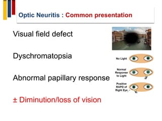
Slides include
Basic anatomy of optic nerves
Background & epidemiology of optic neuritis
Classification of optic neuritis
Clinical features
Investigations
Diagnosis
Differential diagnosis
Managements
Prognosis
Ischemic optic neuropathy constitutes one of the major causes of blindness or seriously impaired vision among the middle-aged and elderly population.
Ischemic optic neuropathy is due to acute ischemia of the optic nerve. it can be classified into two, depending upon the part of the optic nerve involved:
1.Anterior ischemic optic neuropathy (AION)
-AION is due to acute ischemia of the front (anterior) part of the optic nerve (also called optic nerve head), which is supplied mainly by the posterior ciliary arteries.
-AION is divided into two types, depending on what causes it:
1.Arteritic AION: This is the most serious type and is due to a disease called giant cell arteritis or temporal arteritis.
2. Non-arteritic AION: This is the usual, most common type, with many different causes but not associated with giant cell arteritis.
2.Posterior ischemic optic neuropathy (PION). -
-PION is a much less common type. It is due to acute ischemia of the back (posterior) part of the optic nerve, located some distance behind the eyeball; this part of the optic nerve is NOT supplied by the posterior ciliary arteries
(Hayreh, 2009)
Retinal vasculitis refers to the inflammation of the retinal vessel resulting in evident clinical manifestations i.e. vascular sheathing, leakage and occlusion. This presentation covers the etiology, pathogenesis, clinical features, diagnosis and management of this spectrum of retinal disease.
Ischemic condition affecting the eye.
The ischemia can occur secondary to systemically problem [or] particulary the eye.
Many retinal vascular disorders {like CRAO,CRVO,Diabetic retinopathy,Hypertensive Retinopathy} shows ischemic signs.
Metabolic optic neuropathies
The three subcategories of metabolic optic neuropathies are
•Heredodegenerative (such as leber's hereditary optic neuropathy).
•Nutritional deficiencies (such as vitamins B12 or folic acid).
•Toxicities (such as ethambutol or cyanide).
A neuromuscular disorder that leads to weakness of skeletal muscles.
Symptoms
Causes
Prevention
Complications
Common tests & procedures
Neurological examination:
Repetitive nerve stimulation test:
Antibody test:
Pulmonary function tests (PFTs): To check any breathing difficulty.
CT scan: To rule out a presence of tumor in thymus.
Magnetic resonance imaging (MRI): MRI of the chest is performed to rule out a presence of tumor in thymus.
Edrophonium (Tensilon) test:
Medication
Procedures
Nutrition
Parkinsonism is a clinical syndrome and, typically, when the condition
appears to be idiopathic and responsive to levodopa therapy, is referred
to as Parkinson’s disease1
• The four cardinal features of the parkinsonian syndrome are:2
– Bradykinesia
– Muscular rigidity
– Resting tremor
– Postural instability (and gait impairment)
• These features are not always observed in every patient, at any given
time
To make a diagnosis of PD, the physician must distinguish between
different forms of parkinsonism:1
– Parkinson’s disease
– Secondary parkinsonism
– Parkinsonism as part of another neurodegenerative disorder (e.g., multiple
system atrophy, progressive supranuclear palsy, corticobasal degeneration, or
Lewy body dementia)
SCALES COMMONLY USED IN
PARKINSON’S DISEASE
RESEARCH
SCALES COMMONLY USED IN
PARKINSON’S DISEASE
RESEARCH
Progressive multifocal leukoencephalopathy (PML) is a disease of the white matter of the brain, caused by a virus infection that targets cells that make myelin--the material that insulates nerve cells (neurons). Polyomavirus JC (often called JC virus) is carried by a majority of people and is harmless except among those with lowered immune defenses. The disease is rare and occurs in patients undergoing chronic corticosteroid or immunosuppressive therapy for organ transplant, or individuals with cancer (such as Hodgkin’s disease or lymphoma). Individuals with autoimmune conditions such as multiple sclerosis, rheumatoid arthritis, and systemic lupus erythematosus -- some of whom are treated with biological therapies that allow JC virus reactivation -- are at risk for PML as well. PML is most common among individuals with HIV-1 infection / acquired immune deficiency syndrome (AIDS). Currently, the best available therapy is reversal of the immune-deficient state, since there are no effective drugs that block virus infection without toxicity. Reversal may be achieved by using plasma exchange to accelerate the removal of the therapeutic agents that put patients at risk for PML. In the case of HIV-associated PML, immediately beginning anti-retroviral therapy will benefit most individuals. Several new drugs that laboratory tests found effective against infection are being used in PML patients with special permission of the U.S. Food and Drug Administration. Hexadecyloxypropyl-Cidofovir (CMX001) is currently being studied as a treatment option for JVC because of its ability to suppress JVC by inhibiting viral DNA replication.
In general, PML has a mortality rate of 30-50 percent in the first few months following diagnosis but depends on the severity of the underlying disease and treatment received. Those who survive PML can be left with severe neurological disabilities.
Sequencing in management of Multiple sclerosisAmr Hassan
Sequencing of DMTs for individual multiple sclerosis patients should be designed in such a way to maximize disease control and minimize risk based on the mechanism of action, pharmacokinetic and pharmacodynamic properties of each therapy. This includes the DMT patients are being switched from to those they are being switched to. The reversibility of immune system effects should be a key consideration for DMT sequence selection. This feature varies across DMTs and should factor more prominently in decision making as newer treatments become available for the prevention of disability accumulation in patients with progressive MS. In this short review, we discuss the landscape of existing therapies with an eye to the future when planning for optimal DMT sequencing. While no cure exists for MS, efforts are being directed toward research in neuroregeneration with the hope for positive outcomes.
Prediction of outcome of Multiple sclerosisAmr Hassan
Prediction of outcome of Multiple sclerosis
An understanding of the natural history of multiple sclerosis(MS) in a patient is important to begin proper treatment at the correct time, especially when there is a high risk for poor prognosis. Factors that predict unfavorable prognosis are a primary or secondary progressive course, older age at disease onset, short interval between first and second attacks, initial cerebellar or pyramidal symptoms, a large number of functional systems involved at onset, moderate to severe disability within the first 2 years, and the presence of typical plaques or greater lesion volume shown by magnetic resonance imaging results during the first 5 years. However, there are no established laboratory tests able to predict long-term prognosis.
Lifestyle modification in epilepsy
Lifestyle Modifications
Lifestyle modifications can include:
Adequate sleep: Fatigue is one of the most common seizure triggers, and disrupted sleep can make the brain more vulnerable to misfiring.
Avoiding drugs and alcohol: These can be triggers for seizures in patients with epilepsy. Even one or two drinks can provoke seizures.
Minimizing emotional stress: Although there is not definitive proof that stress causes seizures, those who maintain healthy stress levels have reported that they believe it reduces their risk.
Frequency of exercise: In addition to a range of health benefits, regular exercise can help reduce risk of seizure. However, you should consult your physician before starting a new exercise routine, as some exercise can, rarely, cause seizures.
Childhood demyelinating syndromes
In the past decade, the number of studies related to demyelinating diseases in children has exponentially increased. Demyelinating disease in children may be monophasic or chronic. Typical monophasic disorders in children are acute disseminated encephalomyelitis and clinically isolated syndromes, including optic neuritis and transverse myelitis. However, some cases of acute disseminated encephalomyelitis or clinically isolated syndrome progress to become chronic disorders, including multiple sclerosis and neuromyelitis optica. This review summarizes the current knowledge on monophasic and chronic demyelinating disorders in children, focusing on an approach to diagnosis and management.
Diabetic polyneuropathy
Diabetic polyneuropathy (DPN) is defined as peripheral nerve dysfunction. There are three main alterations involved in the pathologic changes of DPN: inflammation, oxidative stress, and mitochondrial dysfunction.
Excessive daytime sleepiness
The most common causes of excessive daytime sleepiness are sleep deprivation, obstructive sleep apnea, and sedating medications. Other potential causes of excessive daytime sleepiness include certain medical and psychiatric conditions and sleep disorders, such as narcolepsy.
Vagal Nerve stimulation
Vagus nerve stimulation (VNS) is a medical treatment that involves delivering electrical impulses to the vagus nerve. It is used as an add-on treatment for certain types of intractable epilepsy and treatment-resistant depression. Frequent side effects include coughing and shortness of breath. Serious side effects may include trouble talking and cardiac arrest.
Dystonia
Dystonia is a movement disorder in which your muscles contract involuntarily, causing repetitive or twisting movements.
The condition can affect one part of your body (focal dystonia), two or more adjacent parts (segmental dystonia) or all parts of your body (general dystonia). The muscle spasms can range from mild to severe. They may be painful, and they can interfere with your performance of day-to-day tasks.
Dystonia: Causes, Types, Symptoms, and Treatments
Trigeminal neuralgia is sudden, severe facial pain. It's often described as a sharp shooting pain or like having an electric shock in the jaw, teeth or gums.
Trigeminal neuralgia
Contents
Overview
Symptoms
Causes
Diagnosis
Treatment
Nootropics and smart drugs are natural or synthetic substances that can be taken to improve mental performance in healthy people.
They have gained popularity in today’s highly competitive society and are most often used to boost memory, focus, creativity, intelligence and motivation.
Here’s a look at the ]best nootropics and how they enhance performance.
Nystagmus is a condition of involuntary (or voluntary, in some cases)eye movement, acquired in infancy or later in life, that in extremely rare cases may result in reduced or limited vision. Due to the involuntary movement of the eye, it has been called "dancing eyes"Contents
1 Causes
1.1 Early-onset nystagmus
1.2 Acquired nystagmus
1.3 Other causes
2 Diagnosis
2.1 Pathologic nystagmus
2.2 Physiological nystagmus
3 Treatment
4 Epidemiology
Basics of Neuroradiology
Neuroradiology is an essential tool in management of patients with neurological and neurosurgical disorders. The aim of this presentation will be to acquaint the reader to understand how images are formed on a computed tomography (CT) and magnetic resonance imaging (MRI) along with a review of the relevant neuroanatomy. This understanding will be helpful to the reader in interpretation of images and diagnosis of various neurological disorders.
Ethanol (CH3CH2OH), or beverage alcohol, is a two-carbon alcohol
that is rapidly distributed in the body and brain. Ethanol alters many
neurochemical systems and has rewarding and addictive properties. It
is the oldest recreational drug and likely contributes to more morbidity,
mortality, and public health costs than all illicit drugs combined. The
5th edition of the Diagnostic and Statistical Manual of Mental Disorders
(DSM-5) integrates alcohol abuse and alcohol dependence into a single
disorder called alcohol use disorder (AUD), with mild, moderate,
and severe subclassifications (American Psychiatric Association, 2013).
In the DSM-5, all types of substance abuse and dependence have been
combined into a single substance use disorder (SUD) on a continuum
from mild to severe. A diagnosis of AUD requires that at least two of
the 11 DSM-5 behaviors be present within a 12-month period (mild
AUD: 2–3 criteria; moderate AUD: 4–5 criteria; severe AUD: 6–11 criteria).
The four main behavioral effects of AUD are impaired control over
drinking, negative social consequences, risky use, and altered physiological
effects (tolerance, withdrawal). This chapter presents an overview
of the prevalence and harmful consequences of AUD in the U.S.,
the systemic nature of the disease, neurocircuitry and stages of AUD,
comorbidities, fetal alcohol spectrum disorders, genetic risk factors, and
pharmacotherapies for AUD.
Prix Galien International 2024 Forum ProgramLevi Shapiro
June 20, 2024, Prix Galien International and Jerusalem Ethics Forum in ROME. Detailed agenda including panels:
- ADVANCES IN CARDIOLOGY: A NEW PARADIGM IS COMING
- WOMEN’S HEALTH: FERTILITY PRESERVATION
- WHAT’S NEW IN THE TREATMENT OF INFECTIOUS,
ONCOLOGICAL AND INFLAMMATORY SKIN DISEASES?
- ARTIFICIAL INTELLIGENCE AND ETHICS
- GENE THERAPY
- BEYOND BORDERS: GLOBAL INITIATIVES FOR DEMOCRATIZING LIFE SCIENCE TECHNOLOGIES AND PROMOTING ACCESS TO HEALTHCARE
- ETHICAL CHALLENGES IN LIFE SCIENCES
- Prix Galien International Awards Ceremony
263778731218 Abortion Clinic /Pills In Harare ,sisternakatoto
263778731218 Abortion Clinic /Pills In Harare ,ABORTION WOMEN’S CLINIC +27730423979 IN women clinic we believe that every woman should be able to make choices in her pregnancy. Our job is to provide compassionate care, safety,affordable and confidential services. That’s why we have won the trust from all generations of women all over the world. we use non surgical method(Abortion pills) to terminate…Dr.LISA +27730423979women Clinic is committed to providing the highest quality of obstetrical and gynecological care to women of all ages. Our dedicated staff aim to treat each patient and her health concerns with compassion and respect.Our dedicated group ABORTION WOMEN’S CLINIC +27730423979 IN women clinic we believe that every woman should be able to make choices in her pregnancy. Our job is to provide compassionate care, safety,affordable and confidential services. That’s why we have won the trust from all generations of women all over the world. we use non surgical method(Abortion pills) to terminate…Dr.LISA +27730423979women Clinic is committed to providing the highest quality of obstetrical and gynecological care to women of all ages. Our dedicated staff aim to treat each patient and her health concerns with compassion and respect.Our dedicated group of receptionists, nurses, and physicians have worked together as a teamof receptionists, nurses, and physicians have worked together as a team wwww.lisywomensclinic.co.za/
Explore natural remedies for syphilis treatment in Singapore. Discover alternative therapies, herbal remedies, and lifestyle changes that may complement conventional treatments. Learn about holistic approaches to managing syphilis symptoms and supporting overall health.
- Video recording of this lecture in English language: https://youtu.be/lK81BzxMqdo
- Video recording of this lecture in Arabic language: https://youtu.be/Ve4P0COk9OI
- Link to download the book free: https://nephrotube.blogspot.com/p/nephrotube-nephrology-books.html
- Link to NephroTube website: www.NephroTube.com
- Link to NephroTube social media accounts: https://nephrotube.blogspot.com/p/join-nephrotube-on-social-media.html
ARTIFICIAL INTELLIGENCE IN HEALTHCARE.pdfAnujkumaranit
Artificial intelligence (AI) refers to the simulation of human intelligence processes by machines, especially computer systems. It encompasses tasks such as learning, reasoning, problem-solving, perception, and language understanding. AI technologies are revolutionizing various fields, from healthcare to finance, by enabling machines to perform tasks that typically require human intelligence.
Flu Vaccine Alert in Bangalore Karnatakaaddon Scans
As flu season approaches, health officials in Bangalore, Karnataka, are urging residents to get their flu vaccinations. The seasonal flu, while common, can lead to severe health complications, particularly for vulnerable populations such as young children, the elderly, and those with underlying health conditions.
Dr. Vidisha Kumari, a leading epidemiologist in Bangalore, emphasizes the importance of getting vaccinated. "The flu vaccine is our best defense against the influenza virus. It not only protects individuals but also helps prevent the spread of the virus in our communities," he says.
This year, the flu season is expected to coincide with a potential increase in other respiratory illnesses. The Karnataka Health Department has launched an awareness campaign highlighting the significance of flu vaccinations. They have set up multiple vaccination centers across Bangalore, making it convenient for residents to receive their shots.
To encourage widespread vaccination, the government is also collaborating with local schools, workplaces, and community centers to facilitate vaccination drives. Special attention is being given to ensuring that the vaccine is accessible to all, including marginalized communities who may have limited access to healthcare.
Residents are reminded that the flu vaccine is safe and effective. Common side effects are mild and may include soreness at the injection site, mild fever, or muscle aches. These side effects are generally short-lived and far less severe than the flu itself.
Healthcare providers are also stressing the importance of continuing COVID-19 precautions. Wearing masks, practicing good hand hygiene, and maintaining social distancing are still crucial, especially in crowded places.
Protect yourself and your loved ones by getting vaccinated. Together, we can help keep Bangalore healthy and safe this flu season. For more information on vaccination centers and schedules, residents can visit the Karnataka Health Department’s official website or follow their social media pages.
Stay informed, stay safe, and get your flu shot today!
3. O C D
Distribution: Uni/Bilateral.
Symm/Asymmetrical
Simultaneous/ Sequential
Severity
Painful or not.
Limitation of ocular motility( double vision)
Ptosis
Field defect
Local eye manifestations: (photophobia,
lacrimation, exophthalmos, red eye)
Optic Neuritis : History taking
12. • Uhthoff described 3
patients in whom exertion
and fatigue caused a
desaturation in colour
vision
• Patient XVIII had
decreased acuity after
walking around the room
Uhthoff’s Symptom
13. Movement phosphenes in optic neuritis: A new clinical sign
(Davis F, Bergen D, Schauf C, McDonald I, Deutsch W)
Neurology 1976; 26: 1100-1104.
• Bright flashes in dark
• Eye movement
• Differentiate from Lightning Streaks of Moore
• Eye equivalent of L’hermittes symptom
Flashes
16. Mental map for diagnosis of ON
16
Optic Neuritis (ON)
ON Typical for MS
MRI Fulfills Criteria
ON Atypical for MS
Red Flags Present
Work Up for
Alternative Diagnoses
Clinical/Imaging Follow Up
Alternative Diagnosis
Established
Further clinical/imaging
typical for MS
MS Diagnosis
17. 17
Typical
• Acute or subacute attack
• Mostly young adults (age 20-55 y)
• Unilateral visual acuity loss
• Improvement with/without treatment
• Continued improvement after corticosteroid
withdrawn
• Mild pain that worsens with eye movement
• The optic disc appears normal or mildly swollen
• Variable visual field defects may occur
• Altered perception of motion (the pulfrich
phenomenon)
• Vision blurs when body temperature rises (Uhthoff’s
phenomenon)
• Bright, fleeting flashes of light (phosphenes)
Atypical
• Progressive disease
• Age group < 12 and > 50 y
• Bilateral visual loss
• No spontaneous visual improvement
• Deterioration after corticosteroid are
discontinued
• Following loss of vision, painless to severe pain
• Severe swelling and hemorrhage in optic disc
• Variable signs and symptoms, depending on etiology
Optic Neuritis : Red Flags
24. Dissemination in space
DIS is established by detecting involvement of at
least two of the five following areas of the CNS:
• Periventricular: ≥3 lesions
• Cortical-juxtacortical: ≥1 lesions
• Infratentorial: ≥1 lesions
• Spinal cord: ≥1 lesions
• Optic nerve: ≥1 lesions
24
New 2016 MAGNIMS MRI criteria
27. Mental map for diagnosis of ON
27
Optic Neuritis
ON Typical for MS
MRI Fulfills Criteria
ON Atypical for MS
Red Flags Present
Work Up for
Alternative Diagnoses
Clinical/Imaging Follow
Up
Alternative Diagnosis
Established
Further clinical/imaging
typical for MS
MS Diagnosis
ON Typical for MS
MRI not Fulfilling Criteria
Clinical/Imaging
Follow Up
CIS
29. 29
The ONTT was a prospective, randomized, multicenter
placebo-controlled clinical trial designed to compare the
benefits of treatment with
(1) intravenous methylprednisolone (IVMP) (250 mg
administered every 6 h for 3 days followed by oral
prednisone [1 mg/kg/day] for 11 days);
(2) oral prednisone (1 mg/kg/day); or
(3) oral placebo in 457 patients with acute optic neuritis.
Beck RW, Cleary PA, Anderson MM Jr, Keltner JL, Shults WT, Kaufman DI. A randomized, controlled trial of corticosteroids in the treatment of acute
optic neuritis. The Optic Neuritis Study Group. N Engl J Med. 1992 Feb 27. 326(9):581-8.
PLEASE
30. • The ONTT showed that treatment with standard-dose
oral prednisone was associated with an increased rate
of new attacks of optic neuritis.
• After 5 years, the recurrence rate was found to be higher
in the oral prednisone (1 mg/kg) group (41%) than in
those who received IVMP or oral placebo (25%).
30
1. Costello F, Burton JM. An approach to optic neuritis: the initial presentation. Expert Rev Ophthalmol. 2013. 8(6):539–551.
2. Shams PN, Plant GT. Optic neuritis: a review. Int MS J. 2009 Sep. 16(3):82-9.
PLEASE
33. • Some of the features that should raise the suspicion of
an inflammatory optic neuritis:
Lack of spontaneous improvement of visual function
after 30 days
Exquisite steroid-responsiveness
Steroid-dependency.
Optic Neuritis: Inflammatory causes
35. • Optic disc swelling frequently occurs with posterior
uveitis and retinitis.
• Optic neuropathy can also occur in the context orbital
inflammatory disease (orbital pseudotumor).
• MRI of the orbit will show inflammation of the optic
nerve sheath (optic perineuritis)
Optic Neuritis: Inflammatory causes
36. • Chronic relapsing inflammatory optic neuropathy is
another entity characterized by:
Recurrences and steroid-responsiveness.
Can behave as granulomatous optic neuropathy
May require long-term immunosuppressive therapy
(kidd et al 2003).
Optic Neuritis: CRION
44. PARAMETER FINDING
Visual Acuity Often better than 20/100.
Visual Field Typically Inferior Altitudinal defect.
Colour Vision May be severely impaired when VA is good.
Ophthalmic Exam Findings Diffuse OR sectorial hyperaemic disc swelling
associated with FEW peripapillary splinter-
shaped haemorrhages.
Small OR cupless disc in fellow eye.
Swelling gradually resolves and pallor in 3-6
weeks after onset.
FA Finding Acute Stage: localized disc hyperfluorescence,
intense, eventually involves entire disc.
Laboratory Evaluation No associated laboratory abnormalities.
Ischemic Optic Neuropathy:NAION
46. Treatment
• No definitive treatment.
• Aspirin is effective in reducing systemic vascular events.
• Aspirin does not reduce risk of involvement of the fellow
eye.
Ischemic Optic Neuropathy:NAION
47. Condition Prognosis
No further loss of vision Very small percentage
Recurrences in the same eye ~6%
Involvement of the FELLOW eye ~10% after 2 years
~15% after 5 years
2 Important Factors caused
involvement of FELLOW eye
Poor VA in 1st eye
DM
Signs of FELLOW eye involvement 1 eye optic atrophy
Ischemic Optic Neuropathy:NAION
52. Posterior ciliary artery is the main
source of blood supply to the optic
nerve head
Occlusion of the posterior ciliary artery
results in infarction of a segment or the
entire optic nerve head
Depending upon the area of the optic
nerve head supplied by the occluded
posterior ciliary artery
Results in development of A-AION and
in massive visual loss.
Ischemic Optic Neuropathy: AAION
53. Age: Fifty years of age or older at
onset.
Gender : 3 times more common in
women than in men
Race: Common among Caucasians
> other races
Giant cell arteritis
New onset of localized headache.
Temporal artery pulse.
Elevated ESR.
Positive temporal artery biopsy.
Ischemic Optic Neuropathy: AAION
55. •Sudden
•Unilateral visual loss (↓ VAthat is typically severe (<6/60) in
over 60% of the patients
VA
• PositiveRAPD
• Visual field defect (altitudinal & inferior), more
extensive than NAIONVisual Field
• DyschromatopsiaColour vision
• Periocular pain + flashingPain
•Pale & swollen optic disc (swollen diffusely)
•Chalky mark
•Hyperemic swelling
Optic Disc
Ischemic Optic Neuropathy: AAION
56. Fundus photographs of right eye with
A-AION:
(A) Before developing A-AION
(B) One week after developing A-
AION with chalky white optic disc
edema and
(C) 4 months later showing optic disc
cupping with a cup/disc ratio of
0.8 (note no cup in A)
Hayreh SS (2009) Ischemic optic neuropathy.
Progress in retinal and eye research 28: 34-62
Ischemic Optic Neuropathy: AAION
57. WORK UP: FFA finding
Fundus photograph (A) and fluorescein fundus angiogram (B) of right eye with A-AION and cilioretinal
artery occlusion during the initial stages. (A) Fundus photograph shows chalky white optic disc edema
with retinal infarct in the distribution of occluded cilioretinal artery. (B) Fluorescein fundus angiogram
shows evidence of occlusion of the medial posterior ciliary artery and no filling of the cilioretinal artery.
Hayreh SS (2009) Ischemic optic neuropathy.
Progress in retinal and eye research 28: 34-62
Ischemic Optic Neuropathy: AAION
58. Pale optic disc edema with adjacent
retina infarcted
Chalky white pale,swollen and
hyperemic optic disc
Ischemic Optic Neuropathy: AAION
60. Diagnostic work up
1. Erythrocyte sedimentation rate (ESR) >47mm
2. C-reactive protein >2.45 MG/DL
3. Fluorescein fundus angiographic (FFA): Critical diagnostic
test for A-AION during the early stages shows
thrombosis and occlusion of the posterior ciliary artery in
GCA
4. Jaw Claudication
5. Neck pain
(Kanski, 2003 & Hayreh, 2011)
Ischemic Optic Neuropathy: AAION
61. • AAION is EMERGENCY case.
• Early treatment is essential.
• Aim of treatment: To prevent blindness of the fellow
eye.
• Treatment: (steroid therapy)
• High dose systemic corticosteroid (IV
methylprednisolone & oral prednisolone) for
several months.
• Temporal artery biopsy- within 3 days of treatment
• Duration of treatment: ~1 to 2 years
Ischemic Optic Neuropathy: AAION
62. • Prognosis-POOR
• Visual loss is usually permanent.
• Visual recovery of the affected eye that has
treatment is poor with a 15-34% improvement
rate.
Ischemic Optic Neuropathy: AAION
63. 1. Systemic symptoms GCA : Patients with NA-AION have no systemic symptoms
of giant cell arteritis.
2. Visual symptoms: Amaurosis fugax is highly suggestive of AAION and is
extremely rare in NA-AION.
3. Hematologic abnormalities:Elevated ESR and CRP, particularly CRP, is helpful in
the diagnosis of GCA. Patients with NA-AION do not show any of these
abnormalities.
4. Early massive visual loss: Extremely suggestive of A-AION.
5. Chalky white optic disc edema: This is almost diagnostic of A-AION and is seen
in 69% of AAION eyes. In NAAION, chalky white optic disc edema occurs only
very rarely with embolic occlusion of the posterior ciliary artery.
6. AAION associated with cilioretinal artery occlusion . This is almost diagnostic of
AAION.
7. Fluorescein fundus angiography: Evidence of posterior ciliary artery occlusion in
AAION.
8. Temporal artery biopsy.
DIFFERENTIATION :
AAION FROM NAAION
66. • Patients usually had suffered craniofacial
trauma but occasionally mild orbital or eye
injury.
• A RAPD is the main clue to the diagnosis.
Traumatic Optic Neuropathy
67. • CT scan of the orbit is recommended to detect
any bony fractures, fractures of the optic canal,
and acute orbital hemorrhages.
Traumatic Optic Neuropathy
68. High-dose steroid therapy
• Was adopted as a treatment because of their
beneficial effect in studies on spinal cord injuries.
• May be harmful to the optic nerve if started 8
hours after the injury
Traumatic Optic Neuropathy
71. • Systemic malignancies such as lymphoma, leukemia,
multiple myeloma, and carcinoma.
• The optic disc can be swollen or normal in appearance.
• MRI of the brain and orbit may show meningeal and optic
nerve enhancement.
Infilterative Optic Neuropathy
72. • Spinal tap is recommended in cases of suspected CNS
malignancy but more than one spinal tap may be needed
to detect malignant cells.
• In case of localized optic nerve infiltration with no
evidence of systemic disease, histopathological diagnosis
may require direct optic nerve sheath biopsy.
Infilterative Optic Neuropathy
75. • Common causes include orbital and intracranial
meningiomas, pituitary adenomas, intracranial
aneurysms, craniopharyngiomas, and gliomas of the
anterior visual pathway.
• Vision loss, however, can be fast and dramatic in pituitary
apoplexy, or ruptured aneurysm.
• Visual field testing aids in the localization of the lesion.
Compressive Optic Neuropathy
76. • Neuroimaging study
(MRI of brain and
orbits) revealed an
extensive meningioma
involving the left orbital
apex (arrow)
Compressive Optic Neuropathy
77. • Thyroid eye disease
and can present as
asymmetric progressive
visual loss.
• This will require prompt
therapy (orbital
radiation, orbital
decompression, high-
dose systemic steroids).
Compressive Optic Neuropathy
81. 81
Hereditary Optic Neuropathy: AD (Kjers’ type)
• There various responsible genetic mutations occur in the
OPA1 gene located on the chromosome 3 q region
• 1st decade of life
• Bilateral symmetric visual loss.
• Bilateral central or cecocentral scotomas.
• Color vision deficit along the tritan (blue-yellow) axis.
82. 82
Hereditary Optic Neuropathy: AD (Kjers’ type)
• The optic disc : temporal pallor and in some cases severe
excavation and cupping.
83. 83
Hereditary Optic Neuropathy
AR (Wolfram syndrome)
• 1st year of life
• It can be associated with diabetes mellitus, diabetes
inspidus, and deafness.
XL
• Xp11.4–Xp11.2 (OPA2)
84. 84
Hereditary Optic Neuropathy
Optic neuropathy can also occur in neurological conditions
such as:
Spinocerebellar degeneration
Olivo-ponto-cerebellar atrophy.
85. 85
Hereditary Optic Neuropathy: LHON
• LHON has 4 primary mitochondrial
genome mutations; G11778A,
G3460A and T14484C and
T10663C.
• MF
• The frequencies of mutation may
vary across different countries
• Newer mutations have been
described worldwide
86. 86
Hereditary Optic Neuropathy: LHON
• Acute unilateral, painless, visual loss.
• some cases may stay asymptomatic or have a chronic
course
• Sequential bilateral involvement may occur weeks or
months later.
• Some patients may demonstrate neurological
manifestations such as peripheral neuropathy, ataxia,
dystonia and cardiac conduction defects
87. 87
Hereditary Optic Neuropathy: LHON
• Occasionally, optic nerve pallor can be seen initially.
Because of the wide age range (6–80 years old) at
which LHON may present, it is frequently
misdiagnosed
• Young patients are often diagnosed as optic neuritis
and older patients as ischemic or infiltrative optic
neuropathy.
88. 88
Hereditary Optic Neuropathy: LHON
• Visual filed defects tend to be central or cecocentral
as the papillo-macular bundle is first and most
severely affected
89. 89
Hereditary Optic Neuropathy: LHON
• Fundoscopy may show disk swelling, thickening of
the peripapillary retinal nerve fiber layer
90. 90
Hereditary Optic Neuropathy: LHON
• MRI may show optic nerve enhancement and white
matter lesions, which may add to the diagnostic
difficulties
93. 93
Radiation-induced Optic Neuropathy
• Patients with RON can present with vision loss, months or
years following history of radiation exposure to the brain or
orbit.
• The risk +chemotherapy
• The mechanism of RON is ischemia caused by endothelial
cell injury from radiation.
• MRI of the orbit may show optic nerve enhancement with
gadolinium
94. 94
Radiation-induced Optic Neuropathy
• The optic disc is usually normal but can be swollen.
• Patients may also have radiation retinopathy with retinal
hemorrhages, cotton wool spots, exudates and macular
edema.
• There is no treatment of proven efficacy for RON.
• Hyperbaric oxygen, steroids, antiplatelet drugs, and
anticoagulants have all been used with limited success .
• The visual prognosis is poor with 45% of eyes ending with
no light perception visual acuity
97. • Cancer-associated retinopathy (CAR)
• Melanoma-associated retinopathy
(MAR)
• Paraneoplastic optic neuropathy (PON)
• Patients with car typically present with
progressive loss of vision and photopsia
97
Paraneoplastic Optic Neuropathy
98. CAR
• Small-cell lung cancer (SCLC),gynecologic tumors
• Autoantibodies against calcium-binding photoreceptor protein that
participates in the transduction of light.
MAR
• Photopsias
• Autoantibodies against the bipolar cells of the retina
PON
• Progressive visual loss and optic disc edema,
• Autoantibodies against
• collapsin-responsive mediator protein-5
• (CRMP-5, also called anti-CV2)98
Paraneoplastic Optic Neuropathy
101. The three subcategories of metabolic optic
neuropathies are
• Heredodegenerative (such as leber's
hereditary optic neuropathy).
• Nutritional deficiencies (such as vitamins B12
or folic acid).
• Toxicities (such as ethambutol or cyanide).
Metabolic Optic Neuropathies
104. • Visual acuity may vary from
minimal reduction to no light
perception (NLP) in rare cases.
• Most patients have 20/200 vision
or better.
• Color vision should be assessed
because dyschromatopsia is a
constant feature in these
conditions.
Toxic Optic Neuropathies
105. • In the early stages of toxic optic neuropathies,
most patients also have normal-appearing
optic nerves, but disc edema and hyperemia
may be seen in some intoxications, especially
in acute poisonings.
• Papillomacular bundle loss and optic atrophy
develop after a variable interval depending on
the responsible toxin.
Toxic Optic Neuropathies
108. • In the workplace, industrial locations, and related
to intentional or accidental poisonings, optic
nerve toxicity has been reported to result from
Methanol
Ethylene glycol (antifreeze)
Lead
Mercury
Thallium
Carbon monoxide.
Toxic Optic Neuropathies
109. • Among the many causes of TON,
the top 10 toxins include:
Medications
– Ethambutol, rifampin, isoniazid,
streptomycin
– Linezolid
– Chloramphenicol
– Isoretinoin
– Cyclosporin
Acute Toxins
– Methanol
– Ethylene glycol
Toxic Optic Neuropathies
110. The clinical presentation and basic pathophysiology
are similar to TON.
Most often, they present as a non-specific
retrobulbar optic neuropathy.
Currently, the treatment is limited to the intensive
use of vitamins with variable results in individual
cases, and to the implementation of preventive
measures, when feasible.
Nutritional Optic Neuropathies
111. • Optic disc may be normal or slightly
hyperemic in the early stages.
• In a small group of patients with hyperemic
discs, small splinter hemorrhages on or off the
disc.
• Several months to years later , papillomacular
bundle dropout and temporal disc pallor,
followed by optic atrophy.
Nutritional Optic Neuropathies
113. Tobacco Alcohol Ambylopia (TAA)
• TAA is an old term for a constellation of
elements that can lead to a mitochondrial
optic neuropathy.
• The classic patient is a man with a history of
heavy alcohol and tobacco consumption.
Nutritional Optic Neuropathies
114. Tobacco Alcohol Ambylopia (TAA)
• Combined nutritional mitochondrial impairment,
from vitamin deficiencies (folate and B-12)
classically seen in alcoholics, with tobacco-
derived products, such as cyanide and ROS.
• It has been suggested that the additive effect of
the cyanide toxicity, ROS, and deficiencies of
thiamine, riboflavin, pyridoxine, and b12 result in
TAA
Nutritional Optic Neuropathies
115. • Hypovitaminosis A – night blindness
(nyctalopia), keratomalacia.
• Hypervitaminosis A – yellow skin and
conjunctiva, pseudotumor cerebri
(papilledema), retinal hemorrhage.
Toxic Optic Neuropathies: Other agents
118. Type of optic
neuropathy
Onset Pattern of
visual field loss
Ophthalmoscopic
findings
Additional features
Demyelinating Acute Central, cecocentral,
arcuate
75% normal disc
(retrobulbar)
Neurological signs of brain
stem (diplopia, ataxia,
weakness) or spinal cord
involvement (leg weakness,
bladder symptoms,
paresthesias, abnormal MRI)
Non-arteritic
Ischemic
Acute Arcuate, altitudinal Swollen disc (usually
sectoral) with disc
hemorrhages
Crowded (anamalous) disk,
systemic vascular risk factors
(diabetes, hypertension,
hyerlipedemia)
Arteritic Ischemic Acute Arcuate Pallid swelling of the
disc
Headache, jaw
claudications, transient
visual loss or diplopia,
myalgias, fever, weight loss,
High ESR and CRP
Inflammatory Acute, sub-
acute
Arcuate, central,
cecocentral
Swollen disc Features of auto-immune
diseases (skin rash, arthritis,
Raynaud’s phenomenon),
exquisite responsiveness to
systemic steroids 118
119. Hereditary Chronic
(dominant
and
recessive),
acute or
subacute
(Leber’s)
Central, cecocentral Pale (dominant and
recessive) or mildly
swollen with
peripapillary
telengiectatic vessels
(Leber’s)
Onset in childhood with
positive family history,
Mitochondrial DNA
testing may reveal Leber’s
mutataion
Traumatic Acute Arcaute, central or
hemianopic
Normal Head or facial trauma
Radiation Acute Arcuate, hemianopic Normal History of radiation to the
brain or orbit, MRI may
show enhancement of the
optic nerve with
Gadolinium
Paraneoplastic Subacute,
chronic
Central Swollen disc Associated small-cell lung
carcinoma, CRMP-5
marker may be positive,
paraneoplastic cerebellar
syndrome 119
Type of optic
neuropathy
Onset Pattern of visual
field loss
Ophthalmoscopic
findings
Additional features
120. Infiltrative Acute,
subacute
Arcuate, hemianopic Normal or swollen disc Systemic malignancy may
be present, MRI may
show optic nerve or
meningeal infiltration
Compressive Chronic Arcuate, hemianopic Normal or pale disc MRI will show a
compressive mass
Toxic/nutritional Acute,
subacute or
chronic
Central, cecocentral Normal or mildly
swollen disc
History of drug use
(ethambutol, alcohol)
120
Type of optic
neuropathy
Onset Pattern of visual
field loss
Ophthalmoscopic
findings
Additional features