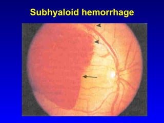
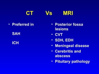
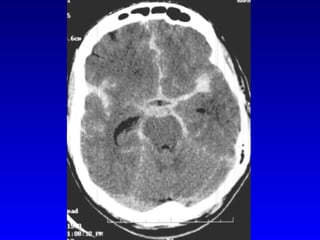
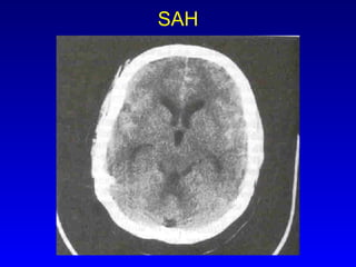
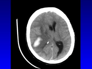
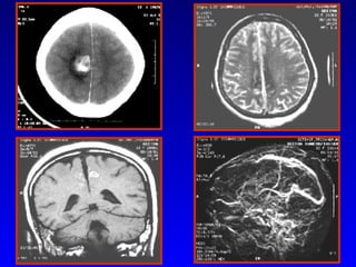
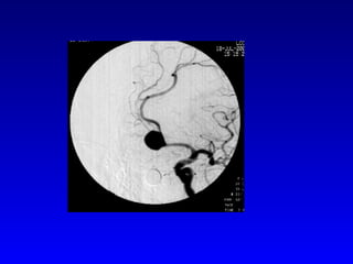
Sudden-onset headache Clinical Approach Joseph Cherian P. Asst Professor of Neurology, Sree Chitra Tirunal Institute for Medical Sciences and Technology, Thiruvananthapuram.
Epidemiology 1-2% of visits to the emergency department 4% of visits to the physician’s office Most have primary headache disorders Among  all patients with headache in an ED, 1% will have SAH In patients with the worst ever headache of their life, and normal neurological exam, 12%  will have SAH
Pain-sensitive structures in the head Blood vessels Meninges Bone  Cranial nerves- V, VII, IX, and X Scalp and muscles Nerve roots, sinus mucosa and teeth
Working classification of headache Migraine (10% prevalence) Tension-type headache(30-80% prevalence) (CTH-2%) Other headache (includes cluster HA and secondary headaches)
Secondary headache disorders Stroke, SAH Tumour Infection Systemic disorders- thyroid disease, HT, pheochromocytoma. Temporal arteritis Ophthalmological and ENT causes. Traumatic
Danger signals First or worst headaches Headache on exertion, early morning, or nocturnal Progressive headache New onset headache in adult >50 years old Abnormal physical or neurological findings (fever, stiff neck)
Sudden onset headache-causes Crash migraine Cluster Benign exertional  Posttraumatic Vascular disorders-stroke, SAH, TA, dissection, CVT, acute HT
Sudden-onset headache -causes(contd) Nonvascular IC disorders- hydrocephalus, IIH, IC hypotension, tumour, pit.apoplexy Acute intoxications Noncephalic infections Cephalic infections Disorders of eyes Cervicogenic
History taking When did the headache start? How long before it reaches maximum intensity? Have you had similar headaches before? Where does the head hurt? Do you have other symptoms? What makes it worse? What makes it better?
Examination Fever, lymphadenopathy, elevated BP Skin- rash, neurocut markers Tenderness-sinuses, TM joint Temporal and carotid arteries Neurological exam: pupils, eye signs, papilloedema, pronator drift, s/o meningeal irritation
Subhyaloid hemorrhage
Physical findings in SAH Nuchal rigidity  Altered consciousness,  Papilloedema, retinal and subhyaloid hemorrhage, 3rd and 6th nerve palsy, Bilateral leg weakness, abulia,  Nystagmus, ataxia,  Aphasia, hemiparesis, left-sided visual neglect
Diagnosis of SAH 25-51% of patients receive an incorrect diagnosis 91% of those with correct diagnosis have a favorable outcome at 6 weeks Vs 53% with an incorrect diagnosis Median delay in diagnosis(4 studies):  3 - 14 days
Reasons for misdiagnosis of SAH Failure to appreciate the spectrum of clinical presentation Failure to understand the limitations of CT Failure to perform and correctly interpret the results of LP
Indications for neuroimaging First or worst headache Progressive or CDH Side-locked headache Headaches not responding to treatment New onset headache in patients with cancer, HIV infection, or age >50 yrs Associated fever, stiff neck, neurological deficits
CT  Vs  MRI Preferred in  SAH ICH Posterior fossa lesions CVT SDH, EDH Meningeal disease Cerebritis and abscess Pituitary pathology
 
SAH
 
 
Imaging in pts with headache and normal neurological exam Benefits- CT MRI Migraine 0.3% 0.4% Any HA 2.4% 2.4% Relief of anxiety 30% Harms-iodine reaction Mild 10% Death 0.002% Claustrophobia Cost Frishberg 1994
Probability of detection of SAH on CT after the initial event Day 0 95% Day 3 75% 1 week 50% 2 weeks 30% 3 weeks almost 0% Evans RW 1999
L.P in evaluation of headache Suspected SAH if CT is negative (Deterioration after LP in patients with  clots on CT or a dilated pupil) Start antibiotics in patients with suspected meningitis, while waiting for CT CSF pressure should be measured Distinguish traumatic tap from true hemorrhage
L.P in evaluation of headache First or worst headache -   SAH, meningitis Headache with features s/o infection -  meningitis /encephalitis CVT, IIH - elevated CSF opening pressure Orthostatic headache with diffuse meningeal enhancement on MRI -   Low CSF pressure syndrome
Probability of detecting xanthochromia in CSF with spectrophotometry after SAH 12 hours 100% 1 week 100% 2 weeks 100% 3 weeks >70% 4 weeks >40%
 
Angiography In proven SAH- 4 vessel angio(DSA) to identify source and r/o multiple aneurysms Initial arteriogram negative in upto 16% of  SAH MRA detects 90% of saccular aneurysms of >5mm Spiral CT angio detects 85% of saccular aneurysms
Thunderclap headache Sudden severe headache with max intensity within 1 minute Normal CT scan Normal CSF study 180 patients followed up for 1- 3 years. None developed SAH. Wijdicks 1988, Markus 1991,  Linn 1994
Thunderclap headache Primary causes-  Migraine, benign thunderclap headache, benign orgasmic headache Secondary- unruptured saccular aneurysm, cerebral vasospasm, CVT, arterial dissection, pituitary apoplexy, occipital neuralgia Evans RW 2000
Thank You

Headache Jc

  • 1.
    Sudden-onset headache ClinicalApproach Joseph Cherian P. Asst Professor of Neurology, Sree Chitra Tirunal Institute for Medical Sciences and Technology, Thiruvananthapuram.
  • 2.
    Epidemiology 1-2% ofvisits to the emergency department 4% of visits to the physician’s office Most have primary headache disorders Among all patients with headache in an ED, 1% will have SAH In patients with the worst ever headache of their life, and normal neurological exam, 12% will have SAH
  • 3.
    Pain-sensitive structures inthe head Blood vessels Meninges Bone Cranial nerves- V, VII, IX, and X Scalp and muscles Nerve roots, sinus mucosa and teeth
  • 4.
    Working classification ofheadache Migraine (10% prevalence) Tension-type headache(30-80% prevalence) (CTH-2%) Other headache (includes cluster HA and secondary headaches)
  • 5.
    Secondary headache disordersStroke, SAH Tumour Infection Systemic disorders- thyroid disease, HT, pheochromocytoma. Temporal arteritis Ophthalmological and ENT causes. Traumatic
  • 6.
    Danger signals Firstor worst headaches Headache on exertion, early morning, or nocturnal Progressive headache New onset headache in adult >50 years old Abnormal physical or neurological findings (fever, stiff neck)
  • 7.
    Sudden onset headache-causesCrash migraine Cluster Benign exertional Posttraumatic Vascular disorders-stroke, SAH, TA, dissection, CVT, acute HT
  • 8.
    Sudden-onset headache -causes(contd)Nonvascular IC disorders- hydrocephalus, IIH, IC hypotension, tumour, pit.apoplexy Acute intoxications Noncephalic infections Cephalic infections Disorders of eyes Cervicogenic
  • 9.
    History taking Whendid the headache start? How long before it reaches maximum intensity? Have you had similar headaches before? Where does the head hurt? Do you have other symptoms? What makes it worse? What makes it better?
  • 10.
    Examination Fever, lymphadenopathy,elevated BP Skin- rash, neurocut markers Tenderness-sinuses, TM joint Temporal and carotid arteries Neurological exam: pupils, eye signs, papilloedema, pronator drift, s/o meningeal irritation
  • 11.
  • 12.
    Physical findings inSAH Nuchal rigidity Altered consciousness, Papilloedema, retinal and subhyaloid hemorrhage, 3rd and 6th nerve palsy, Bilateral leg weakness, abulia, Nystagmus, ataxia, Aphasia, hemiparesis, left-sided visual neglect
  • 13.
    Diagnosis of SAH25-51% of patients receive an incorrect diagnosis 91% of those with correct diagnosis have a favorable outcome at 6 weeks Vs 53% with an incorrect diagnosis Median delay in diagnosis(4 studies): 3 - 14 days
  • 14.
    Reasons for misdiagnosisof SAH Failure to appreciate the spectrum of clinical presentation Failure to understand the limitations of CT Failure to perform and correctly interpret the results of LP
  • 15.
    Indications for neuroimagingFirst or worst headache Progressive or CDH Side-locked headache Headaches not responding to treatment New onset headache in patients with cancer, HIV infection, or age >50 yrs Associated fever, stiff neck, neurological deficits
  • 16.
    CT Vs MRI Preferred in SAH ICH Posterior fossa lesions CVT SDH, EDH Meningeal disease Cerebritis and abscess Pituitary pathology
  • 17.
  • 18.
  • 19.
  • 20.
  • 21.
    Imaging in ptswith headache and normal neurological exam Benefits- CT MRI Migraine 0.3% 0.4% Any HA 2.4% 2.4% Relief of anxiety 30% Harms-iodine reaction Mild 10% Death 0.002% Claustrophobia Cost Frishberg 1994
  • 22.
    Probability of detectionof SAH on CT after the initial event Day 0 95% Day 3 75% 1 week 50% 2 weeks 30% 3 weeks almost 0% Evans RW 1999
  • 23.
    L.P in evaluationof headache Suspected SAH if CT is negative (Deterioration after LP in patients with clots on CT or a dilated pupil) Start antibiotics in patients with suspected meningitis, while waiting for CT CSF pressure should be measured Distinguish traumatic tap from true hemorrhage
  • 24.
    L.P in evaluationof headache First or worst headache - SAH, meningitis Headache with features s/o infection - meningitis /encephalitis CVT, IIH - elevated CSF opening pressure Orthostatic headache with diffuse meningeal enhancement on MRI - Low CSF pressure syndrome
  • 25.
    Probability of detectingxanthochromia in CSF with spectrophotometry after SAH 12 hours 100% 1 week 100% 2 weeks 100% 3 weeks >70% 4 weeks >40%
  • 26.
  • 27.
    Angiography In provenSAH- 4 vessel angio(DSA) to identify source and r/o multiple aneurysms Initial arteriogram negative in upto 16% of SAH MRA detects 90% of saccular aneurysms of >5mm Spiral CT angio detects 85% of saccular aneurysms
  • 28.
    Thunderclap headache Suddensevere headache with max intensity within 1 minute Normal CT scan Normal CSF study 180 patients followed up for 1- 3 years. None developed SAH. Wijdicks 1988, Markus 1991, Linn 1994
  • 29.
    Thunderclap headache Primarycauses- Migraine, benign thunderclap headache, benign orgasmic headache Secondary- unruptured saccular aneurysm, cerebral vasospasm, CVT, arterial dissection, pituitary apoplexy, occipital neuralgia Evans RW 2000
  • 30.