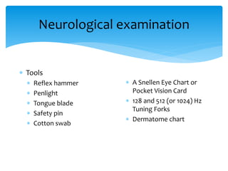
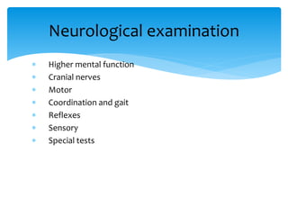
Dr Amit Agrawal, MCh
Neurological Examination
 First step in clinical encounter
 Will provide baseline information
 Information about the course and characteristics of the
present illness
Patient History
 Personal profile (Age, sex, address, occupation, socio-
economic status)
History
 Symptom onset (acute, sub-acute, chronic, insidious)
 Duration of symptoms
 Course of the condition (static, progressive, or exacerbating
and remitting)
 Associated symptoms such as seizures, headache, nausea,
vomiting, and pain
 Type of accident
 Mechanism of injury
 High or low velocity injury
 Loss or alteration of consciousness
 Post-traumatic amnesia and retrograde amnesia
 Vomiting
 Epileptic fits or seizures
Head Injury
 Past history
 Past accidents and injuries
 Operations
 Previous illnesses and hospitalizations
 Personnel history
 Family history
 Treatment history
 General examination
 Inspection of the skin and neck
 Pulse
 Blood pressure
 Temperature
 Respiratory rate
 Pallor, icterus ,cyanosis etc.
Physical Examination
 Chest
 CVS
 Abdomen
 Skeletal system
Systemic Examination
Neurological examination
 Tools
 Reflex hammer
 Penlight
 Tongue blade
 Safety pin
 Cotton swab
 A Snellen Eye Chart or
Pocket Vision Card
 128 and 512 (or 1024) Hz
Tuning Forks
 Dermatome chart
 Higher mental function
 Cranial nerves
 Motor
 Coordination and gait
 Reflexes
 Sensory
 Special tests
Neurological examination
 Glasgow Coma Scale
 Highest score = 15
 Lowest score = 3
Level of consciousness
Eye opening
4 Spontaneous
3 To voice
2 To pain
1 No response
Best verbal response
5 Oriented, converses
4 Disoriented, converses
3 Inappropriate words
2 Incomprehensible sounds
1 No response or intubated
Best motor response
6 Follows commands
5 Localizes response
4 Withdraws
3 Abnormal flexion
2 Abnormal extension
1 No response
 Vegetative state- No cerebral cortical function that can be
judged by behavior
 Severe disability- Conscious but dependent
 Moderate disability- Independent but disabled
 Good recovery- Able to participate in normal social life and
able to return to work
Glasgow Outcome Scale
 Speech and language
 Memory
 Immediate
 Recent
 Past
 Meningeal signs
Cranial nerves
 CN I - The olfactory nerve
 CN II - The optic nerve
 CN III-The oculomotor
 CN IV-The trochlear
 CN VI – The abducens
nerves
 CN V - The trigeminal
nerve
 CN VII - The facial nerve
 CN VIII - The
vestibulocochlear nerve
 CN IX-The glossopharyngeal
 CN X - The vagus nerves
 CN XI - The spinal accessory
nerve
 CN XII - The hypoglossal
nerve
 Tone
 Muscle Strength
 Muscle Symmetry
 Left to Right
 Proximal vs. Distal
 Atrophy
 Pay particular attention to the hands, shoulders, and
thighs.
 Involuntary Movements
Motor Exam
Muscle Strength
Grade Strength
0 No muscle movement
1 Visible muscle movement, but no movement at
the joint
2 Movement at the joint, but not against gravity
3 Movement against gravity, but not against added
resistance
4 Movement against resistance, but less than normal
5 Normal strength
 The stretch or deep tendon reflexes
 The superficial or cutaneous reflexes
Reflexes
 Biceps reflex (C5, C6)
 Triceps reflex (C6, C7)
 Brachioradialis reflex (C5, C6)
 Patellar reflex (L2,3,4)
 Achilles reflex (S1)
Stretch or deep tendon reflexes
 Abdominal reflex
 Cremasteric reflex
 Anal wink
 Pharyngeal or gag reflex
 Plantar or Babinski reflex (L5, S1)
Superficial or cutaneous reflexes
 Primary sensory modalities such as pain, light touch,
temperature, vibration and joint position sense.
 Cortical sensory modalities such as stereognosis, two-point
discrimination, graphesthesia and double simultaneous
stimulation.
Sensory Exam
 Finger tap
 Finger to nose
 Rapid alternating hand movement
Coordination Exam
 Nuchal rigidity or neck stiffness
 Brudzinski sign
 Kernig sign
Meningeal signs
Head injuries
 Trauma-leading cause of death in individuals under
45 years of age
 Road traffic accidents account for 70% of all brain
injuries and 50% of trauma related deaths
 Brain injury is 10 times more common than spinal
injury
 Majority of deaths in severe head injury occurs
before reaching to hospital.
Epidemiology
 Head injuries are twice more common among males
compared with females.
 Pedestrian injuries account for about 15% of brain
injuries.
 Motorcycle-related deaths occur at a rate that is 15
times greater than the rate for occupants of
passenger cars.
 Alcohol intoxication- in at least 30% to 50% of head
injuries
Epidemiology
Classification
 May be localized or diffuse
 Scalp-laceration, avulsion, contusion
 Bone-different type of fractures
 Membranes- extradural haematoma, subdural
haematoma acute/chronic
 Brain- contusion, laceration, intracerebral haematoma,
diffuse axonal injury
 Intracranial- intraventricular haemorrhage
Mechanism
Coup injuries-
 Sustaining an injury directly
underneath the point of
impact
 Scalp injury (bruise, abrasion
or laceration) at the point of
impact
 Contusion or laceration of
the brain surface at the site
of a depressed fracture
Contre-coup injuries-
 Injury to the brain on the
diametrically opposite side
of point of impact
 Sudden deceleration
 Brain glides over the
irregular, jagged contours of
the skull bones
 Frontal pole
 Temporal pole
 Occipital pole
Pathophysiology
Primary head injury-
 Mechanical trauma that occurs
at the moment of impact at
trauma site
 Can be focal or diffuse
 Direct insult to the brain and
skull
 Difficult to treat
 Decreased tissue perfusion,
ischemia and infarction.
Secondary head injury-
 Insult imposed after initial
injury
 Can occur within minutes,
hours, or days after the initial
injury
 Early treatment is aimed at
the prevention of secondary
injury
Secondary Brain Injury
Systemic Causes
 Hypoxemia
 Anaemia
 Hypotension
 Hyperthermia
 Hypercapnoea
 Hypocapnoea
 Respiratory complications
 Electrolyte imbalances
Intracranial Causes
 Increased ICP
 Cerebral oedema
 Mass lesions (e.g. haematoma)
 Infection
 Seizures
Severity of head
injury
Characteristics
Mild (80%)
GCS-13-15
Transient loss of consciousness
Generally followed by recovery over several weeks or
months
Moderate (10%)
GCS-9-12
Injury resulting in an impairment of consciousness
Recovery is more prolonged and focal deficits are
more common
Severe (10%)
GCS-3-8
Patient in coma for 24 hours or more
Mortality rate among those reaching the hospital alive
is about one-third
Although most will recover with some permanent
deficit, a few will remain in a persistent vegetative
state
 Primary survey and resuscitation
 A = Airway and cervical spine
 B = Breathing
 C = Circulation and haemorrhage control
 D = Dysfunction of the central nervous system
 E = Exposure
 Secondary survey
 Definitive treatment
ATLS methodology
 A - History of patient’s allergies
 M - Patient’s medication history
 P - Past medical and surgical history
 L - Time of last meal
 E - Full description of events leading to injury and hospital
admission
AMPLE History
Parameter Score Response
Eye opening 4
3
2
1
Spontaneous
To voice
To pain
No response
Best verbal response 5
4
3
2
1
Oriented, converses
Disoriented, converses
Inappropriate words
Incomprehensible sounds
No response or intubated
Best motor response 6
5
4
3
2
1
Follows commands
Localizes response
Withdraws
Abnormal flexion
Abnormal extension
No response
 A: Patient is awake, alert, and appropriate.
 V: Patient responds to voice.
 P: Patient responds to pain.
 U: Patient is unresponsive.
Australasian College of Surgeons
 Test Pupillary Reactions to Light
 Dim the room lights as necessary
 Ask the patient to look into the distance
 Shine a bright light obliquely into each pupil in turn
 Look for both the direct (same eye) and consensual
(other eye) reactions
 Record pupil size in mm and any asymmetry or
irregularity.
Pupillary Examination
 Head examination
 Chest examination
 Abdomen and pelvis examination
 Extremity evaluation
 Detail neurological examination
Secondary Survey
 Routine investigations
 Skull x-ray
 CT scan
 MRI
Investigations in Head Injuries
 Not much of help
 If CT scan is available do CT scan
Skull X-rays
 Lateral - right or left according to site of injury
 Antero-posterior
 Half axial (Towne's) view
 PNS view
Views
Possibility of Intracranial Haematoma
Following Head Injury
Children Adults
No fracture Fully
conscious
1:25000 1:7006
Impaired 1:589 1:1160
comatose 1:65 1:27
Skull
fracture
Fully
conscious
1:157 1:45
Impaired 1:25 1:6
comatose 1:12 1:4
 Loss of consciousness for longer than 5 minutes
 Confusion (GCS <14) persisting after initial assessment and
resuscitation
 GCS score <15 at 2 hours after injury
 Amnesia before impact >30 minutes
Indications For CT Scan
 Unreliable history or examination (alcohol or drug
ingestion)
 Dangerous mechanism (pedestrian struck by motor vehicle,
occupant ejected from motor vehicle, fall from height >3
feet or 5 stairs)
 Age > 65 years
 Repeated vomiting or vomiting for more than 8 hours after
injury
Indications For CT Scan
 Post-traumatic seizures
 Progressive headache
 Deterioration in level of consciousness
 Progressive neurological deficit
 Tense fontanelle in child
 Skull fracture with neurological signs
 Skull fracture with epileptic fit
 Depressed skull fracture
Indications For CT Scan
 Physical signs of basilar skull fracture
 Haemotympanum
 “Raccoon" eyes
 CSF otorrhoea or rhinorrhoea
 Battle's sign
 Instability following multiple traumas
 Penetrating injury
Indications For CT Scan
Approach To CT ScanCT scan
Bone window
Look for fractures-
•Cranial vault
•Skull base
•Facial bones
Tissue window
•EDH
•SDH
•ICH
•Contusions
•Pneumocephalus
•Hydrocephalus
•Cerebral edema
Subdural window
•Any hemorrhage
•Soft tissue and bone
•Fontanel
•Suture lines
•Foreign bodies
 Appearance of the normal structures
CT Scan
 More sensitive
 Provides more detailed information regarding the anatomic
and vascular structures
 Detects small hemorrhages
 May not be possible in emergency situation
MRI
 Criteria for hospitalization should be directed on an
individual basis
 Severe injuries
 Any surgical intervention
 No responsible carer
 ICU admission should be based upon the severity of the
trauma and associated injuries
Criteria For Admission
Specific Lesions
 Between the dura mater and inner table of the
skull; usually in the temporal or temporoparietal
region
 Tearing of the middle meningeal artery or, less
commonly, secondary to venous bleeding
 Directly related to the status of the patient before
surgery
 Close to 0 for patients not in a coma, 9% for
obtunded patients, and 20% for patients in deep
coma
Extradural Haematoma
 Between the dura mater and arachnoid membrane
 Tearing of bridging veins
 Severe brain damage more likely than with epidural
haematomas
 Approximately 60%, but can be lowered with rapid surgical
intervention and aggressive medical management
Subdural Haematomas
 Primarily frontal and temporal lobes, but other sites are
possible
 Impact at the time of injury
 Most patients with subdural haematoma have associated
contusions of the brain
 Greatly dependent on size and location
Cerebral Contusions
Skull
Fractures
Vault
Linear
Simple Compound
Depressed
Simple Compound
Skull base
Closed Compound
Gun shot injuries
 Diffuse axonal injury (DAI) possible major contributing
factor
 The most common injury, but pathophysiology not
completely understood
 The term DAI also describes prolonged post-traumatic coma
not caused by mass lesions or ischemic insults
Diffuse Injuries
Subarachnoid hemorrhage
Brain stem injury
Prognosis
Glasgow coma
scale at 24 hours
Good recovery
or moderate
disability
Vegetative or
dead
11-15 91% 6%
8-10 59% 27%
5-7 28% 54%
3-4 13% 80%
Causes of Mortality
 According to the type of lesion
Management
Neurological examination

Neurological examination

  • 1.
    Dr Amit Agrawal,MCh Neurological Examination
  • 2.
     First stepin clinical encounter  Will provide baseline information  Information about the course and characteristics of the present illness Patient History
  • 3.
     Personal profile(Age, sex, address, occupation, socio- economic status) History
  • 4.
     Symptom onset(acute, sub-acute, chronic, insidious)  Duration of symptoms  Course of the condition (static, progressive, or exacerbating and remitting)  Associated symptoms such as seizures, headache, nausea, vomiting, and pain
  • 5.
     Type ofaccident  Mechanism of injury  High or low velocity injury  Loss or alteration of consciousness  Post-traumatic amnesia and retrograde amnesia  Vomiting  Epileptic fits or seizures Head Injury
  • 6.
     Past history Past accidents and injuries  Operations  Previous illnesses and hospitalizations  Personnel history  Family history  Treatment history
  • 7.
     General examination Inspection of the skin and neck  Pulse  Blood pressure  Temperature  Respiratory rate  Pallor, icterus ,cyanosis etc. Physical Examination
  • 8.
     Chest  CVS Abdomen  Skeletal system Systemic Examination
  • 9.
    Neurological examination  Tools Reflex hammer  Penlight  Tongue blade  Safety pin  Cotton swab  A Snellen Eye Chart or Pocket Vision Card  128 and 512 (or 1024) Hz Tuning Forks  Dermatome chart
  • 10.
     Higher mentalfunction  Cranial nerves  Motor  Coordination and gait  Reflexes  Sensory  Special tests Neurological examination
  • 11.
     Glasgow ComaScale  Highest score = 15  Lowest score = 3 Level of consciousness
  • 12.
    Eye opening 4 Spontaneous 3To voice 2 To pain 1 No response
  • 13.
    Best verbal response 5Oriented, converses 4 Disoriented, converses 3 Inappropriate words 2 Incomprehensible sounds 1 No response or intubated
  • 14.
    Best motor response 6Follows commands 5 Localizes response 4 Withdraws 3 Abnormal flexion 2 Abnormal extension 1 No response
  • 15.
     Vegetative state-No cerebral cortical function that can be judged by behavior  Severe disability- Conscious but dependent  Moderate disability- Independent but disabled  Good recovery- Able to participate in normal social life and able to return to work Glasgow Outcome Scale
  • 16.
     Speech andlanguage  Memory  Immediate  Recent  Past  Meningeal signs
  • 17.
    Cranial nerves  CNI - The olfactory nerve  CN II - The optic nerve  CN III-The oculomotor  CN IV-The trochlear  CN VI – The abducens nerves  CN V - The trigeminal nerve  CN VII - The facial nerve  CN VIII - The vestibulocochlear nerve  CN IX-The glossopharyngeal  CN X - The vagus nerves  CN XI - The spinal accessory nerve  CN XII - The hypoglossal nerve
  • 18.
     Tone  MuscleStrength  Muscle Symmetry  Left to Right  Proximal vs. Distal  Atrophy  Pay particular attention to the hands, shoulders, and thighs.  Involuntary Movements Motor Exam
  • 19.
    Muscle Strength Grade Strength 0No muscle movement 1 Visible muscle movement, but no movement at the joint 2 Movement at the joint, but not against gravity 3 Movement against gravity, but not against added resistance 4 Movement against resistance, but less than normal 5 Normal strength
  • 20.
     The stretchor deep tendon reflexes  The superficial or cutaneous reflexes Reflexes
  • 21.
     Biceps reflex(C5, C6)  Triceps reflex (C6, C7)  Brachioradialis reflex (C5, C6)  Patellar reflex (L2,3,4)  Achilles reflex (S1) Stretch or deep tendon reflexes
  • 22.
     Abdominal reflex Cremasteric reflex  Anal wink  Pharyngeal or gag reflex  Plantar or Babinski reflex (L5, S1) Superficial or cutaneous reflexes
  • 23.
     Primary sensorymodalities such as pain, light touch, temperature, vibration and joint position sense.  Cortical sensory modalities such as stereognosis, two-point discrimination, graphesthesia and double simultaneous stimulation. Sensory Exam
  • 24.
     Finger tap Finger to nose  Rapid alternating hand movement Coordination Exam
  • 25.
     Nuchal rigidityor neck stiffness  Brudzinski sign  Kernig sign Meningeal signs
  • 26.
  • 27.
     Trauma-leading causeof death in individuals under 45 years of age  Road traffic accidents account for 70% of all brain injuries and 50% of trauma related deaths  Brain injury is 10 times more common than spinal injury  Majority of deaths in severe head injury occurs before reaching to hospital. Epidemiology
  • 28.
     Head injuriesare twice more common among males compared with females.  Pedestrian injuries account for about 15% of brain injuries.  Motorcycle-related deaths occur at a rate that is 15 times greater than the rate for occupants of passenger cars.  Alcohol intoxication- in at least 30% to 50% of head injuries Epidemiology
  • 29.
  • 30.
     May belocalized or diffuse  Scalp-laceration, avulsion, contusion  Bone-different type of fractures  Membranes- extradural haematoma, subdural haematoma acute/chronic  Brain- contusion, laceration, intracerebral haematoma, diffuse axonal injury  Intracranial- intraventricular haemorrhage
  • 31.
  • 32.
    Coup injuries-  Sustainingan injury directly underneath the point of impact  Scalp injury (bruise, abrasion or laceration) at the point of impact  Contusion or laceration of the brain surface at the site of a depressed fracture Contre-coup injuries-  Injury to the brain on the diametrically opposite side of point of impact  Sudden deceleration  Brain glides over the irregular, jagged contours of the skull bones  Frontal pole  Temporal pole  Occipital pole
  • 33.
  • 34.
    Primary head injury- Mechanical trauma that occurs at the moment of impact at trauma site  Can be focal or diffuse  Direct insult to the brain and skull  Difficult to treat  Decreased tissue perfusion, ischemia and infarction. Secondary head injury-  Insult imposed after initial injury  Can occur within minutes, hours, or days after the initial injury  Early treatment is aimed at the prevention of secondary injury
  • 35.
    Secondary Brain Injury SystemicCauses  Hypoxemia  Anaemia  Hypotension  Hyperthermia  Hypercapnoea  Hypocapnoea  Respiratory complications  Electrolyte imbalances Intracranial Causes  Increased ICP  Cerebral oedema  Mass lesions (e.g. haematoma)  Infection  Seizures
  • 36.
    Severity of head injury Characteristics Mild(80%) GCS-13-15 Transient loss of consciousness Generally followed by recovery over several weeks or months Moderate (10%) GCS-9-12 Injury resulting in an impairment of consciousness Recovery is more prolonged and focal deficits are more common Severe (10%) GCS-3-8 Patient in coma for 24 hours or more Mortality rate among those reaching the hospital alive is about one-third Although most will recover with some permanent deficit, a few will remain in a persistent vegetative state
  • 37.
     Primary surveyand resuscitation  A = Airway and cervical spine  B = Breathing  C = Circulation and haemorrhage control  D = Dysfunction of the central nervous system  E = Exposure  Secondary survey  Definitive treatment ATLS methodology
  • 38.
     A -History of patient’s allergies  M - Patient’s medication history  P - Past medical and surgical history  L - Time of last meal  E - Full description of events leading to injury and hospital admission AMPLE History
  • 39.
    Parameter Score Response Eyeopening 4 3 2 1 Spontaneous To voice To pain No response Best verbal response 5 4 3 2 1 Oriented, converses Disoriented, converses Inappropriate words Incomprehensible sounds No response or intubated Best motor response 6 5 4 3 2 1 Follows commands Localizes response Withdraws Abnormal flexion Abnormal extension No response
  • 40.
     A: Patientis awake, alert, and appropriate.  V: Patient responds to voice.  P: Patient responds to pain.  U: Patient is unresponsive. Australasian College of Surgeons
  • 41.
     Test PupillaryReactions to Light  Dim the room lights as necessary  Ask the patient to look into the distance  Shine a bright light obliquely into each pupil in turn  Look for both the direct (same eye) and consensual (other eye) reactions  Record pupil size in mm and any asymmetry or irregularity. Pupillary Examination
  • 42.
     Head examination Chest examination  Abdomen and pelvis examination  Extremity evaluation  Detail neurological examination Secondary Survey
  • 43.
     Routine investigations Skull x-ray  CT scan  MRI Investigations in Head Injuries
  • 44.
     Not muchof help  If CT scan is available do CT scan Skull X-rays
  • 45.
     Lateral -right or left according to site of injury  Antero-posterior  Half axial (Towne's) view  PNS view Views
  • 46.
    Possibility of IntracranialHaematoma Following Head Injury Children Adults No fracture Fully conscious 1:25000 1:7006 Impaired 1:589 1:1160 comatose 1:65 1:27 Skull fracture Fully conscious 1:157 1:45 Impaired 1:25 1:6 comatose 1:12 1:4
  • 47.
     Loss ofconsciousness for longer than 5 minutes  Confusion (GCS <14) persisting after initial assessment and resuscitation  GCS score <15 at 2 hours after injury  Amnesia before impact >30 minutes Indications For CT Scan
  • 48.
     Unreliable historyor examination (alcohol or drug ingestion)  Dangerous mechanism (pedestrian struck by motor vehicle, occupant ejected from motor vehicle, fall from height >3 feet or 5 stairs)  Age > 65 years  Repeated vomiting or vomiting for more than 8 hours after injury Indications For CT Scan
  • 49.
     Post-traumatic seizures Progressive headache  Deterioration in level of consciousness  Progressive neurological deficit  Tense fontanelle in child  Skull fracture with neurological signs  Skull fracture with epileptic fit  Depressed skull fracture Indications For CT Scan
  • 50.
     Physical signsof basilar skull fracture  Haemotympanum  “Raccoon" eyes  CSF otorrhoea or rhinorrhoea  Battle's sign  Instability following multiple traumas  Penetrating injury Indications For CT Scan
  • 51.
    Approach To CTScanCT scan Bone window Look for fractures- •Cranial vault •Skull base •Facial bones Tissue window •EDH •SDH •ICH •Contusions •Pneumocephalus •Hydrocephalus •Cerebral edema Subdural window •Any hemorrhage •Soft tissue and bone •Fontanel •Suture lines •Foreign bodies
  • 52.
     Appearance ofthe normal structures CT Scan
  • 53.
     More sensitive Provides more detailed information regarding the anatomic and vascular structures  Detects small hemorrhages  May not be possible in emergency situation MRI
  • 54.
     Criteria forhospitalization should be directed on an individual basis  Severe injuries  Any surgical intervention  No responsible carer  ICU admission should be based upon the severity of the trauma and associated injuries Criteria For Admission
  • 55.
  • 56.
     Between thedura mater and inner table of the skull; usually in the temporal or temporoparietal region  Tearing of the middle meningeal artery or, less commonly, secondary to venous bleeding  Directly related to the status of the patient before surgery  Close to 0 for patients not in a coma, 9% for obtunded patients, and 20% for patients in deep coma Extradural Haematoma
  • 58.
     Between thedura mater and arachnoid membrane  Tearing of bridging veins  Severe brain damage more likely than with epidural haematomas  Approximately 60%, but can be lowered with rapid surgical intervention and aggressive medical management Subdural Haematomas
  • 60.
     Primarily frontaland temporal lobes, but other sites are possible  Impact at the time of injury  Most patients with subdural haematoma have associated contusions of the brain  Greatly dependent on size and location Cerebral Contusions
  • 62.
  • 70.
  • 76.
     Diffuse axonalinjury (DAI) possible major contributing factor  The most common injury, but pathophysiology not completely understood  The term DAI also describes prolonged post-traumatic coma not caused by mass lesions or ischemic insults Diffuse Injuries
  • 77.
  • 80.
  • 82.
    Prognosis Glasgow coma scale at24 hours Good recovery or moderate disability Vegetative or dead 11-15 91% 6% 8-10 59% 27% 5-7 28% 54% 3-4 13% 80%
  • 83.
  • 84.
     According tothe type of lesion Management