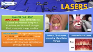
FRENECTOMY
Dr. Rinisha Sinha
MDS Part I
CONTENTS
 Gaining an Insight
 What is a Frenum ?
 Etiology of an Aberrant Frenum
 Classification
 Diagnosing an Abnormal Frenum
 Syndromes and Frenal Attachments
 Indications for Frenal Correction
 Let’s Understand : Frenectomy or Frenotomy
 Conventional Frenectomy
 Electrosurgery
 Lasers
 References
References : American Academy of Periodontology; 1989, 1996
Surgical Procedures performed to correct or
eliminate anatomic, developmental, or
traumatic deformities of the gingiva or
alveolar mucosa
Surgical Procedures for the
correction of relationships between
the gingiva and the oral mucous
membrane with reference to three
specific problem areas
GAINING AN INSIGHT
Mucogingival Surgery Periodontal Plastic Surgery
FRIEDMAN
Attached
Gingiva
Shallow
Vestibules
Aberrant
Frenum
MILLER
• Esthetic Surgical Corrections
• Crown Lengthening
• Ridge Augmentation
• Reconstruction of papilla
A Frenum is a mucous
membrane fold which
contains muscle and
connective tissue fibers
that attach the lip and
the cheek to the
alveolar mucosa, the
gingiva and the
underlying periosteum.
WHAT IS A FRENUM !?
References : Jhaveri H. The Aberrant Frenum; 2006
Normal Frenal Attachment
Apical to the Free
Gingival Margin
ETIOLOGY OF ABERRANT FRENUM
MAXILLARY LABIAL FRENUM MANDIBULAR FRENUM
When the two central incisors erupt widely
separated, no bone is deposited inferior to the
frenum A V-shaped bony cleft between
the two central incisors and an abnormal frenum
attachment results.
A decreased vestibular depth and an
inadequate width of the attached gingiva
Aberrant Mandibular Frenum
References : Huang WJ, Creath CJ –
1995; Jhaveri H - 2006
Sewerin’s Classification
[ 1971 ]
CLASSIFICATION
Merko et al’s Classification
[ 1974 ]
Papilla – Penetrating Frenal
AttachmentPapillary Frenal Attachment
Mucosal Frenal Attachment Gingival Frenal Attachment
References : Sewerin I – 1997; Mirko P, Miroslav S, Lubor M - 1974
Normal Frenum Persistent Tectolabial Frenum With Appendix Frenum With Nodule
Duplication of Frenum Recess of the frenum Bifid Frenum
Blanch Test
DIAGNOSING AN ABNORMAL FRENA
Tension Test
Detected visually by applying
tension over the frenum to see the
movement of the Papillary tip
The lip is pulled superiorly and anteriorly
(For maxillary frenum) and (For mandible)
the lip is pulled outward and downward
Midline
Diastema
The frenum is
pathogenic when
• unusually wide
• no apparent
zone of
attached
gingiva along
the midline
• the interdental
papilla shifts
when the
frenum is
extended
Reference : Miller - 1985
References : Da Felice C, Toti P, Maggio G, Parinmi S, Bagnoli F; Martin RA, Jones
KL; Kusiak A, Sadiak-Nowicka J, Limon J, Kochanska B; Ichida M, Komuro Y et al
SYNDROMES
AND FRENAL
ATTACHMENTS
Ehlers-
Danlos
Syndrome
Infantile
hypertrophic
pyloric
stenosis
Holoprosencephaly
Ellis-van
Creveld
Syndrome
Oro-facial
Digital
syndrome
Pallister-hall
Syndrome
INDICATIONS
Aberrant Frenal
Attachment
causing Midline
Diastema
Aberrant Frenal
Attachment
causing Gingival
recession
Aberrant Frenal
Attachment causing
hindrance in
maintaining oral
hygiene
Aberrant Frenal
Attachment
interfering with
Speech
ANKYLOGLOSSIA
Aberrant Frenal
Attachment
indicating
Shallow vestibule
Aberrant Frenal
Attachment visible as
a pendulous piece of
tissue in the midline
of the upper lip
FRENECTOMY FRENOTOMY
• FRENECTOMY
The complete removal of the
frenum, including its
attachment to the underlying
bone.
LET’S UNDERSTAND
• FRENOTOMY
The incision and the
relocation of the frenal
attachment.
References : Dibart S, Karima M
FRENECTOMY
Scalpel
Technique
LasersElectrosurgery
• Safe and effective
• Shorter duration of
the surgery
• Simplicity of the
procedure
• Absence of
postoperative
infections
• Lesser pain, swelling
• Small or no scar
• Efficient
• Mild bleeding
• Absence of
postoperative
complications
Disadvantages
• Bleeding
• Patient
compliance
• Conventional
technique
• Excision of the frenum
through scalpel
• Cost-effective
Argon Beam Coagulation References : Dibart S, Karima M; Cunha RF, Silva JZ, Faria MD
CONVENTIONAL ( CLASSICAL )
FRENECTOMY
Archer (1961) and Kruger (1964)Indication
For midline diastema cases
Armamentarium
• Haemostat
• BP handle
• Scalpel blade no.15
• Gauze sponges
• 3-0 black silk sutures
• Suture pliers
• Scissors
• A Periodontal dressing
(Coe-pak)
References : Jhaveri H. The Aberrant Frenum – 2006; Archer WH. Oral surgery for a dental prosthesis - 1975
MILLER’S TECHNIQUE
Miller PD - 1985Indication : For the post-orthodontic diastema cases
References : Miller PD. Frenectomy combined with a laterally positioned pedicle graft -functional and esthetic considerations. J Periodont, 1985
A Horizontal
incision to
separate the
frenulum from the
interdental papilla
Excision of the
frenulum and
exposure of labial
alveolar bone in
the midline
Laterally
positioned
pedicle graft
taken to obtain
primary closure
across midline
Gingivoplasty
of any excessive
interdental
tissue labially or
palatally
Z PLASTY TECHNIQUE
Indication : Hypertrophic labial frenum with a low insertion, which is associated with an
inter-incisor diastema and also in cases of a short vestibule
Length of the
frenum was incised
with scalpel
The sub - muscosal
tissue were
dissected beyond
the base of each
flap by using fine
tissue forceps
The resultant flaps
were mobilized
and transposed
through 90º to
close vertical
incisions
horizontally
Absorbable 5-0
vicryl sutures were
placed and a
periodontal
dressing was
placed. No
hypertrophic
scar formation
References : Howe GL; Puig JR, Lefebvre E, Landat F. The Z-plasty technique – 1977; Langdon JD, Patel MF. Reconstructive surgery - 1998
1 cm long Double
rotation flaps
were obtained
V-Y PLASTY TECHNIQUE
Indication : For lengthening the localized area, like the broad frenum in the premolar-molar area
A V-shaped
Incision on the
undersurface of the
frenal attachment
The frenum was
relocated at an
apical position
Sutured with 4-0
silk suture and a
periodontal
pack was placed.
After 1 month -
Frenal attachment
was found to be
relocated at an
apical position,
with an
uneventful
healing
References : Kruger GO. Acquired defects of the hard and soft tissues of the face
The V-shaped
incision was
coverted into a Y
ELECTROSURGERY
Armamentarium
• An Electrocautery unit
with the loop electrode
• A Haemostat
Indication
 In cases of Patients with bleeding disorders
 In non-compliant Patients
The frenum was held with the haemostat
It was excised by using a loop electrode tip
Advantages
 minimal procedural bleeding
 there was no need of sutures
The healing was by Secondary intention, as the
wound edges were not approximated with
sutures
References : Cunha RF, Silva JZ, Faria MD – 2008; Verco PJW. “A case report and a
clinical technique: argon beam electrosurgery for the tongue ties and maxillary
frenectomies in infants and children - 2010
LASERS
940 nm Diode Laser
removing Mandibular
Frenum
Robert N. Hall – 1962
DIODE LASER uses a combination of
Gallium and Arsenide along with
Aluminium and Indium  changes
Electro-magnetic energy into Heat
DIODE LASER are highly absorbable by
MELANIN and HAEMOGLOBIN, hence
allows soft-tissue manipulations;
accompanied by improved epithelization
and wound healing.
Carbon dioxide Laser
REFERENCES
 Carranza’s Clinical
Periodontology :
10th Edition
 Periobasics : A
textbook of
Periodontics and
Implantology
 Journal of
Pharmaceuticals and
Scientific Innovation
 Devi Shree, Sheela Kumar Gujjari,
ShubhaShini P.v. – 2012
Frenectomy: A Review with the
Reports of Surgical Techniques
 Puneet Sharma, Sanjeev Kumar
Salaria, Ravi Kiran N Gowda,
Sameer Ahuja, Sidharth Joshi,
Deepak Kumar Bansal – 2016
Frenectomy- A Brief Review
 Dr. Manish Ashtankar, Dr. Mala
Dixit Baburaj and Dr. Abhishek
Singh – 2018
LABIAL FRENECTOMY- A REVIEW
AND CASE REPORTS

Frenectomy

  • 1.
  • 2.
    CONTENTS  Gaining anInsight  What is a Frenum ?  Etiology of an Aberrant Frenum  Classification  Diagnosing an Abnormal Frenum  Syndromes and Frenal Attachments  Indications for Frenal Correction  Let’s Understand : Frenectomy or Frenotomy  Conventional Frenectomy  Electrosurgery  Lasers  References
  • 3.
    References : AmericanAcademy of Periodontology; 1989, 1996 Surgical Procedures performed to correct or eliminate anatomic, developmental, or traumatic deformities of the gingiva or alveolar mucosa Surgical Procedures for the correction of relationships between the gingiva and the oral mucous membrane with reference to three specific problem areas GAINING AN INSIGHT Mucogingival Surgery Periodontal Plastic Surgery FRIEDMAN Attached Gingiva Shallow Vestibules Aberrant Frenum MILLER • Esthetic Surgical Corrections • Crown Lengthening • Ridge Augmentation • Reconstruction of papilla
  • 4.
    A Frenum isa mucous membrane fold which contains muscle and connective tissue fibers that attach the lip and the cheek to the alveolar mucosa, the gingiva and the underlying periosteum. WHAT IS A FRENUM !? References : Jhaveri H. The Aberrant Frenum; 2006 Normal Frenal Attachment Apical to the Free Gingival Margin
  • 5.
    ETIOLOGY OF ABERRANTFRENUM MAXILLARY LABIAL FRENUM MANDIBULAR FRENUM When the two central incisors erupt widely separated, no bone is deposited inferior to the frenum A V-shaped bony cleft between the two central incisors and an abnormal frenum attachment results. A decreased vestibular depth and an inadequate width of the attached gingiva Aberrant Mandibular Frenum References : Huang WJ, Creath CJ – 1995; Jhaveri H - 2006
  • 6.
    Sewerin’s Classification [ 1971] CLASSIFICATION Merko et al’s Classification [ 1974 ] Papilla – Penetrating Frenal AttachmentPapillary Frenal Attachment Mucosal Frenal Attachment Gingival Frenal Attachment References : Sewerin I – 1997; Mirko P, Miroslav S, Lubor M - 1974 Normal Frenum Persistent Tectolabial Frenum With Appendix Frenum With Nodule Duplication of Frenum Recess of the frenum Bifid Frenum
  • 7.
    Blanch Test DIAGNOSING ANABNORMAL FRENA Tension Test Detected visually by applying tension over the frenum to see the movement of the Papillary tip The lip is pulled superiorly and anteriorly (For maxillary frenum) and (For mandible) the lip is pulled outward and downward Midline Diastema The frenum is pathogenic when • unusually wide • no apparent zone of attached gingiva along the midline • the interdental papilla shifts when the frenum is extended Reference : Miller - 1985
  • 8.
    References : DaFelice C, Toti P, Maggio G, Parinmi S, Bagnoli F; Martin RA, Jones KL; Kusiak A, Sadiak-Nowicka J, Limon J, Kochanska B; Ichida M, Komuro Y et al SYNDROMES AND FRENAL ATTACHMENTS Ehlers- Danlos Syndrome Infantile hypertrophic pyloric stenosis Holoprosencephaly Ellis-van Creveld Syndrome Oro-facial Digital syndrome Pallister-hall Syndrome
  • 9.
    INDICATIONS Aberrant Frenal Attachment causing Midline Diastema AberrantFrenal Attachment causing Gingival recession Aberrant Frenal Attachment causing hindrance in maintaining oral hygiene Aberrant Frenal Attachment interfering with Speech ANKYLOGLOSSIA Aberrant Frenal Attachment indicating Shallow vestibule Aberrant Frenal Attachment visible as a pendulous piece of tissue in the midline of the upper lip
  • 10.
  • 11.
    • FRENECTOMY The completeremoval of the frenum, including its attachment to the underlying bone. LET’S UNDERSTAND • FRENOTOMY The incision and the relocation of the frenal attachment. References : Dibart S, Karima M
  • 12.
    FRENECTOMY Scalpel Technique LasersElectrosurgery • Safe andeffective • Shorter duration of the surgery • Simplicity of the procedure • Absence of postoperative infections • Lesser pain, swelling • Small or no scar • Efficient • Mild bleeding • Absence of postoperative complications Disadvantages • Bleeding • Patient compliance • Conventional technique • Excision of the frenum through scalpel • Cost-effective Argon Beam Coagulation References : Dibart S, Karima M; Cunha RF, Silva JZ, Faria MD
  • 13.
    CONVENTIONAL ( CLASSICAL) FRENECTOMY Archer (1961) and Kruger (1964)Indication For midline diastema cases Armamentarium • Haemostat • BP handle • Scalpel blade no.15 • Gauze sponges • 3-0 black silk sutures • Suture pliers • Scissors • A Periodontal dressing (Coe-pak) References : Jhaveri H. The Aberrant Frenum – 2006; Archer WH. Oral surgery for a dental prosthesis - 1975
  • 14.
    MILLER’S TECHNIQUE Miller PD- 1985Indication : For the post-orthodontic diastema cases References : Miller PD. Frenectomy combined with a laterally positioned pedicle graft -functional and esthetic considerations. J Periodont, 1985 A Horizontal incision to separate the frenulum from the interdental papilla Excision of the frenulum and exposure of labial alveolar bone in the midline Laterally positioned pedicle graft taken to obtain primary closure across midline Gingivoplasty of any excessive interdental tissue labially or palatally
  • 15.
    Z PLASTY TECHNIQUE Indication: Hypertrophic labial frenum with a low insertion, which is associated with an inter-incisor diastema and also in cases of a short vestibule Length of the frenum was incised with scalpel The sub - muscosal tissue were dissected beyond the base of each flap by using fine tissue forceps The resultant flaps were mobilized and transposed through 90º to close vertical incisions horizontally Absorbable 5-0 vicryl sutures were placed and a periodontal dressing was placed. No hypertrophic scar formation References : Howe GL; Puig JR, Lefebvre E, Landat F. The Z-plasty technique – 1977; Langdon JD, Patel MF. Reconstructive surgery - 1998 1 cm long Double rotation flaps were obtained
  • 16.
    V-Y PLASTY TECHNIQUE Indication: For lengthening the localized area, like the broad frenum in the premolar-molar area A V-shaped Incision on the undersurface of the frenal attachment The frenum was relocated at an apical position Sutured with 4-0 silk suture and a periodontal pack was placed. After 1 month - Frenal attachment was found to be relocated at an apical position, with an uneventful healing References : Kruger GO. Acquired defects of the hard and soft tissues of the face The V-shaped incision was coverted into a Y
  • 17.
    ELECTROSURGERY Armamentarium • An Electrocauteryunit with the loop electrode • A Haemostat Indication  In cases of Patients with bleeding disorders  In non-compliant Patients The frenum was held with the haemostat It was excised by using a loop electrode tip Advantages  minimal procedural bleeding  there was no need of sutures The healing was by Secondary intention, as the wound edges were not approximated with sutures References : Cunha RF, Silva JZ, Faria MD – 2008; Verco PJW. “A case report and a clinical technique: argon beam electrosurgery for the tongue ties and maxillary frenectomies in infants and children - 2010
  • 18.
    LASERS 940 nm DiodeLaser removing Mandibular Frenum Robert N. Hall – 1962 DIODE LASER uses a combination of Gallium and Arsenide along with Aluminium and Indium  changes Electro-magnetic energy into Heat DIODE LASER are highly absorbable by MELANIN and HAEMOGLOBIN, hence allows soft-tissue manipulations; accompanied by improved epithelization and wound healing. Carbon dioxide Laser
  • 19.
    REFERENCES  Carranza’s Clinical Periodontology: 10th Edition  Periobasics : A textbook of Periodontics and Implantology  Journal of Pharmaceuticals and Scientific Innovation  Devi Shree, Sheela Kumar Gujjari, ShubhaShini P.v. – 2012 Frenectomy: A Review with the Reports of Surgical Techniques  Puneet Sharma, Sanjeev Kumar Salaria, Ravi Kiran N Gowda, Sameer Ahuja, Sidharth Joshi, Deepak Kumar Bansal – 2016 Frenectomy- A Brief Review  Dr. Manish Ashtankar, Dr. Mala Dixit Baburaj and Dr. Abhishek Singh – 2018 LABIAL FRENECTOMY- A REVIEW AND CASE REPORTS