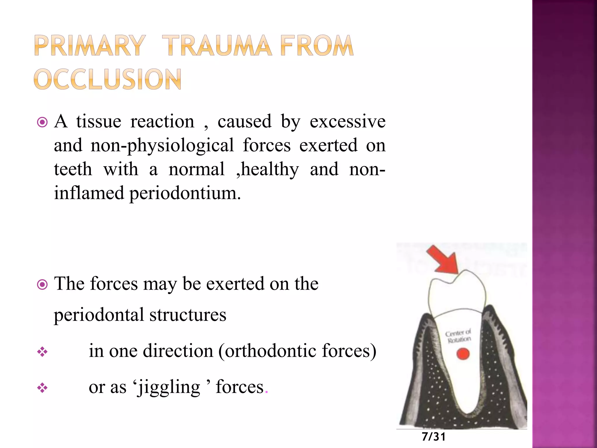
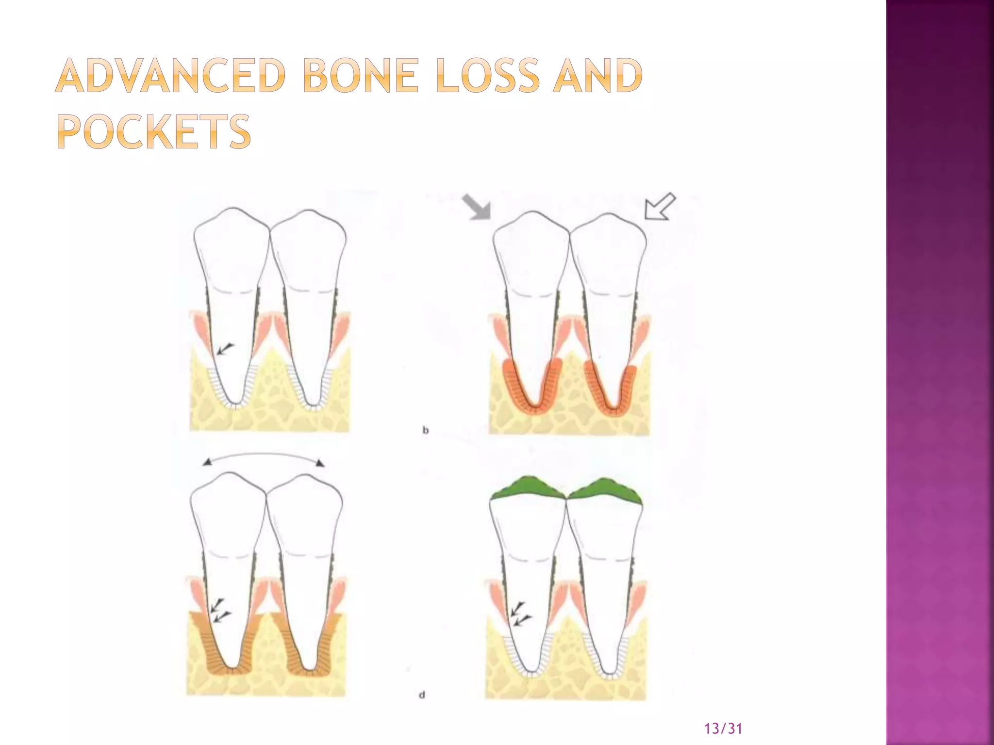
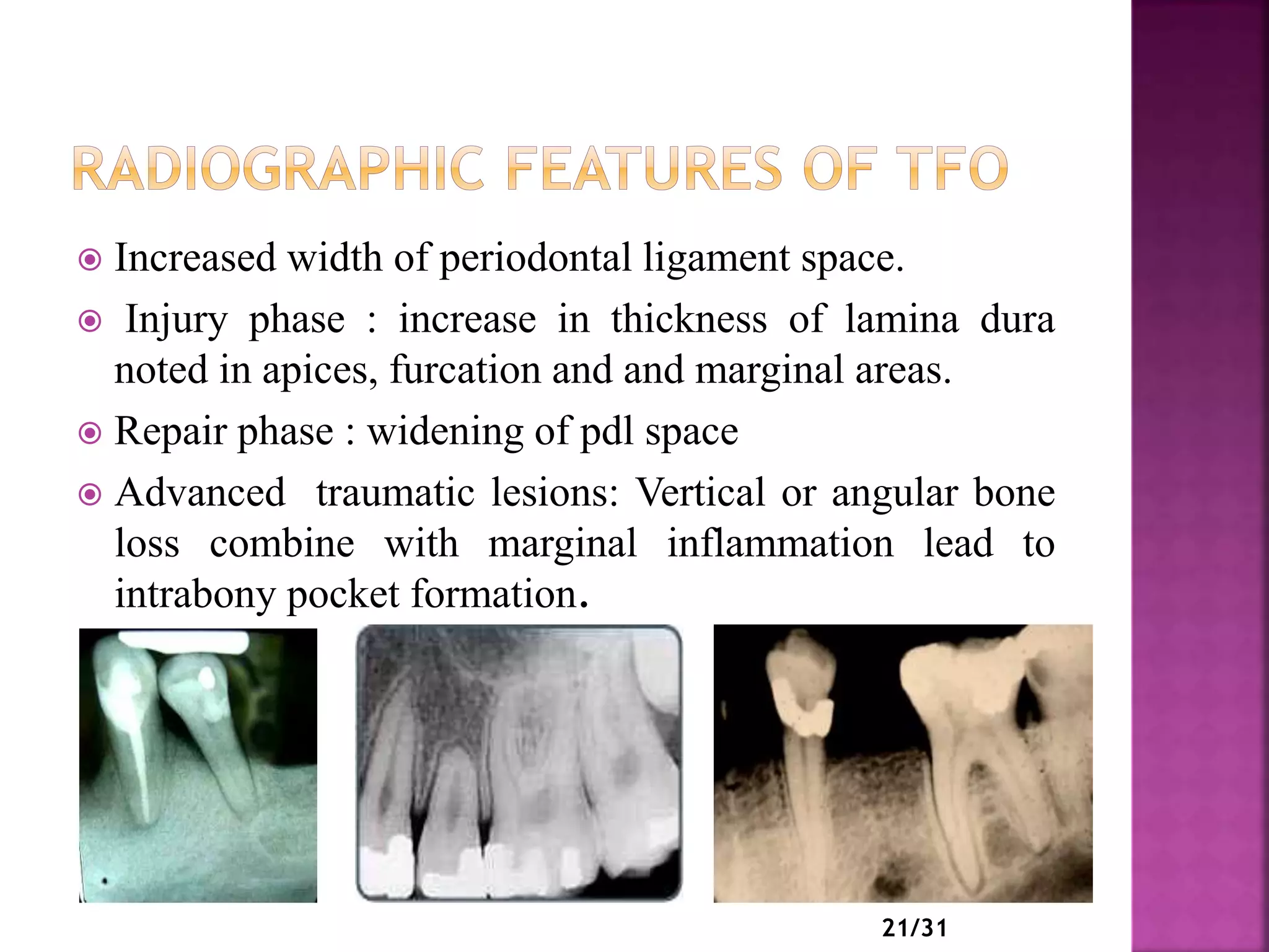
GUIDED BY: PRESENTED BY:
DR DIVYA JAGGI DR ANKITA DADWAL
MDS 2ND YR
1
 INTRODUCTION
 DEFINITION
 TYPES OF TRAUMA FROM OCCLUSION
 GLICKMAN CONCEPT
 WAERHAUG CONCEPT
 STAGES OF TISSUE RESPONSE TO INJURY
 CLINICAL AND RADIOGRAPHIC FEATURES OF TFO
 CLINICAL DIAGNOSIS OF TFO
 TFO AND IMPLANTS
 TREATMENT OF TFO
 CONCLUSION
 REFRENCES
1/31
 Trauma from occlusion is a term used to describe
pathologic alterations or adaptive changes which
develop in the periodontium as a result of undue
force produced by the masticatory muscles. It is
only one of many terms that have been used to
describe such alterations in the periodontium.
2/31
 “A condition where injury results to the supporting structures of
the teeth by the act of bringing the jaws into a closed position”.
(Stillman 1917)
 When occlusal forces exceed the adaptive capacity of the tissues,
tissue injury results. this resultant injury is termed TFO.
(Glickman 1962)
 “Damage in the periodontium caused by stress on the teeth
produced directly or indirectly by teeth of the opposing jaw”.
( WHO1978)
 An injury to the attachment apparatus as a result of excessive
occlusal force.
(Glossary of periodontic terms ,1992)
3/31
 Traumatizing occlusion
 Occlusal trauma
 Traumatogenic occlusion
 Periodontal traumatism
 Overload
 Occlusal disharmony
 Functional imbalance
 Occlusal dystrophy
4/31
 results from an abrupt occlusal impact, such as that
produced by biting on a hard object (e.g., an olive pit).
 In addition, restorations or prosthetic appliances that
interfere with the direction of occlusal forces on the
teeth may induce acute trauma
Clinical features :
1. Tooth pain.
2. Sensitivity to percussion.
3. Tooth mobility.
5/31
 develops from gradual changes in
a) occlusion produced by tooth wear,
b) drifting movement,
c) extrusion of teeth,
d) combined with parafunctional habits such as bruxism
and clenching .
6/31
 A tissue reaction , caused by excessive
and non-physiological forces exerted on
teeth with a normal ,healthy and non-
inflamed periodontium.
 The forces may be exerted on the
periodontal structures
 in one direction (orthodontic forces)
 or as ‘jiggling ’ forces.
7/31
 In the periodontal ligament,
zone of compression
zone of tension
 temporary increased mobility
(functional adaptation ).
- no changes in the supracrestal
fibres,
- no loss of periodontal attachment,
- no increased probing pocket depth.
 Forces too high (above the adaptation
level),
- aseptic necrosis of the PDL.
- root resorption occurs
- resulting in shorter roots.
8/31
9/31
11
 It is related to situations in which occlusal forces cause
damage in a periodontium of reduced height
(attachment loss present).
 In his original studies in the
1960s, Glickman formulated the hypothesis that
premature contacts and excessive occlusal forces could
be a cofactor in the progression of periodontal disease
by changing the pathway and spread of inflammation
into the deeper periodontal tissues.
11/31
12/31
13/31
 Glickman (1965, 1967)
claimed that the pathway
of the spread of a
plaque‐associated gingival
lesion can be changed if
forces of an abnormal
magnitude are acting on
the contaminated tooth.
 ZONE OF IRRITATION
 ZONE OF CO
DESTRUCTION
14/31
 The zone of irritation includes the marginal and
interdental gingiva.
 The soft tissue of this zone is bordered by hard tissue
(the tooth) only on one side and cannot therefore be
affected by forces of occlusion.
 The gingival lesion is the tissue response to products
from microbial plaque.
 This gingival lesion at a “non‐traumatized” tooth
propagates, in the apical direction by first involving the
alveolar bone and only later the periodontal ligament
area.
 The progression of this lesion results in an even
(horizontal) bone destruction.
15/31
 It was claimed that the fiber
bundles which separate the
zone of co‐destruction from
the zone of irritation can be
affected from two different
directions:
1. From the inflammatory
gingival lesion maintained in
the zone of irritation
2. From trauma‐induced
changes in the zone of
co‐destruction
16/31
 The loss of connective attachment and bone around
teeth is, according to Waerhaug, exclusively the result
of inflammatory lesions associated with subgingival
plaque.
 Waerhaug concluded that angular bony defects and
infrabony pockets occur when the subgingival plaque
of one tooth has reached a more apical level than the
plaque on the neighbouring tooth, and when the
volume of the alveolar bone surrounding the roots is
comparatively large.
17/31
 STAGE –I: INJURY
- slightly excessive pressure and tension
-increased pressure and tension
-severe tension
 STAGE II: REPAIR
- central buttressing
- peripheral buttressing
 STAGE III: ADAPTIVE REMODELLING
18/31
19/31
1)Tooth mobility
2) Pain on chewing or percussion
3) Fremitus
4) Occlusal prematurities/discrepancies
5) Wear facets
6) Tooth migration
7) Chipped or fractured tooth (teeth)
8) Thermal sensitivity
20/31
 Increased width of periodontal ligament space.
 Injury phase : increase in thickness of lamina dura
noted in apices, furcation and and marginal areas.
 Repair phase : widening of pdl space
 Advanced traumatic lesions: Vertical or angular bone
loss combine with marginal inflammation lead to
intrabony pocket formation.
21/31
TOOTH MOBILITY :
A clinical diagnosis of occlusal
trauma can only be confirmed where progressive
mobility can be identified through a series of repeated
measurements over an extended period .
HOW TO MEASURE TOOTH MOBILITY???
1. MANUAL EXAMINATION
2. FRMITUS TEST
3. PERIOTEST
4. PERIODONTOMETRE
22/31
2. WEAR PATTERNS:
a) BRUXISM- wear facets on occlusal and incisal
surfaces which can be identified as shiny and irregular
surfaces.
b) ABFRACTIONS- excessive lateral forces,
abfractions can be seen on pre molars.
3. THERMAL SENSITIVITY
4. MUSCLE HYPERTONOCITY
5. T- SCAN
23/31
 An osseointegrated implant is in direct contact with
bone.
 No periodontal ligament space
 Functional load enhance osseointegration and doesnot
result in marginal bone loss.
 Occlusal overload adversely affect osseointegration
 Lead to implant failure
24/31
Key factors controlling bone remodelling at molecular
level are:
1. strain: change in length to the original length
2. modulus of elasticity: stiffness of material
25/31
A goal of periodontal therapy in the treatment of
occlusal traumatism should be to maintain the
periodontium in comfort and function.
PROPOSED BY AAP(1996)
1. Reduce /eliminate tooth mobility
2. Eliminate occlusal prematurities and fremitus
3. Eliminate parafunctional habits
4. Prevent further tooth migration
5. Decrease / stablize radiographic changes
26/31
Indications (1989 WORLD WORKSHOP IN PERIODONTICS)
1) To reduce traumatic forces to teeth that exhibit:
• Increasing mobility or fremitus .
• Discomfort during occlusal contact or function.
2) To achieve functional relationships and masticatory efficiency in
conjunction with restorative treatment, orthodontic, orthognathic
surgery or jaw trauma when indicated.
3) As adjunctive therapy that may reduce the damage from parafunctional
habits.
4) To reshape teeth contributing to soft tissue injury.
5) To adjust marginal ridge relationships and cusps that are contributing
to food impaction 27/31
INDICATIONS : (1989 WORLD WORKSHOP)
1) Stabilize teeth with increasing mobility that have not responded to
occlusal adjustment and periodontal treatment.
2) Stabilize teeth with advanced mobility that have not responded to
occlusal adjustment and treatment when there is interference with
normal function and patient comfort.
3) Facilitate treatment of extremely mobile teeth by splinting them prior
to periodontal instrumentation and occlusal adjustment procedures.
4) Prevent tipping or drifting of teeth and extrusion of unopposed teeth.
5) Stabilize teeth, when indicated, following orthodontic movement.
6) Create adequate occlusal stability when replacing missing teeth.
7) Stabilize teeth following acute trauma. 28/31
 BRUXISM : night guard
29/31
 Periodontal structures depend on functional occlusal
forces to activate the periodontal mechanoreceptors in
the neuromuscular physiology of the masticatory
system. A traumatic occlusion on a healthy
periodontium leads to an increased mobility but not to
attachment loss. In inflamed periodontal structures
traumatic occlusion contributes to a further and faster
spread of the inflammation apically and to more bone
loss.
30/31
1) Carranza’s Clinical Periodontology; 4th edition
2) Carranza’s Clinical Periodontology; 9th edition
3) Carranza’s Clinical Periodontology; 10th edition
4) Clinical Periodontology and Implantology;6th edition; Jan
Lindhe
5) Trauma from occlusion: a review- Commander R. “Dave”
Rupprecht, DC, US. CLINICAL UPDATES VOL26,NO.1
JANUARY 2004.
31/31
33

Trauma from occlusion

  • 1.
    GUIDED BY: PRESENTEDBY: DR DIVYA JAGGI DR ANKITA DADWAL MDS 2ND YR 1
  • 2.
     INTRODUCTION  DEFINITION TYPES OF TRAUMA FROM OCCLUSION  GLICKMAN CONCEPT  WAERHAUG CONCEPT  STAGES OF TISSUE RESPONSE TO INJURY  CLINICAL AND RADIOGRAPHIC FEATURES OF TFO  CLINICAL DIAGNOSIS OF TFO  TFO AND IMPLANTS  TREATMENT OF TFO  CONCLUSION  REFRENCES 1/31
  • 3.
     Trauma fromocclusion is a term used to describe pathologic alterations or adaptive changes which develop in the periodontium as a result of undue force produced by the masticatory muscles. It is only one of many terms that have been used to describe such alterations in the periodontium. 2/31
  • 4.
     “A conditionwhere injury results to the supporting structures of the teeth by the act of bringing the jaws into a closed position”. (Stillman 1917)  When occlusal forces exceed the adaptive capacity of the tissues, tissue injury results. this resultant injury is termed TFO. (Glickman 1962)  “Damage in the periodontium caused by stress on the teeth produced directly or indirectly by teeth of the opposing jaw”. ( WHO1978)  An injury to the attachment apparatus as a result of excessive occlusal force. (Glossary of periodontic terms ,1992) 3/31
  • 5.
     Traumatizing occlusion Occlusal trauma  Traumatogenic occlusion  Periodontal traumatism  Overload  Occlusal disharmony  Functional imbalance  Occlusal dystrophy 4/31
  • 6.
     results froman abrupt occlusal impact, such as that produced by biting on a hard object (e.g., an olive pit).  In addition, restorations or prosthetic appliances that interfere with the direction of occlusal forces on the teeth may induce acute trauma Clinical features : 1. Tooth pain. 2. Sensitivity to percussion. 3. Tooth mobility. 5/31
  • 7.
     develops fromgradual changes in a) occlusion produced by tooth wear, b) drifting movement, c) extrusion of teeth, d) combined with parafunctional habits such as bruxism and clenching . 6/31
  • 8.
     A tissuereaction , caused by excessive and non-physiological forces exerted on teeth with a normal ,healthy and non- inflamed periodontium.  The forces may be exerted on the periodontal structures  in one direction (orthodontic forces)  or as ‘jiggling ’ forces. 7/31
  • 9.
     In theperiodontal ligament, zone of compression zone of tension  temporary increased mobility (functional adaptation ). - no changes in the supracrestal fibres, - no loss of periodontal attachment, - no increased probing pocket depth.  Forces too high (above the adaptation level), - aseptic necrosis of the PDL. - root resorption occurs - resulting in shorter roots. 8/31
  • 10.
  • 11.
  • 12.
     It isrelated to situations in which occlusal forces cause damage in a periodontium of reduced height (attachment loss present).  In his original studies in the 1960s, Glickman formulated the hypothesis that premature contacts and excessive occlusal forces could be a cofactor in the progression of periodontal disease by changing the pathway and spread of inflammation into the deeper periodontal tissues. 11/31
  • 13.
  • 14.
  • 15.
     Glickman (1965,1967) claimed that the pathway of the spread of a plaque‐associated gingival lesion can be changed if forces of an abnormal magnitude are acting on the contaminated tooth.  ZONE OF IRRITATION  ZONE OF CO DESTRUCTION 14/31
  • 16.
     The zoneof irritation includes the marginal and interdental gingiva.  The soft tissue of this zone is bordered by hard tissue (the tooth) only on one side and cannot therefore be affected by forces of occlusion.  The gingival lesion is the tissue response to products from microbial plaque.  This gingival lesion at a “non‐traumatized” tooth propagates, in the apical direction by first involving the alveolar bone and only later the periodontal ligament area.  The progression of this lesion results in an even (horizontal) bone destruction. 15/31
  • 17.
     It wasclaimed that the fiber bundles which separate the zone of co‐destruction from the zone of irritation can be affected from two different directions: 1. From the inflammatory gingival lesion maintained in the zone of irritation 2. From trauma‐induced changes in the zone of co‐destruction 16/31
  • 18.
     The lossof connective attachment and bone around teeth is, according to Waerhaug, exclusively the result of inflammatory lesions associated with subgingival plaque.  Waerhaug concluded that angular bony defects and infrabony pockets occur when the subgingival plaque of one tooth has reached a more apical level than the plaque on the neighbouring tooth, and when the volume of the alveolar bone surrounding the roots is comparatively large. 17/31
  • 19.
     STAGE –I:INJURY - slightly excessive pressure and tension -increased pressure and tension -severe tension  STAGE II: REPAIR - central buttressing - peripheral buttressing  STAGE III: ADAPTIVE REMODELLING 18/31
  • 20.
  • 21.
    1)Tooth mobility 2) Painon chewing or percussion 3) Fremitus 4) Occlusal prematurities/discrepancies 5) Wear facets 6) Tooth migration 7) Chipped or fractured tooth (teeth) 8) Thermal sensitivity 20/31
  • 22.
     Increased widthof periodontal ligament space.  Injury phase : increase in thickness of lamina dura noted in apices, furcation and and marginal areas.  Repair phase : widening of pdl space  Advanced traumatic lesions: Vertical or angular bone loss combine with marginal inflammation lead to intrabony pocket formation. 21/31
  • 23.
    TOOTH MOBILITY : Aclinical diagnosis of occlusal trauma can only be confirmed where progressive mobility can be identified through a series of repeated measurements over an extended period . HOW TO MEASURE TOOTH MOBILITY??? 1. MANUAL EXAMINATION 2. FRMITUS TEST 3. PERIOTEST 4. PERIODONTOMETRE 22/31
  • 24.
    2. WEAR PATTERNS: a)BRUXISM- wear facets on occlusal and incisal surfaces which can be identified as shiny and irregular surfaces. b) ABFRACTIONS- excessive lateral forces, abfractions can be seen on pre molars. 3. THERMAL SENSITIVITY 4. MUSCLE HYPERTONOCITY 5. T- SCAN 23/31
  • 25.
     An osseointegratedimplant is in direct contact with bone.  No periodontal ligament space  Functional load enhance osseointegration and doesnot result in marginal bone loss.  Occlusal overload adversely affect osseointegration  Lead to implant failure 24/31
  • 26.
    Key factors controllingbone remodelling at molecular level are: 1. strain: change in length to the original length 2. modulus of elasticity: stiffness of material 25/31
  • 27.
    A goal ofperiodontal therapy in the treatment of occlusal traumatism should be to maintain the periodontium in comfort and function. PROPOSED BY AAP(1996) 1. Reduce /eliminate tooth mobility 2. Eliminate occlusal prematurities and fremitus 3. Eliminate parafunctional habits 4. Prevent further tooth migration 5. Decrease / stablize radiographic changes 26/31
  • 28.
    Indications (1989 WORLDWORKSHOP IN PERIODONTICS) 1) To reduce traumatic forces to teeth that exhibit: • Increasing mobility or fremitus . • Discomfort during occlusal contact or function. 2) To achieve functional relationships and masticatory efficiency in conjunction with restorative treatment, orthodontic, orthognathic surgery or jaw trauma when indicated. 3) As adjunctive therapy that may reduce the damage from parafunctional habits. 4) To reshape teeth contributing to soft tissue injury. 5) To adjust marginal ridge relationships and cusps that are contributing to food impaction 27/31
  • 29.
    INDICATIONS : (1989WORLD WORKSHOP) 1) Stabilize teeth with increasing mobility that have not responded to occlusal adjustment and periodontal treatment. 2) Stabilize teeth with advanced mobility that have not responded to occlusal adjustment and treatment when there is interference with normal function and patient comfort. 3) Facilitate treatment of extremely mobile teeth by splinting them prior to periodontal instrumentation and occlusal adjustment procedures. 4) Prevent tipping or drifting of teeth and extrusion of unopposed teeth. 5) Stabilize teeth, when indicated, following orthodontic movement. 6) Create adequate occlusal stability when replacing missing teeth. 7) Stabilize teeth following acute trauma. 28/31
  • 30.
     BRUXISM :night guard 29/31
  • 31.
     Periodontal structuresdepend on functional occlusal forces to activate the periodontal mechanoreceptors in the neuromuscular physiology of the masticatory system. A traumatic occlusion on a healthy periodontium leads to an increased mobility but not to attachment loss. In inflamed periodontal structures traumatic occlusion contributes to a further and faster spread of the inflammation apically and to more bone loss. 30/31
  • 32.
    1) Carranza’s ClinicalPeriodontology; 4th edition 2) Carranza’s Clinical Periodontology; 9th edition 3) Carranza’s Clinical Periodontology; 10th edition 4) Clinical Periodontology and Implantology;6th edition; Jan Lindhe 5) Trauma from occlusion: a review- Commander R. “Dave” Rupprecht, DC, US. CLINICAL UPDATES VOL26,NO.1 JANUARY 2004. 31/31
  • 33.

Editor's Notes

  • #18 The zone of co‐destruction includes the root cementum (mineralized tissue), the periodontal ligament, and the alveolar bone (mineralized tissue), and is coronally demarcated by the trans‐septal (interdental) and the dentoalveolar collagen fiber bundles.
  • #20 thickened periodontal ligament (funnel shaped) angular defects, no pocket formation. loose teeth Increased vascularization.
  • #22 Injury stage: pdl destroy Adaptive stage: widening of pdl It is nt pathologic as it is body adaptive response not a disease.
  • #24 Increased tooth mobility is not always indicative of trauma from occlusion. It is important, however, that hypermobility which does occur as a result of trauma from occlusion is detected in patients with reduced periodontal attachment. The reason for this is that trauma from occlusion may accelerate further reduction in attachment in a patient with active periodontitis.
  • #27 Titanium modulus of elasticity is 5-10 times more than the that of bone.
  • #29 Occlusal adjustment or selective grinding is defined as reshaping the occluding surfaces of teeth by grinding to create harmonious contact relationships between the upper and lower teeth