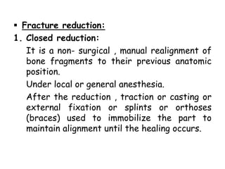
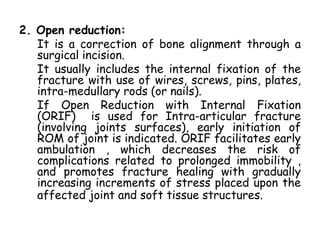
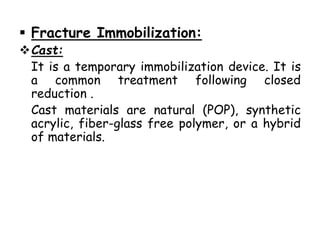
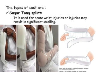
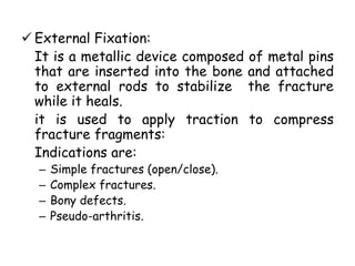
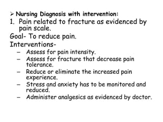
FRACTURES
By :
Abhishek Yadav
M Sc (N) 1st Year
OUTLINES
• Introduction of Fracture.
• Definition of Fracture.
• Incidence of Fracture.
• Etiology & Risk factors of Fracture.
• Pathophysiology of Fracture.
• Process of Bone Healing.
• Clinical manifestations of Fracture.
• Classification of Fracture.
• Diagnostic Evaluation of Fracture.
• Management of Fracture.
INTRODUCTION
• Any one who has experienced a broken bone
or a ligament strain can appreciate the
challenges facing a client who is recovering
from musculoskeletal trauma or overuse .
• Activity restrictions and assistive devices
both complicate & facilitate the healing
process.
DEFINITION
• A Fracture is any disruption in the
normal continuity of a bone. When
fracture occurs, surrounding soft
tissues are often damaged as well.
INCIDENCE
• Trauma is the leading cause of death in the
USA for those b/w the ages of 1 and 37, and
the 4th leading cause of death for all age
groups.
• Fractures account for high traumatic injuries.
They can create significant changes in one’s
quality of life by causing activity restricts,
disability & economic loss.
• The total incidence rate of fractures is 53.4
per 1000 per year in women, and 24.9 per
1000 per year in men.
• Rate of hip and wrist fractures tended to be
higher in women.
ETIOLOGY & RISK FACTORS
 Mechanical overload of bone:
– When more stress is placed on the bone
than it can absorb.
 Metabolic bone disease:
– In the diseases like osteoporosis the bone
may fracture with even minor trauma
because of its weakening by pre-existing
disorder.
 Direct force:
– It may occur when a moving object strikes the
body area over the bone.
 Indirect force:
– It may occur when a powerful muscle
contraction pulls against the bone .
 Biological conditions:
Pre-disposition to fracture results from
biological conditions such as :
– Osteopenia (caused by steroid use or Cushing’s
syndrome).
– Osteogenesis Imperfecta (a congenital bone
disease characterized by defective collagen
production by osteoblasts).
 Neoplasms:
– It also weaken the bone and contribute to
fracture (pathological fracture).
– It can be due to benign or malignant tumour.
 Post menopausal estrogen loss:
– Release of estrogen stops after menopause , it
can make bone weaken.
 Malnutrition:
– Due to protein malnutrition the strength and
stiffness of bone become lower, which lead to
decreased bone mass and increased risk for
fracture
 High risk recreation or employment
related activities:
– Skateboarding, rock climbing, etc.
 Domestic violence:
– Victims of domestic violence are also among
people treated for traumatic injuries.
 Road traffic accidents:
– Due to high traffic and high speed vehicle,
road condtions, etc.
PATHOPHYSIOLOGY
Bone formation
& reabsorption
Bone balance
(remodeling)
Perforation or
fracture
of trabecular plates
Degree of
mineralization
Collagen structure
& bone proteins
Loss of micro
-architecture
Abilty to
Repair damage
Crystal size
& structure
Marrow & fat
composition
Bone quality
Growth
(modeling)
Bone
volume
Bone
Shape
Bone
strength
Falls Soft-tissue
padding
Force of impact FRACTURE
BONE HEALING
• Few tissues in human body that heal through
regeneration rather than formation of scar.
• Bone is one of these tissues.
• Fracture repair mechanism is same as bone
formation during normal growth.
• Fracture healing may not occur in expected
time (delayed union) or may not occur at all
(non- union).
• The ossification process is arrested by causes :
– Inadequate reduction & immobilization,
– Excessive movement of the fracture
fragments,
– Infection,
– Poor nutrition,
– Systemic disease.
• Healing time for fractures increases with age.
• Electrical stimulation and pulsed
electromagnetic fields (PEMF’s) can be used
to stimulate bone healing in some situations of
non-union or delayed union. The electric
current acts by modifying cell mechanisms,
causing bone remodeling.
Fracture healing occurs in 5 stages:
1.Hematoma / inflammatory stage (1-3 day):
It includes the bleeding at fractured ends of
the bone with subsequent hematoma
formation.
2.Fibrocartilage formation (3 days –2 weeks):
It includes the organization of hematoma into
fibrous network.
3.Callus formation (3 weeks- 6 months):
It includes the invasion of osteoblasts,
lengthening of collagen strands, and
deposition of calcium. The new bone built up
as osteoclasts destroy dead bone.
4. Ossification (3 weeks – 6 months):
A permanent callus of rigid bone crosses the
fracture gap b/w the periosteum and cortex
to join the fragments.
5.Consolidation & Remodeling (6 weeks-1
year):
Unnecessary callus is reabsorbed or chiseled
away from the healing bone. The process of
bone resorption and the deposition along
stress lines allows bone to withstand the
loads applied to it.
• Complications of fracture healing
/bone healing:
– Delayed union.
– Non-union.
– Mal-union.
– Angulation.
– Pseudo-arthritis.
– Refracture.
– Myositis Ossificans.
CLINICAL MANIFESTATIONS
OF FRACTURE
The physical assessment may reveal any of
the following clinical manifestations:
 Deformity
( due to fracture limb shortening, rotational
deformity , or angulation may be shown).
 Swelling
(edema may appear as a result of accumulation
of serous fluid at fracture site &
extravasation of blood into surroundings ).
 bruising / Ecchymosis
(develops from subcutaneous bleeding).
 Muscle spasm
( involuntary spasm acts as a natural splint to
decrease further motion of fracture
fragments ).
 Pain.
 Tenderness.
 Loss of function
( paralysis may be due to nerve damage ).
 Abnormal mobility & crepitus
(due to motion in the middle of bone or by
fracture fragments rubbing together to
create sounds).
 Neurovascular changes
(due to damage to peripheral nerves or to
associated vascular structures . Client may
complain of numbness and tingling or have
no palpable pulse distal to fracture).
 Shock
(Bony fragments may lacerate blood
vessels. Frank or occult hemorrhage can
lead to shock).
CLASSIFICATION OF
FRACTURE
(A)The simplest classification method is
based on weather the fracture is
closed or open to environment:
1. Closed Fracture:
Has intact skin over the site of injury.
2. Open fracture:
• Characterized by a break in skin over the
bone injury.
• Tissue damage can be extensive with open
fractures, which are graded according to
their severity:
Grade 1st : the wound is smaller than 1 cm;
contamination is minimal.
Grade 2nd : wound is larger than 1 cm;
contamination is moderate.
Grade 3rd : wound exceeds 6 to 8 cm ;there
is extensive damage to soft tissues, nerves,
tendons; and there is high degree of
contamination.
(B) Fractures are also described as
stable & unstable:
1. Stable fracture:
When a piece of the periosteum is intact
across the fracture and either external or
internal fixation has rendered the fragments
stationary.
EX: Transverse, Spiral, or Greenstick.
2. Unstable Fracture:
It is grossly displaced during injury and is a
site of a poor fixation.
Ex: Comminuted or Oblique.
(C) Fracture have many descriptors. In
fact more than 150 types of
fractures are labeled according to
various classification methods.
the classification of fractures are as
follows:
a) On the basis of
appearance:
1. Burst:
It is characterized by
multiple pieces of bone ;
often occurs at bone ends
or in vertebrae
2. Comminuted Fractures:
More than one fracture line ;more than two
bone fragments may be splintered or
crushed.
3. Complete Fracture:
Break across entire section of bone, dividing
it into distinct fragments ; often displaced.
4. Displaced fracture:
Fragments out of normal position at fracture
site.
5. Incomplete fracture:
Fracture occurs through only one cortex of
bone ; usually non- displaced.
6. Linear fracture:
Fracture line is intact ; fracture is caused by
minor to moderate force applied directly to bone.
7. Longitudinal fracture:
Fracture line extends in direction of bone’s
longitudinal axis.
8. Non-displaced fracture:
Fragments aligned at fracture site.
9.Oblique fracture:
Fracture line occurs at approximately 45
degree angle across longitudinal axis of bone.
10. Spiral fracture:
Fracture line results from twisting force ;
forms a spiral encircling bone.
11. Stellate fracture:
Fracture line radiate from one central point.
12. Transverse fracture:
Fracture line occurs at a 90 degree angle to
the longitudinal axis of bone.
b) On the basis of general description:
1. Avulsion fracture:
Bone fragments are torn away from body of
bone at site of attachment of a ligament or
tendon.
2. Compression fracture:
Bone buckles and eventually cracks as a
result of unusual loading force applied to its
longitudinal axis
3. Greenstick fracture:
Incomplete fracture in which one side of
cortex is broken and other side is flexed but
intact.
4. Impacted fracture:
Telescoped fracture, with one fragment
driven into another.
c) On the basis of anatomic location:
1. Colle’s fracture:
Fracture within last inch of distal radius ;
distal fragment is displaced in a position of
dorsal and medial deviation.
2. Pott’s fracture:
Fracture of distal fibula, seriously disrupting
tibio-fibular articulation ; a piece of medial
malleolus may be chipped off as a result of
rupture of internal lateral ligament.
DIAGNOSTIC EVALUATION
• History collection.
• Physical examination.
• X-ray ( commonly taken before and after the
fracture reduction and then periodically
during bone healing).
• CT scan ( an advantage of other structures
like blood vessels and abnormalities like
hematoma can be seen)
MANAGEMENT OF FRACTURES
 MEDICAL MANAGEMENT:
The goal of medical management include
prompt and thorough assessment of client
to discover all injuries, reduction and
stabilization of fracture with
immobilization , observation for
complications and eventual remobilization &
rehabilitation.
Collaborative care: Fracture-
Diagnostic:
History & physical examination.
X-ray.
CT scan, MRI.
Collaborative therapy:
Fracture reduction:
• Manipulation.
• Closed reduction.
• Traction devices.
• Open reduction / internal fixation.
Fracture immobilization:
• Casting or splinting.
• Traction.
• External fixation.
• Internal fixation
Open fractures:
• Surgical debridement and irrigation.
• Tetanus and diphtheria immunization.
• Prophylactic antibiotic therapy.
• Immobilization.
 Fracture reduction:
1. Closed reduction:
It is a non- surgical , manual realignment of
bone fragments to their previous anatomic
position.
Under local or general anesthesia.
After the reduction , traction or casting or
external fixation or splints or orthoses
(braces) used to immobilize the part to
maintain alignment until the healing occurs.
2. Open reduction:
It is a correction of bone alignment through a
surgical incision.
It usually includes the internal fixation of the
fracture with use of wires, screws, pins, plates,
intra-medullary rods (or nails).
If Open Reduction with Internal Fixation
(ORIF) is used for Intra-articular fracture
(involving joints surfaces), early initiation of
ROM of joint is indicated. ORIF facilitates early
ambulation , which decreases the risk of
complications related to prolonged immobility ,
and promotes fracture healing with gradually
increasing increments of stress placed upon the
affected joint and soft tissue structures.
3. Traction:
It is the application of a pulling force to an
injured or diseased part of the body or an
extremity while counter pulls in the oposite
direction. Its purpose are :
– to prevent or reduce muscle spasm.
– To immobilize a joint or part of body.
– To reduce a fracture or dislocation.
– To treat a pathologic joint condition.
The main two types of tractions are skin &
skeletal traction:
o Skin Traction is used for short term
treatment (48-72 hrs) until skeletal traction
or surgery is possible.
o Skeletal traction It is generally placed for
longer periods than skin traction, is used to
align injured bones & joints or to treat joint
contractures and congenital hip dysplasia.
To apply this traction , the doctor surgically
inserts the pin or wire into the bone, either
partially or completely, to align and immobilize
the injured body part. Weight for skeletal
traction ranges from 5- 45 lbs (2.3- 20.4 kg).
o Cervical traction includes a metal brace
which is placed around the client’s neck . The
brace is then attached to a body harness or
weights, which are used to help correct the
affected area.
It is performed under general anesthesia.
 Fracture Immobilization:
Cast:
It is a temporary immobilization device. It is
a common treatment following closed
reduction .
Cast materials are natural (POP), synthetic
acrylic, fiber-glass free polymer, or a hybrid
of materials.
The types of cast are :
 Sugar Tong splint:
– It is used for acute wrist injuries or injuries may
result in significant swelling.
 Body jacket cast or
brace: It is often used
for immobilization and
support for stable spine
injuries of thoracic or
lumbar spine.
Applied around chest &
abdomen and extends
from above the nipple line
to pubis.
 Hip Spica cast :It is used
for treatment of femoral
fractures.
 Injuries to lower limbs:
 External Fixation:
It is a metallic device composed of metal pins
that are inserted into the bone and attached
to external rods to stabilize the fracture
while it heals.
it is used to apply traction to compress
fracture fragments:
Indications are:
– Simple fractures (open/close).
– Complex fractures.
– Bony defects.
– Pseudo-arthritis.
 Internal fixation:
These devices (pins, plates, intra-medullary rods,
and metal, screws) are surgically inserted at time
of realignment.
Biologically insert metal devices such as stainless
steel, vitallium, or titanium are used to realign and
maintain bony fragments. Proper alignment is
evaluated by X-rays study at regular intervals.
 Drug therapy:
 Central and peripheral muscle relaxants may
be advised to relieve pain and muscle spasm
Ex: Carisoprodol, Cyclobenzaprine,
methocaramol.
 Immunization with tetanus& diphtheria, or
tetanus immunoglobulin for patients with open
fracture.
 Antibiotics (Cephalosporins).
 Nutritional Therapy:
 For better reparative process, client
should take:
– Ample protein ( 1gm/kg body weight)
– Vitamins (B,C, D)
– Minerals for soft tissue and bone healing
(Calcium, phosphorus, magnesium).
 Adequate fluid & high fiber diet with
fruits and vegetables to prevent
constipation.
 Advice to avoid overeating , to prevent
from abdominal pressure and cramping.
 Surgery:
Surgical debridement and irrigation can be
done in which the surgeon removes
damaged tissues from body to promote
healing. Along with this procedure the
ascetic washing of the wound / fracture is
to be done to irrigate the contamination.
This combination of procedure is called as
Irrigation & Debridement ( I& D), mainly
done in case of open fracture.
NURSING MANAGEMENT:
 Assessment:
– History taking.
– Assess the client & perform physical
examination.
– Record subjective and objective data.
 Nursing Diagnosis with intervention:
1. Pain related to fracture as evidenced by
pain scale.
Goal- To reduce pain.
Interventions-
– Assess for pain intensity.
– Assess for fracture that decrease pain
tolerance.
– Reduce or eliminate the increased pain
experience.
– Stress and anxiety has to be monitored and
reduced.
– Administer analgesics as evidenced by doctor.
2. Stress and anxiety related to treatment
regimen as evidenced by verbally expressed
feelings.
Goal- To reduce stress.
Interventions-
– Assist the person to reduce his present
level of anxiety.
– Provide resources and comfort.
– Decrease sensory stimulation.
– Reduce or eliminate problematic coping
mechanism.
3. Disturbed body image related to amputation
as evidenced by low self esteem .
Goal- To improve self esteem.
Interventions-
– Establish a trusting nurse and client
relationship.
– Promote social interaction.
– Initiate health teaching.
4. Activity intolerance related to immobilization
as evidenced by decreased activity.
Goal- To improve activity intolerance.
Interventions-
– Assess the knowledge of activity level.
– Monitor the individual’s response to
activity.
– Promote ambulation with or without
assistive devices.
– Provide sufficient support to ensure safety
and present feeling.
Complications of fracture:
• Nerve injury .
• Compartment syndrome.
• Fat embolism syndrome.
• Volkmann’s contracture.
• DVT / pulmonary embolism.
• Infection.
• Cast syndrome.
CONCLUSION
A Fracture is any disruption in the normal
continuity of a bone. When fracture
occurs, surrounding soft tissues are
often damaged as well.
It is a problem which needs immediate
collaborative care and management, if
the management is being delayed than
the complications can be lead…..
REFRENCES
• Lewis, Heitkemper, et al. (2011) A
Textbook of Medical Surgical Nursing ,
Elsevier India pvt. Ltd. 1599-1612.
• Black J.M., et al. A Textbook of Medical
Surgical Nursing , Reed Elsevier India
pvt. Ltd. 8(1), 507- 524.
• http://
www.bmj,com/content/349/bmj.g6015
Thankyou

Fractures

  • 1.
  • 2.
    OUTLINES • Introduction ofFracture. • Definition of Fracture. • Incidence of Fracture. • Etiology & Risk factors of Fracture. • Pathophysiology of Fracture. • Process of Bone Healing. • Clinical manifestations of Fracture. • Classification of Fracture. • Diagnostic Evaluation of Fracture. • Management of Fracture.
  • 3.
    INTRODUCTION • Any onewho has experienced a broken bone or a ligament strain can appreciate the challenges facing a client who is recovering from musculoskeletal trauma or overuse . • Activity restrictions and assistive devices both complicate & facilitate the healing process.
  • 4.
    DEFINITION • A Fractureis any disruption in the normal continuity of a bone. When fracture occurs, surrounding soft tissues are often damaged as well.
  • 5.
    INCIDENCE • Trauma isthe leading cause of death in the USA for those b/w the ages of 1 and 37, and the 4th leading cause of death for all age groups. • Fractures account for high traumatic injuries. They can create significant changes in one’s quality of life by causing activity restricts, disability & economic loss. • The total incidence rate of fractures is 53.4 per 1000 per year in women, and 24.9 per 1000 per year in men. • Rate of hip and wrist fractures tended to be higher in women.
  • 6.
    ETIOLOGY & RISKFACTORS  Mechanical overload of bone: – When more stress is placed on the bone than it can absorb.  Metabolic bone disease: – In the diseases like osteoporosis the bone may fracture with even minor trauma because of its weakening by pre-existing disorder.
  • 7.
     Direct force: –It may occur when a moving object strikes the body area over the bone.  Indirect force: – It may occur when a powerful muscle contraction pulls against the bone .  Biological conditions: Pre-disposition to fracture results from biological conditions such as : – Osteopenia (caused by steroid use or Cushing’s syndrome). – Osteogenesis Imperfecta (a congenital bone disease characterized by defective collagen production by osteoblasts).
  • 8.
     Neoplasms: – Italso weaken the bone and contribute to fracture (pathological fracture). – It can be due to benign or malignant tumour.  Post menopausal estrogen loss: – Release of estrogen stops after menopause , it can make bone weaken.  Malnutrition: – Due to protein malnutrition the strength and stiffness of bone become lower, which lead to decreased bone mass and increased risk for fracture
  • 9.
     High riskrecreation or employment related activities: – Skateboarding, rock climbing, etc.  Domestic violence: – Victims of domestic violence are also among people treated for traumatic injuries.  Road traffic accidents: – Due to high traffic and high speed vehicle, road condtions, etc.
  • 10.
    PATHOPHYSIOLOGY Bone formation & reabsorption Bonebalance (remodeling) Perforation or fracture of trabecular plates Degree of mineralization Collagen structure & bone proteins Loss of micro -architecture Abilty to Repair damage Crystal size & structure Marrow & fat composition Bone quality Growth (modeling) Bone volume Bone Shape Bone strength Falls Soft-tissue padding Force of impact FRACTURE
  • 11.
    BONE HEALING • Fewtissues in human body that heal through regeneration rather than formation of scar. • Bone is one of these tissues. • Fracture repair mechanism is same as bone formation during normal growth. • Fracture healing may not occur in expected time (delayed union) or may not occur at all (non- union).
  • 12.
    • The ossificationprocess is arrested by causes : – Inadequate reduction & immobilization, – Excessive movement of the fracture fragments, – Infection, – Poor nutrition, – Systemic disease. • Healing time for fractures increases with age.
  • 13.
    • Electrical stimulationand pulsed electromagnetic fields (PEMF’s) can be used to stimulate bone healing in some situations of non-union or delayed union. The electric current acts by modifying cell mechanisms, causing bone remodeling.
  • 14.
    Fracture healing occursin 5 stages: 1.Hematoma / inflammatory stage (1-3 day): It includes the bleeding at fractured ends of the bone with subsequent hematoma formation. 2.Fibrocartilage formation (3 days –2 weeks): It includes the organization of hematoma into fibrous network. 3.Callus formation (3 weeks- 6 months): It includes the invasion of osteoblasts, lengthening of collagen strands, and deposition of calcium. The new bone built up as osteoclasts destroy dead bone.
  • 15.
    4. Ossification (3weeks – 6 months): A permanent callus of rigid bone crosses the fracture gap b/w the periosteum and cortex to join the fragments. 5.Consolidation & Remodeling (6 weeks-1 year): Unnecessary callus is reabsorbed or chiseled away from the healing bone. The process of bone resorption and the deposition along stress lines allows bone to withstand the loads applied to it.
  • 17.
    • Complications offracture healing /bone healing: – Delayed union. – Non-union. – Mal-union. – Angulation. – Pseudo-arthritis. – Refracture. – Myositis Ossificans.
  • 18.
    CLINICAL MANIFESTATIONS OF FRACTURE Thephysical assessment may reveal any of the following clinical manifestations:  Deformity ( due to fracture limb shortening, rotational deformity , or angulation may be shown).  Swelling (edema may appear as a result of accumulation of serous fluid at fracture site & extravasation of blood into surroundings ).
  • 19.
     bruising /Ecchymosis (develops from subcutaneous bleeding).  Muscle spasm ( involuntary spasm acts as a natural splint to decrease further motion of fracture fragments ).  Pain.  Tenderness.  Loss of function ( paralysis may be due to nerve damage ).
  • 20.
     Abnormal mobility& crepitus (due to motion in the middle of bone or by fracture fragments rubbing together to create sounds).  Neurovascular changes (due to damage to peripheral nerves or to associated vascular structures . Client may complain of numbness and tingling or have no palpable pulse distal to fracture).  Shock (Bony fragments may lacerate blood vessels. Frank or occult hemorrhage can lead to shock).
  • 21.
  • 22.
    (A)The simplest classificationmethod is based on weather the fracture is closed or open to environment: 1. Closed Fracture: Has intact skin over the site of injury.
  • 24.
    2. Open fracture: •Characterized by a break in skin over the bone injury. • Tissue damage can be extensive with open fractures, which are graded according to their severity: Grade 1st : the wound is smaller than 1 cm; contamination is minimal. Grade 2nd : wound is larger than 1 cm; contamination is moderate. Grade 3rd : wound exceeds 6 to 8 cm ;there is extensive damage to soft tissues, nerves, tendons; and there is high degree of contamination.
  • 25.
    (B) Fractures arealso described as stable & unstable: 1. Stable fracture: When a piece of the periosteum is intact across the fracture and either external or internal fixation has rendered the fragments stationary. EX: Transverse, Spiral, or Greenstick.
  • 26.
    2. Unstable Fracture: Itis grossly displaced during injury and is a site of a poor fixation. Ex: Comminuted or Oblique.
  • 27.
    (C) Fracture havemany descriptors. In fact more than 150 types of fractures are labeled according to various classification methods. the classification of fractures are as follows: a) On the basis of appearance: 1. Burst: It is characterized by multiple pieces of bone ; often occurs at bone ends or in vertebrae
  • 28.
    2. Comminuted Fractures: Morethan one fracture line ;more than two bone fragments may be splintered or crushed.
  • 29.
    3. Complete Fracture: Breakacross entire section of bone, dividing it into distinct fragments ; often displaced.
  • 30.
    4. Displaced fracture: Fragmentsout of normal position at fracture site.
  • 31.
    5. Incomplete fracture: Fractureoccurs through only one cortex of bone ; usually non- displaced.
  • 32.
    6. Linear fracture: Fractureline is intact ; fracture is caused by minor to moderate force applied directly to bone.
  • 33.
    7. Longitudinal fracture: Fractureline extends in direction of bone’s longitudinal axis.
  • 34.
    8. Non-displaced fracture: Fragmentsaligned at fracture site.
  • 35.
    9.Oblique fracture: Fracture lineoccurs at approximately 45 degree angle across longitudinal axis of bone.
  • 36.
    10. Spiral fracture: Fractureline results from twisting force ; forms a spiral encircling bone.
  • 37.
    11. Stellate fracture: Fractureline radiate from one central point.
  • 38.
    12. Transverse fracture: Fractureline occurs at a 90 degree angle to the longitudinal axis of bone.
  • 39.
    b) On thebasis of general description: 1. Avulsion fracture: Bone fragments are torn away from body of bone at site of attachment of a ligament or tendon.
  • 40.
    2. Compression fracture: Bonebuckles and eventually cracks as a result of unusual loading force applied to its longitudinal axis
  • 41.
    3. Greenstick fracture: Incompletefracture in which one side of cortex is broken and other side is flexed but intact.
  • 42.
    4. Impacted fracture: Telescopedfracture, with one fragment driven into another.
  • 43.
    c) On thebasis of anatomic location: 1. Colle’s fracture: Fracture within last inch of distal radius ; distal fragment is displaced in a position of dorsal and medial deviation.
  • 44.
    2. Pott’s fracture: Fractureof distal fibula, seriously disrupting tibio-fibular articulation ; a piece of medial malleolus may be chipped off as a result of rupture of internal lateral ligament.
  • 45.
    DIAGNOSTIC EVALUATION • Historycollection. • Physical examination. • X-ray ( commonly taken before and after the fracture reduction and then periodically during bone healing). • CT scan ( an advantage of other structures like blood vessels and abnormalities like hematoma can be seen)
  • 46.
    MANAGEMENT OF FRACTURES MEDICAL MANAGEMENT: The goal of medical management include prompt and thorough assessment of client to discover all injuries, reduction and stabilization of fracture with immobilization , observation for complications and eventual remobilization & rehabilitation.
  • 47.
    Collaborative care: Fracture- Diagnostic: History& physical examination. X-ray. CT scan, MRI. Collaborative therapy: Fracture reduction: • Manipulation. • Closed reduction. • Traction devices. • Open reduction / internal fixation.
  • 48.
    Fracture immobilization: • Castingor splinting. • Traction. • External fixation. • Internal fixation Open fractures: • Surgical debridement and irrigation. • Tetanus and diphtheria immunization. • Prophylactic antibiotic therapy. • Immobilization.
  • 49.
     Fracture reduction: 1.Closed reduction: It is a non- surgical , manual realignment of bone fragments to their previous anatomic position. Under local or general anesthesia. After the reduction , traction or casting or external fixation or splints or orthoses (braces) used to immobilize the part to maintain alignment until the healing occurs.
  • 50.
    2. Open reduction: Itis a correction of bone alignment through a surgical incision. It usually includes the internal fixation of the fracture with use of wires, screws, pins, plates, intra-medullary rods (or nails). If Open Reduction with Internal Fixation (ORIF) is used for Intra-articular fracture (involving joints surfaces), early initiation of ROM of joint is indicated. ORIF facilitates early ambulation , which decreases the risk of complications related to prolonged immobility , and promotes fracture healing with gradually increasing increments of stress placed upon the affected joint and soft tissue structures.
  • 51.
    3. Traction: It isthe application of a pulling force to an injured or diseased part of the body or an extremity while counter pulls in the oposite direction. Its purpose are : – to prevent or reduce muscle spasm. – To immobilize a joint or part of body. – To reduce a fracture or dislocation. – To treat a pathologic joint condition.
  • 52.
    The main twotypes of tractions are skin & skeletal traction: o Skin Traction is used for short term treatment (48-72 hrs) until skeletal traction or surgery is possible.
  • 53.
    o Skeletal tractionIt is generally placed for longer periods than skin traction, is used to align injured bones & joints or to treat joint contractures and congenital hip dysplasia. To apply this traction , the doctor surgically inserts the pin or wire into the bone, either partially or completely, to align and immobilize the injured body part. Weight for skeletal traction ranges from 5- 45 lbs (2.3- 20.4 kg).
  • 56.
    o Cervical tractionincludes a metal brace which is placed around the client’s neck . The brace is then attached to a body harness or weights, which are used to help correct the affected area. It is performed under general anesthesia.
  • 59.
     Fracture Immobilization: Cast: Itis a temporary immobilization device. It is a common treatment following closed reduction . Cast materials are natural (POP), synthetic acrylic, fiber-glass free polymer, or a hybrid of materials.
  • 61.
    The types ofcast are :  Sugar Tong splint: – It is used for acute wrist injuries or injuries may result in significant swelling.
  • 62.
     Body jacketcast or brace: It is often used for immobilization and support for stable spine injuries of thoracic or lumbar spine. Applied around chest & abdomen and extends from above the nipple line to pubis.
  • 63.
     Hip Spicacast :It is used for treatment of femoral fractures.
  • 64.
     Injuries tolower limbs:
  • 65.
     External Fixation: Itis a metallic device composed of metal pins that are inserted into the bone and attached to external rods to stabilize the fracture while it heals. it is used to apply traction to compress fracture fragments: Indications are: – Simple fractures (open/close). – Complex fractures. – Bony defects. – Pseudo-arthritis.
  • 67.
     Internal fixation: Thesedevices (pins, plates, intra-medullary rods, and metal, screws) are surgically inserted at time of realignment. Biologically insert metal devices such as stainless steel, vitallium, or titanium are used to realign and maintain bony fragments. Proper alignment is evaluated by X-rays study at regular intervals.
  • 69.
     Drug therapy: Central and peripheral muscle relaxants may be advised to relieve pain and muscle spasm Ex: Carisoprodol, Cyclobenzaprine, methocaramol.  Immunization with tetanus& diphtheria, or tetanus immunoglobulin for patients with open fracture.  Antibiotics (Cephalosporins).
  • 70.
     Nutritional Therapy: For better reparative process, client should take: – Ample protein ( 1gm/kg body weight) – Vitamins (B,C, D) – Minerals for soft tissue and bone healing (Calcium, phosphorus, magnesium).  Adequate fluid & high fiber diet with fruits and vegetables to prevent constipation.  Advice to avoid overeating , to prevent from abdominal pressure and cramping.
  • 71.
     Surgery: Surgical debridementand irrigation can be done in which the surgeon removes damaged tissues from body to promote healing. Along with this procedure the ascetic washing of the wound / fracture is to be done to irrigate the contamination. This combination of procedure is called as Irrigation & Debridement ( I& D), mainly done in case of open fracture.
  • 72.
    NURSING MANAGEMENT:  Assessment: –History taking. – Assess the client & perform physical examination. – Record subjective and objective data.
  • 73.
     Nursing Diagnosiswith intervention: 1. Pain related to fracture as evidenced by pain scale. Goal- To reduce pain. Interventions- – Assess for pain intensity. – Assess for fracture that decrease pain tolerance. – Reduce or eliminate the increased pain experience. – Stress and anxiety has to be monitored and reduced. – Administer analgesics as evidenced by doctor.
  • 74.
    2. Stress andanxiety related to treatment regimen as evidenced by verbally expressed feelings. Goal- To reduce stress. Interventions- – Assist the person to reduce his present level of anxiety. – Provide resources and comfort. – Decrease sensory stimulation. – Reduce or eliminate problematic coping mechanism.
  • 75.
    3. Disturbed bodyimage related to amputation as evidenced by low self esteem . Goal- To improve self esteem. Interventions- – Establish a trusting nurse and client relationship. – Promote social interaction. – Initiate health teaching.
  • 76.
    4. Activity intolerancerelated to immobilization as evidenced by decreased activity. Goal- To improve activity intolerance. Interventions- – Assess the knowledge of activity level. – Monitor the individual’s response to activity. – Promote ambulation with or without assistive devices. – Provide sufficient support to ensure safety and present feeling.
  • 77.
    Complications of fracture: •Nerve injury . • Compartment syndrome. • Fat embolism syndrome. • Volkmann’s contracture. • DVT / pulmonary embolism. • Infection. • Cast syndrome.
  • 78.
    CONCLUSION A Fracture isany disruption in the normal continuity of a bone. When fracture occurs, surrounding soft tissues are often damaged as well. It is a problem which needs immediate collaborative care and management, if the management is being delayed than the complications can be lead…..
  • 79.
    REFRENCES • Lewis, Heitkemper,et al. (2011) A Textbook of Medical Surgical Nursing , Elsevier India pvt. Ltd. 1599-1612. • Black J.M., et al. A Textbook of Medical Surgical Nursing , Reed Elsevier India pvt. Ltd. 8(1), 507- 524. • http:// www.bmj,com/content/349/bmj.g6015
  • 80.