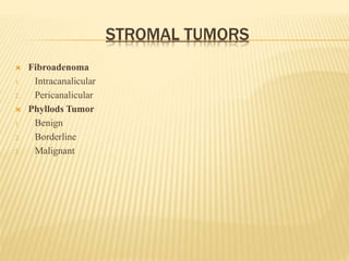
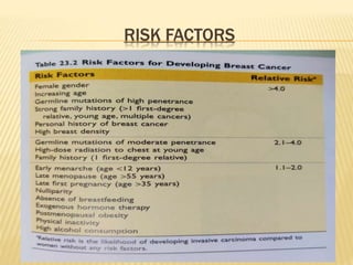
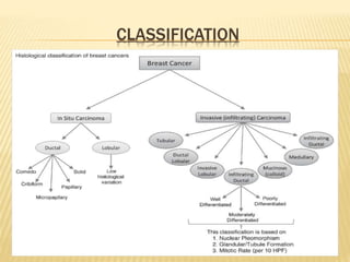
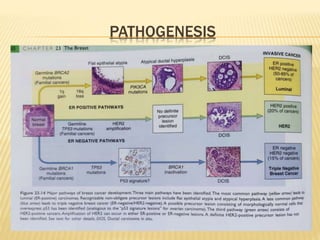
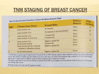
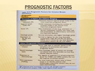
BREAST CARCINOMA
Presented By :-
Dr. Bhavya Singh
PG Resident Pathology department
JLN Medical College, Bhagalpur
INTRODUCTION
 The second most common cancer in females world
wide is breast cancer accounting for about 1.4 million
cases each year, with more than 50% of the deaths
occurring in low and middle income countries.
 The second leading cause of cancer related deaths in
females is breast cancer world wide.
 Breast cancer have doubled in India in the recent
decade.
 Tumor type, histological grading (HG), DNA ploidy,
cell proliferation markers, hormone receptor status
and expression of different oncogenes decide the
prognosis of breast cancer.
 Histological grading (HG) of breast carcinoma
using the Elston-Ellis modification of Scarff
Bloom Richardson (SBR) grading system is a
widely accepted.
 Among all the different cytological grading (CG)
methods, Robinson cytological grading system
is widely used.
 Fine needle aspiration cytology (FNAC) is being
increasingly used to determine the nature of the
lesion (benign or malignant).
STRUCTURE OF THE BREAST
 The mammary gland is a compound tubuloalveolar gland.
 The mammary gland is covered with skin and subcutaneous tissue
and rests on pectoralis muscle, from which it is separated by a
fascia.
 Each breast is composed of 15 to 25 independent lobes, within
each lobe of the breast, the terminal ducts combines to form the
main duct.
 Each terminal duct and its associated lobule is called a terminal
duct lobular unit (TDLU).
HISTOLOGY OF BREAST
 A Glandular lobule consists of intralobular ducts surrounded by loose
intralobular connective tissue.
 Intralobular ducts are lined by a specialized two layered epithelial cells;
cuboidal to low columnar epithelial cells and basal myoepithelial cells.
BENIGN EPITHELIAL LESIONS
 Nonproliferative Breast Changes (Fibrocystic changes) : consist of 3
morphological changes
• Cysts
• Fibrosis
• Adenosis
 Proliferative Breast Disease
1) Without Atypia
• Epithelial Hyperplasia
• Sclerosing Adenosis
• Complex sclerosing lesion
• Papilloma
2) With Atypia
• Atypical ductal hyperplasia
• Atypical lobular hyperplasia
STROMAL TUMORS
 Fibroadenoma
1. Intracanalicular
2. Pericanalicular
 Phyllods Tumor
1. Benign
2. Borderline
3. Malignant
RISK FACTORS
CLASSIFICATION
PATHOGENESIS
DIAGNOSTIC CRITERIA FOR DUCT CARCINOMA
IN CYTOLOGY:
 Cellular smear
 Malignant epithelial cells arranged in three-
dimensional clusters, syncytium, and acini
 Single population of cells
 Absence of myoepithelial cells & bare or bipolar
nuclei
 Moderate to severe nuclear atypia
 Uneven necrosis
 Tumor diathesis may be present
 Some cases demonstrate cytolpasmic vacuolation or
intracellular lumina
ROBINSON CYTOLOGICAL GRADING SYSTEM
SCORE 1 SCORE 2 SCORE 3
Cell Dissociation Mostly in clusters Single cells and clusters Mostly in single cells
Nuclear size 1-2 times the size of
RBC
3-4 times the sizeof RBC >5 times the size of
RBC
Cell uniformity Monomorphic Mildly pleomorphic Pleomorphic
Nucleoli Indistinct/small Noticeable Abnormal
Nuclear margin Smooth Slightly
irregular/folds/grooves
Buds and clefts
Chromatin pattern Vesicular Granular Clumping and
clearing
Grade 1: Score 6- 11
Grade 2: Score 12-14
Grade 3: Score 15-18
MODIFIED BLOOM RICHARDSON HISTOLOGIC
GRADING
1 2 3
Tubule
formation
>75% 10-75% <10%
Nuclear
pleomorphism
Small regular
uniform cells
Moderate increase in
size and variability
Marked
variation
Marked variation <9 10-19 >20
Grade 1: Score 3-5
Grade 2: Score 6-7
Grade 3: Score 8-9
INVASIVE DUCT CARCINOMA NOS
 Microscopy : Tumor arranged in diffuse sheets, well defined nest,
or as individual cells, nuclei may be regular or large pleomorphic
with prominent nucleoli. Mitotic figures may be seen depending
on the grade of the tumor. There is marked fibrosis and stromal
hypercellularity
TUBULAR CARCINOMA
 Histopathology: Well differentiated neoplasm with irregular
angulated tubules with open lumina lined by only one layer of
epithelial cells with mild pleomorphism. The stroma is abundant
and cellular.The nucleus show little pleomorphism with rare
mitoses. For a tumor to be typed as tubular carcinoma , tubular
morphology should comprise >90% of its area
MUCINOUS CARCINOMA
 Histopathology: Small clusters of regular epithelial cells floating
in a pool of extracellular mucin. The cells are small with darkly
staining nuclei which exhibiting mild pleomorphism. Mitoses are
infrequent.
MEDULLARY CARCINOMA
 Histopathology: Three main morphologic criteria for diagnosis of
medullary carcinoma :
1. Epithelial cells arranged in interconnecting sheets forming a syncytial
network. Syncytial pattern of growth comprise >75% of tumor. Tumor cells are
large pleomorphic with prominent nucleoli and high mitotic rate.
2. The intervening stroma contains lymphoplasmacytic infiltrate.
3. Grossly evident circumscription, border of the tumor is pushing rather than
infiltrative.
MICROPAPILLARY CARCINOMA
 Histopathology: Cluster of epithelial cells arranged in a
micropapillary or tubular-alveolar pattern without a fibrovascular
core suspended in a clear space. Cell clusters appear to have an
inside out appearance. This tumor has an increased incidence of
vascular invasion and lymph node involvement.
PAPILLARY CARCINOMA
 Histopathology: Presence of papillary structures with
fibrovascular cores. Cytologic appearance may be varied. Nuclear
pleomorphism and increased mitosis may be seen.
LOBULAR CARCINOMA
 Histopathology: Classic variant: Small uniform cells growing in a
single file and in a concentric fashion around the lobules. The
stroma is abundant and fibrous. The individual cells are small
round to ovoid with little cytoplasm with eccentrically placed
nucleus exhibiting mild pleomorphism and infrequent mitoses
TNM STAGING OF BREAST CANCER
PROGNOSTIC FACTORS
THANK YOU

breast cancer ppt.pptx

  • 1.
    BREAST CARCINOMA Presented By:- Dr. Bhavya Singh PG Resident Pathology department JLN Medical College, Bhagalpur
  • 2.
    INTRODUCTION  The secondmost common cancer in females world wide is breast cancer accounting for about 1.4 million cases each year, with more than 50% of the deaths occurring in low and middle income countries.  The second leading cause of cancer related deaths in females is breast cancer world wide.  Breast cancer have doubled in India in the recent decade.  Tumor type, histological grading (HG), DNA ploidy, cell proliferation markers, hormone receptor status and expression of different oncogenes decide the prognosis of breast cancer.
  • 3.
     Histological grading(HG) of breast carcinoma using the Elston-Ellis modification of Scarff Bloom Richardson (SBR) grading system is a widely accepted.  Among all the different cytological grading (CG) methods, Robinson cytological grading system is widely used.  Fine needle aspiration cytology (FNAC) is being increasingly used to determine the nature of the lesion (benign or malignant).
  • 4.
    STRUCTURE OF THEBREAST  The mammary gland is a compound tubuloalveolar gland.  The mammary gland is covered with skin and subcutaneous tissue and rests on pectoralis muscle, from which it is separated by a fascia.  Each breast is composed of 15 to 25 independent lobes, within each lobe of the breast, the terminal ducts combines to form the main duct.  Each terminal duct and its associated lobule is called a terminal duct lobular unit (TDLU).
  • 5.
    HISTOLOGY OF BREAST A Glandular lobule consists of intralobular ducts surrounded by loose intralobular connective tissue.  Intralobular ducts are lined by a specialized two layered epithelial cells; cuboidal to low columnar epithelial cells and basal myoepithelial cells.
  • 6.
    BENIGN EPITHELIAL LESIONS Nonproliferative Breast Changes (Fibrocystic changes) : consist of 3 morphological changes • Cysts • Fibrosis • Adenosis  Proliferative Breast Disease 1) Without Atypia • Epithelial Hyperplasia • Sclerosing Adenosis • Complex sclerosing lesion • Papilloma 2) With Atypia • Atypical ductal hyperplasia • Atypical lobular hyperplasia
  • 7.
    STROMAL TUMORS  Fibroadenoma 1.Intracanalicular 2. Pericanalicular  Phyllods Tumor 1. Benign 2. Borderline 3. Malignant
  • 8.
  • 9.
  • 10.
  • 11.
    DIAGNOSTIC CRITERIA FORDUCT CARCINOMA IN CYTOLOGY:  Cellular smear  Malignant epithelial cells arranged in three- dimensional clusters, syncytium, and acini  Single population of cells  Absence of myoepithelial cells & bare or bipolar nuclei  Moderate to severe nuclear atypia  Uneven necrosis  Tumor diathesis may be present  Some cases demonstrate cytolpasmic vacuolation or intracellular lumina
  • 12.
    ROBINSON CYTOLOGICAL GRADINGSYSTEM SCORE 1 SCORE 2 SCORE 3 Cell Dissociation Mostly in clusters Single cells and clusters Mostly in single cells Nuclear size 1-2 times the size of RBC 3-4 times the sizeof RBC >5 times the size of RBC Cell uniformity Monomorphic Mildly pleomorphic Pleomorphic Nucleoli Indistinct/small Noticeable Abnormal Nuclear margin Smooth Slightly irregular/folds/grooves Buds and clefts Chromatin pattern Vesicular Granular Clumping and clearing Grade 1: Score 6- 11 Grade 2: Score 12-14 Grade 3: Score 15-18
  • 13.
    MODIFIED BLOOM RICHARDSONHISTOLOGIC GRADING 1 2 3 Tubule formation >75% 10-75% <10% Nuclear pleomorphism Small regular uniform cells Moderate increase in size and variability Marked variation Marked variation <9 10-19 >20 Grade 1: Score 3-5 Grade 2: Score 6-7 Grade 3: Score 8-9
  • 14.
    INVASIVE DUCT CARCINOMANOS  Microscopy : Tumor arranged in diffuse sheets, well defined nest, or as individual cells, nuclei may be regular or large pleomorphic with prominent nucleoli. Mitotic figures may be seen depending on the grade of the tumor. There is marked fibrosis and stromal hypercellularity
  • 15.
    TUBULAR CARCINOMA  Histopathology:Well differentiated neoplasm with irregular angulated tubules with open lumina lined by only one layer of epithelial cells with mild pleomorphism. The stroma is abundant and cellular.The nucleus show little pleomorphism with rare mitoses. For a tumor to be typed as tubular carcinoma , tubular morphology should comprise >90% of its area
  • 16.
    MUCINOUS CARCINOMA  Histopathology:Small clusters of regular epithelial cells floating in a pool of extracellular mucin. The cells are small with darkly staining nuclei which exhibiting mild pleomorphism. Mitoses are infrequent.
  • 17.
    MEDULLARY CARCINOMA  Histopathology:Three main morphologic criteria for diagnosis of medullary carcinoma : 1. Epithelial cells arranged in interconnecting sheets forming a syncytial network. Syncytial pattern of growth comprise >75% of tumor. Tumor cells are large pleomorphic with prominent nucleoli and high mitotic rate. 2. The intervening stroma contains lymphoplasmacytic infiltrate. 3. Grossly evident circumscription, border of the tumor is pushing rather than infiltrative.
  • 18.
    MICROPAPILLARY CARCINOMA  Histopathology:Cluster of epithelial cells arranged in a micropapillary or tubular-alveolar pattern without a fibrovascular core suspended in a clear space. Cell clusters appear to have an inside out appearance. This tumor has an increased incidence of vascular invasion and lymph node involvement.
  • 19.
    PAPILLARY CARCINOMA  Histopathology:Presence of papillary structures with fibrovascular cores. Cytologic appearance may be varied. Nuclear pleomorphism and increased mitosis may be seen.
  • 20.
    LOBULAR CARCINOMA  Histopathology:Classic variant: Small uniform cells growing in a single file and in a concentric fashion around the lobules. The stroma is abundant and fibrous. The individual cells are small round to ovoid with little cytoplasm with eccentrically placed nucleus exhibiting mild pleomorphism and infrequent mitoses
  • 21.
    TNM STAGING OFBREAST CANCER
  • 22.
  • 23.