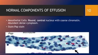
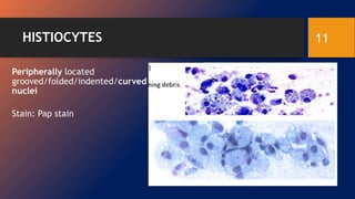
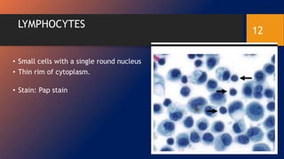
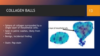
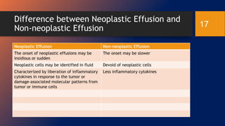
SlideShare a Scribd company logo
EFFUSION CYTOLOGY
Nicholas Afriyie
1
EFFUSION CYTOLOGY
• Effusion is an excess amount of serous fluid in the body cavity
• All serous membranes produces serous fluid but excess amount of the
serous fluid may indicate a pathology and is termed as an effusion.
• Serous effusion can be found in the following body cavities: pleural
cavity, peritoneal cavity and pericardial cavity.
2
TYPES OF EFFUSION
Transudates
• ultrafiltrate of plasma with little protein and few or no cells
• arise from either increased capillary hydrostatic pressure or
decreased oncotic pressure secondary to congestive heart failure,
fluid overload, cirrhosis or hypoalbuminemia.
 Exudates
• extravascular fluid collection that is rich in protein and/or cells.
• result from increased capillary permeability or decreased
lymphatic resorption associated with infection, connective tissue
disease, pancreatitis or cancer
3
SAMPLING TECHNIQUES
• Serous effusion is usually sampled by simply inserting a wide-bore
needle (under local anesthesia) through the body wall into the fluid-
containing cavity.
• Examples:
 Peritoneal fluid from peritoneal cavity- by paracentesis
 Pleural fluid from pleural cavity- by thoracentesis
 pericardial fluid from pericardial cavity- by pericardiocentesis
4
GROSS APPEARANCE OF SEROUS EFFUSION
• Many serous effusions are noticeably bloodstained.
• The cancerous cells in serous effusion form a thick, whitish-yellow layer
at the bottom of container when allowed to stand and sediment
spontaneously.
• Individual particles of cancer in a serous effusion may occasionally be large
enough to be visible to the naked eye. Eg: spheroids, ellipsoids .
• Fluids containing numerous pigmented melanoma cells may be chocolate
brown.
5
CONT’D
• Effusions containing many hemosiderophages may appear lighter brown.
• Effusions from patients who are jaundiced or that are a result of leakage of
bile into the peritoneal cavity may have a rather dark brown-orange or
greenish appearance that remains with the supernatant after the specimen
has been centrifuged
• Serous effusions caused by malignant mesothelioma of epithelial type
often have thin honey consistence due to increased viscosity of the fluid
due to the high concentration of hyaluronic acid contained in the
malignant mesothelioma.
6
CONT’D
• Peritoneal effusion due to pseudomyxoma peritonei has heavy mucoid
consistence and the cells it contains do not sediment during
centrifugation.
•
Effusions containing numerous cholesterol crystals are
yellow and turbid and have a swirling, shimmering, gold paint
appearance when agitated
• Chylous effusions have a
milky white appearance with a creamy topmost layer due to
their high concentration of emulsified lipid.
7
PREPARATION OF SEROUS FLUID
 Remove any clots that may be present and extract all fluid
 Pour off an aliquot (up to 50 mL) of the fluid and centrifuge for 3
mins at 2000rpm
 Pour off the supernatant
 Prepare slide with the sediment and stain
8
Staining techniques
• Three main staining techniques are employed
a toluidine blue-stained wet film
a liquid-based preparation stained with the Papanicolaou stain
and cell blocks stained with hematoxylin and eosin
9
NORMAL COMPONENTS OF EFFUSION
• Mesothelial Cells: Round, central nucleus with coarse chromatin.
Abundant dense cytoplasm.
• Stain:Pap stain
10
HISTIOCYTES
Peripherally located
grooved/folded/indented/curved
nuclei
Stain: Pap stain
11
LYMPHOCYTES
• Small cells with a single round nucleus
• Thin rim of cytoplasm.
• Stain: Pap stain
12
COLLAGEN BALLS
• Sphere of collagen surrounded by a
single layer of mesothelial cells.
• Seen in pelvic washes, likely from
ovary.
• Benign, incidental finding
• Stain: Pap stain
13
LYMPHOMA
• Dominated by lymphocytes
• Single disclosive cells (of varying
size depending on type)
14
MALIGNANT MESOTHELIOMA
• All mesothelial cells (no “foreign” second
population)
Often maintain similar cytologic features (lacy
skirts, etc…)
Can be cytologically malignant or relatively bland
“More and bigger cells, in more and bigger
clusters.”
Large clusters with knobby, flower-like contours
15
BENIGN EFFUSIONS
• Rheumatoid effusions- Granulomas, multinucleated giant cells,
and epithelioid histiocytes.
• Eosinophilic effusion- abundant eosinophils
• Lupus effusion- neutrophils with a phagocytosed nucleus, Necrotic
debris
• Chylous effusion- lymphocytes with some lipophages and mesos
• Tuberculous effusion- Abundant lymphocytes (T-cells) with sparse
mesos and
16
Difference between Neoplastic Effusion and
Non-neoplastic Effusion
Neoplastic Effusion Non-neoplastic Effusion
The onset of neoplastic effusions may be
insidious or sudden
The onset may be slower
Neoplastic cells may be identified in fluid Devoid of neoplastic cells
Characterized by liberation of inflammatory
cytokines in response to the tumor or
damage-associated molecular patterns from
tumor or immune cells
Less inflammatory cytokines
17
References
1. Ducatman, B. S. (2020). Cytology: Diagnostic principles and
clinical correlates. Elsevier.
2. Bibbo, M., & Hoda, R. S. (1998). Comprehensive
Cytopathology. International Journal of Gynecological
Pathology, 17(1), 93.
18

More Related Content

What's hot

AUTOMATION IN HEMATOLOGY
AUTOMATION IN HEMATOLOGYAUTOMATION IN HEMATOLOGY
AUTOMATION IN HEMATOLOGY
Dr. Ajit Surya Singh
 
Technical faults in histopathology lab
Technical faults in histopathology labTechnical faults in histopathology lab
Technical faults in histopathology lab
magdy abdelghany
 
cytology of urinary tract
cytology of urinary tractcytology of urinary tract
cytology of urinary tract
SHRUTHI VASAN
 
THYROID - cytology pptx
THYROID - cytology pptxTHYROID - cytology pptx
THYROID - cytology pptx
KalaivaniGanapathy
 
Quality Assurance in Hematology laboratory
Quality Assurance in Hematology laboratoryQuality Assurance in Hematology laboratory
Quality Assurance in Hematology laboratory
Mohammed Abdalla Elhassan
 
Cell block and its diagnostic utility
Cell block and its diagnostic utilityCell block and its diagnostic utility
Cell block and its diagnostic utility
Gaurav Gupta
 
Cell block in cytology
Cell block in cytologyCell block in cytology
Cell block in cytology
Anam Khurshid
 
Myeloperoxidases Stains
Myeloperoxidases StainsMyeloperoxidases Stains
Automation in hematology
Automation in hematologyAutomation in hematology
Automation in hematology
Dr Siddartha
 
Bone marrow aspirate&biopsy preparation
Bone marrow aspirate&biopsy preparationBone marrow aspirate&biopsy preparation
Bone marrow aspirate&biopsy preparation
Malak Salam
 
Romanowsky stain
Romanowsky stainRomanowsky stain
Romanowsky stain
sandeep singh
 
Liquid based cytology ( l b c)
Liquid  based  cytology ( l b c)Liquid  based  cytology ( l b c)
Liquid based cytology ( l b c)
vikramsaraswat
 
Fluid cytology in serous cavity effusions
Fluid cytology in serous cavity effusionsFluid cytology in serous cavity effusions
Fluid cytology in serous cavity effusions
tashagarwal
 
Liquid based cytology | Abdul Quddus
Liquid based cytology | Abdul QuddusLiquid based cytology | Abdul Quddus
Liquid based cytology | Abdul Quddus
Abdul Quddus
 
Cytotechniques
Cytotechniques  Cytotechniques
Cytotechniques
drtousif
 
diagnostic Cytology introduction , Body fluids cytology
diagnostic Cytology introduction , Body fluids cytologydiagnostic Cytology introduction , Body fluids cytology
diagnostic Cytology introduction , Body fluids cytology
Aayra
 
Utility of cell block in cytology.
Utility of cell block in cytology.Utility of cell block in cytology.
Utility of cell block in cytology.
Manan Shah
 
Apheresis
ApheresisApheresis
Cytologic assessment of bronchopulmonary lesions
Cytologic assessment of bronchopulmonary lesionsCytologic assessment of bronchopulmonary lesions
Cytologic assessment of bronchopulmonary lesions
Aseem Jain
 
Automation histopathology
Automation histopathologyAutomation histopathology
Automation histopathology
Ankita Sain
 

What's hot (20)

AUTOMATION IN HEMATOLOGY
AUTOMATION IN HEMATOLOGYAUTOMATION IN HEMATOLOGY
AUTOMATION IN HEMATOLOGY
 
Technical faults in histopathology lab
Technical faults in histopathology labTechnical faults in histopathology lab
Technical faults in histopathology lab
 
cytology of urinary tract
cytology of urinary tractcytology of urinary tract
cytology of urinary tract
 
THYROID - cytology pptx
THYROID - cytology pptxTHYROID - cytology pptx
THYROID - cytology pptx
 
Quality Assurance in Hematology laboratory
Quality Assurance in Hematology laboratoryQuality Assurance in Hematology laboratory
Quality Assurance in Hematology laboratory
 
Cell block and its diagnostic utility
Cell block and its diagnostic utilityCell block and its diagnostic utility
Cell block and its diagnostic utility
 
Cell block in cytology
Cell block in cytologyCell block in cytology
Cell block in cytology
 
Myeloperoxidases Stains
Myeloperoxidases StainsMyeloperoxidases Stains
Myeloperoxidases Stains
 
Automation in hematology
Automation in hematologyAutomation in hematology
Automation in hematology
 
Bone marrow aspirate&biopsy preparation
Bone marrow aspirate&biopsy preparationBone marrow aspirate&biopsy preparation
Bone marrow aspirate&biopsy preparation
 
Romanowsky stain
Romanowsky stainRomanowsky stain
Romanowsky stain
 
Liquid based cytology ( l b c)
Liquid  based  cytology ( l b c)Liquid  based  cytology ( l b c)
Liquid based cytology ( l b c)
 
Fluid cytology in serous cavity effusions
Fluid cytology in serous cavity effusionsFluid cytology in serous cavity effusions
Fluid cytology in serous cavity effusions
 
Liquid based cytology | Abdul Quddus
Liquid based cytology | Abdul QuddusLiquid based cytology | Abdul Quddus
Liquid based cytology | Abdul Quddus
 
Cytotechniques
Cytotechniques  Cytotechniques
Cytotechniques
 
diagnostic Cytology introduction , Body fluids cytology
diagnostic Cytology introduction , Body fluids cytologydiagnostic Cytology introduction , Body fluids cytology
diagnostic Cytology introduction , Body fluids cytology
 
Utility of cell block in cytology.
Utility of cell block in cytology.Utility of cell block in cytology.
Utility of cell block in cytology.
 
Apheresis
ApheresisApheresis
Apheresis
 
Cytologic assessment of bronchopulmonary lesions
Cytologic assessment of bronchopulmonary lesionsCytologic assessment of bronchopulmonary lesions
Cytologic assessment of bronchopulmonary lesions
 
Automation histopathology
Automation histopathologyAutomation histopathology
Automation histopathology
 

Similar to Effusion cytology.pptx

Friday Presentation.pptx
Friday Presentation.pptxFriday Presentation.pptx
Friday Presentation.pptx
OMJHA20
 
Hematology basics pt 2
Hematology basics pt 2Hematology basics pt 2
Hematology basics pt 2
eyedogtor
 
Hydatid cyst
Hydatid cystHydatid cyst
Hydatid cyst
Rojan Adhikari
 
Approach to a patient with active urinary sediments
Approach to a patient with active urinary sedimentsApproach to a patient with active urinary sediments
Approach to a patient with active urinary sediments
AIIMS, New Delhi, India
 
spleen power point by doctor ahmed .ppt.ppt
spleen power point by doctor ahmed .ppt.pptspleen power point by doctor ahmed .ppt.ppt
spleen power point by doctor ahmed .ppt.ppt
esraa98x
 
Protozoa - overview & Entamoeba histolytica
Protozoa - overview & Entamoeba histolyticaProtozoa - overview & Entamoeba histolytica
Protozoa - overview & Entamoeba histolytica
Neenajoel
 
peripheral smear. DMLT.pptx
peripheral smear. DMLT.pptxperipheral smear. DMLT.pptx
peripheral smear. DMLT.pptx
Priya780950
 
Clinical diagnostic Cytology Central Nervous System.pdf
Clinical diagnostic Cytology Central Nervous System.pdfClinical diagnostic Cytology Central Nervous System.pdf
Clinical diagnostic Cytology Central Nervous System.pdf
222101989
 
Spleen
SpleenSpleen
Microscopic examination of urine
Microscopic examination of urineMicroscopic examination of urine
Microscopic examination of urine
globalsoin
 
Tumors of kidney
Tumors of kidneyTumors of kidney
Tumors of kidney
Venkata Karthik
 
486 qualitative disorders of wbc
486 qualitative disorders of wbc486 qualitative disorders of wbc
486 qualitative disorders of wbc
Nabin Chaudhary
 
examination_of_urinary_system.pptx
examination_of_urinary_system.pptxexamination_of_urinary_system.pptx
examination_of_urinary_system.pptx
ssuser59bb22
 
microscopic examination of urine and other special tests
microscopic examination of urine and other special testsmicroscopic examination of urine and other special tests
microscopic examination of urine and other special tests
regional institute of medical sciences
 
Hematopoiesis: Formation of Blood Cells - An Overview
Hematopoiesis: Formation of Blood Cells - An OverviewHematopoiesis: Formation of Blood Cells - An Overview
Hematopoiesis: Formation of Blood Cells - An Overview
StudyFriend
 
Salivary gland-- cytology .pptx
Salivary gland-- cytology .pptxSalivary gland-- cytology .pptx
Salivary gland-- cytology .pptx
KalaivaniGanapathy
 
Liver lesion by dr muhammad qasim khan
Liver lesion by dr muhammad qasim khanLiver lesion by dr muhammad qasim khan
Liver lesion by dr muhammad qasim khan
Holy Family Hospital Rawalpindi
 
Spleen NMS
Spleen NMSSpleen NMS
Spleen NMS
Genevieve Canas
 
body fluids analysis - corrected - Copy.pptx
body fluids analysis - corrected - Copy.pptxbody fluids analysis - corrected - Copy.pptx
body fluids analysis - corrected - Copy.pptx
SURAJ PANCHAL
 
5. intracellular accumulation.pptx
5. intracellular accumulation.pptx5. intracellular accumulation.pptx
5. intracellular accumulation.pptx
AndulRehman
 

Similar to Effusion cytology.pptx (20)

Friday Presentation.pptx
Friday Presentation.pptxFriday Presentation.pptx
Friday Presentation.pptx
 
Hematology basics pt 2
Hematology basics pt 2Hematology basics pt 2
Hematology basics pt 2
 
Hydatid cyst
Hydatid cystHydatid cyst
Hydatid cyst
 
Approach to a patient with active urinary sediments
Approach to a patient with active urinary sedimentsApproach to a patient with active urinary sediments
Approach to a patient with active urinary sediments
 
spleen power point by doctor ahmed .ppt.ppt
spleen power point by doctor ahmed .ppt.pptspleen power point by doctor ahmed .ppt.ppt
spleen power point by doctor ahmed .ppt.ppt
 
Protozoa - overview & Entamoeba histolytica
Protozoa - overview & Entamoeba histolyticaProtozoa - overview & Entamoeba histolytica
Protozoa - overview & Entamoeba histolytica
 
peripheral smear. DMLT.pptx
peripheral smear. DMLT.pptxperipheral smear. DMLT.pptx
peripheral smear. DMLT.pptx
 
Clinical diagnostic Cytology Central Nervous System.pdf
Clinical diagnostic Cytology Central Nervous System.pdfClinical diagnostic Cytology Central Nervous System.pdf
Clinical diagnostic Cytology Central Nervous System.pdf
 
Spleen
SpleenSpleen
Spleen
 
Microscopic examination of urine
Microscopic examination of urineMicroscopic examination of urine
Microscopic examination of urine
 
Tumors of kidney
Tumors of kidneyTumors of kidney
Tumors of kidney
 
486 qualitative disorders of wbc
486 qualitative disorders of wbc486 qualitative disorders of wbc
486 qualitative disorders of wbc
 
examination_of_urinary_system.pptx
examination_of_urinary_system.pptxexamination_of_urinary_system.pptx
examination_of_urinary_system.pptx
 
microscopic examination of urine and other special tests
microscopic examination of urine and other special testsmicroscopic examination of urine and other special tests
microscopic examination of urine and other special tests
 
Hematopoiesis: Formation of Blood Cells - An Overview
Hematopoiesis: Formation of Blood Cells - An OverviewHematopoiesis: Formation of Blood Cells - An Overview
Hematopoiesis: Formation of Blood Cells - An Overview
 
Salivary gland-- cytology .pptx
Salivary gland-- cytology .pptxSalivary gland-- cytology .pptx
Salivary gland-- cytology .pptx
 
Liver lesion by dr muhammad qasim khan
Liver lesion by dr muhammad qasim khanLiver lesion by dr muhammad qasim khan
Liver lesion by dr muhammad qasim khan
 
Spleen NMS
Spleen NMSSpleen NMS
Spleen NMS
 
body fluids analysis - corrected - Copy.pptx
body fluids analysis - corrected - Copy.pptxbody fluids analysis - corrected - Copy.pptx
body fluids analysis - corrected - Copy.pptx
 
5. intracellular accumulation.pptx
5. intracellular accumulation.pptx5. intracellular accumulation.pptx
5. intracellular accumulation.pptx
 

Recently uploaded

Ear and its clinical correlations By Dr. Rabia Inam Gandapore.pptx
Ear and its clinical correlations By Dr. Rabia Inam Gandapore.pptxEar and its clinical correlations By Dr. Rabia Inam Gandapore.pptx
Ear and its clinical correlations By Dr. Rabia Inam Gandapore.pptx
Dr. Rabia Inam Gandapore
 
TEST BANK For Basic and Clinical Pharmacology, 14th Edition by Bertram G. Kat...
TEST BANK For Basic and Clinical Pharmacology, 14th Edition by Bertram G. Kat...TEST BANK For Basic and Clinical Pharmacology, 14th Edition by Bertram G. Kat...
TEST BANK For Basic and Clinical Pharmacology, 14th Edition by Bertram G. Kat...
rightmanforbloodline
 
CHEMOTHERAPY_RDP_CHAPTER 1_ANTI TB DRUGS.pdf
CHEMOTHERAPY_RDP_CHAPTER 1_ANTI TB DRUGS.pdfCHEMOTHERAPY_RDP_CHAPTER 1_ANTI TB DRUGS.pdf
CHEMOTHERAPY_RDP_CHAPTER 1_ANTI TB DRUGS.pdf
rishi2789
 
Muscles of Mastication by Dr. Rabia Inam Gandapore.pptx
Muscles of Mastication by Dr. Rabia Inam Gandapore.pptxMuscles of Mastication by Dr. Rabia Inam Gandapore.pptx
Muscles of Mastication by Dr. Rabia Inam Gandapore.pptx
Dr. Rabia Inam Gandapore
 
Abortion PG Seminar Power point presentation
Abortion PG Seminar Power point presentationAbortion PG Seminar Power point presentation
Abortion PG Seminar Power point presentation
AksshayaRajanbabu
 
TEST BANK For Community Health Nursing A Canadian Perspective, 5th Edition by...
TEST BANK For Community Health Nursing A Canadian Perspective, 5th Edition by...TEST BANK For Community Health Nursing A Canadian Perspective, 5th Edition by...
TEST BANK For Community Health Nursing A Canadian Perspective, 5th Edition by...
Donc Test
 
Complementary feeding in infant IAP PROTOCOLS
Complementary feeding in infant IAP PROTOCOLSComplementary feeding in infant IAP PROTOCOLS
Complementary feeding in infant IAP PROTOCOLS
chiranthgowda16
 
Journal Article Review on Rasamanikya
Journal Article Review on RasamanikyaJournal Article Review on Rasamanikya
Journal Article Review on Rasamanikya
Dr. Jyothirmai Paindla
 
Best Ayurvedic medicine for Gas and Indigestion
Best Ayurvedic medicine for Gas and IndigestionBest Ayurvedic medicine for Gas and Indigestion
Best Ayurvedic medicine for Gas and Indigestion
Swastik Ayurveda
 
CHEMOTHERAPY_RDP_CHAPTER 3_ANTIFUNGAL AGENT.pdf
CHEMOTHERAPY_RDP_CHAPTER 3_ANTIFUNGAL AGENT.pdfCHEMOTHERAPY_RDP_CHAPTER 3_ANTIFUNGAL AGENT.pdf
CHEMOTHERAPY_RDP_CHAPTER 3_ANTIFUNGAL AGENT.pdf
rishi2789
 
Integrating Ayurveda into Parkinson’s Management: A Holistic Approach
Integrating Ayurveda into Parkinson’s Management: A Holistic ApproachIntegrating Ayurveda into Parkinson’s Management: A Holistic Approach
Integrating Ayurveda into Parkinson’s Management: A Holistic Approach
Ayurveda ForAll
 
CBL Seminar 2024_Preliminary Program.pdf
CBL Seminar 2024_Preliminary Program.pdfCBL Seminar 2024_Preliminary Program.pdf
CBL Seminar 2024_Preliminary Program.pdf
suvadeepdas911
 
Local Advanced Lung Cancer: Artificial Intelligence, Synergetics, Complex Sys...
Local Advanced Lung Cancer: Artificial Intelligence, Synergetics, Complex Sys...Local Advanced Lung Cancer: Artificial Intelligence, Synergetics, Complex Sys...
Local Advanced Lung Cancer: Artificial Intelligence, Synergetics, Complex Sys...
Oleg Kshivets
 
Part II - Body Grief: Losing parts of ourselves and our identity before, duri...
Part II - Body Grief: Losing parts of ourselves and our identity before, duri...Part II - Body Grief: Losing parts of ourselves and our identity before, duri...
Part II - Body Grief: Losing parts of ourselves and our identity before, duri...
bkling
 
Clinic ^%[+27633867063*Abortion Pills For Sale In Tembisa Central
Clinic ^%[+27633867063*Abortion Pills For Sale In Tembisa CentralClinic ^%[+27633867063*Abortion Pills For Sale In Tembisa Central
Clinic ^%[+27633867063*Abortion Pills For Sale In Tembisa Central
19various
 
Efficacy of Avartana Sneha in Ayurveda
Efficacy of Avartana Sneha in AyurvedaEfficacy of Avartana Sneha in Ayurveda
Efficacy of Avartana Sneha in Ayurveda
Dr. Jyothirmai Paindla
 
Ketone bodies and metabolism-biochemistry
Ketone bodies and metabolism-biochemistryKetone bodies and metabolism-biochemistry
Ketone bodies and metabolism-biochemistry
Dhayanithi C
 
Diabetic nephropathy diagnosis treatment
Diabetic nephropathy diagnosis treatmentDiabetic nephropathy diagnosis treatment
Diabetic nephropathy diagnosis treatment
arahmanzai5
 
The Electrocardiogram - Physiologic Principles
The Electrocardiogram - Physiologic PrinciplesThe Electrocardiogram - Physiologic Principles
The Electrocardiogram - Physiologic Principles
MedicoseAcademics
 
CHEMOTHERAPY_RDP_CHAPTER 6_Anti Malarial Drugs.pdf
CHEMOTHERAPY_RDP_CHAPTER 6_Anti Malarial Drugs.pdfCHEMOTHERAPY_RDP_CHAPTER 6_Anti Malarial Drugs.pdf
CHEMOTHERAPY_RDP_CHAPTER 6_Anti Malarial Drugs.pdf
rishi2789
 

Recently uploaded (20)

Ear and its clinical correlations By Dr. Rabia Inam Gandapore.pptx
Ear and its clinical correlations By Dr. Rabia Inam Gandapore.pptxEar and its clinical correlations By Dr. Rabia Inam Gandapore.pptx
Ear and its clinical correlations By Dr. Rabia Inam Gandapore.pptx
 
TEST BANK For Basic and Clinical Pharmacology, 14th Edition by Bertram G. Kat...
TEST BANK For Basic and Clinical Pharmacology, 14th Edition by Bertram G. Kat...TEST BANK For Basic and Clinical Pharmacology, 14th Edition by Bertram G. Kat...
TEST BANK For Basic and Clinical Pharmacology, 14th Edition by Bertram G. Kat...
 
CHEMOTHERAPY_RDP_CHAPTER 1_ANTI TB DRUGS.pdf
CHEMOTHERAPY_RDP_CHAPTER 1_ANTI TB DRUGS.pdfCHEMOTHERAPY_RDP_CHAPTER 1_ANTI TB DRUGS.pdf
CHEMOTHERAPY_RDP_CHAPTER 1_ANTI TB DRUGS.pdf
 
Muscles of Mastication by Dr. Rabia Inam Gandapore.pptx
Muscles of Mastication by Dr. Rabia Inam Gandapore.pptxMuscles of Mastication by Dr. Rabia Inam Gandapore.pptx
Muscles of Mastication by Dr. Rabia Inam Gandapore.pptx
 
Abortion PG Seminar Power point presentation
Abortion PG Seminar Power point presentationAbortion PG Seminar Power point presentation
Abortion PG Seminar Power point presentation
 
TEST BANK For Community Health Nursing A Canadian Perspective, 5th Edition by...
TEST BANK For Community Health Nursing A Canadian Perspective, 5th Edition by...TEST BANK For Community Health Nursing A Canadian Perspective, 5th Edition by...
TEST BANK For Community Health Nursing A Canadian Perspective, 5th Edition by...
 
Complementary feeding in infant IAP PROTOCOLS
Complementary feeding in infant IAP PROTOCOLSComplementary feeding in infant IAP PROTOCOLS
Complementary feeding in infant IAP PROTOCOLS
 
Journal Article Review on Rasamanikya
Journal Article Review on RasamanikyaJournal Article Review on Rasamanikya
Journal Article Review on Rasamanikya
 
Best Ayurvedic medicine for Gas and Indigestion
Best Ayurvedic medicine for Gas and IndigestionBest Ayurvedic medicine for Gas and Indigestion
Best Ayurvedic medicine for Gas and Indigestion
 
CHEMOTHERAPY_RDP_CHAPTER 3_ANTIFUNGAL AGENT.pdf
CHEMOTHERAPY_RDP_CHAPTER 3_ANTIFUNGAL AGENT.pdfCHEMOTHERAPY_RDP_CHAPTER 3_ANTIFUNGAL AGENT.pdf
CHEMOTHERAPY_RDP_CHAPTER 3_ANTIFUNGAL AGENT.pdf
 
Integrating Ayurveda into Parkinson’s Management: A Holistic Approach
Integrating Ayurveda into Parkinson’s Management: A Holistic ApproachIntegrating Ayurveda into Parkinson’s Management: A Holistic Approach
Integrating Ayurveda into Parkinson’s Management: A Holistic Approach
 
CBL Seminar 2024_Preliminary Program.pdf
CBL Seminar 2024_Preliminary Program.pdfCBL Seminar 2024_Preliminary Program.pdf
CBL Seminar 2024_Preliminary Program.pdf
 
Local Advanced Lung Cancer: Artificial Intelligence, Synergetics, Complex Sys...
Local Advanced Lung Cancer: Artificial Intelligence, Synergetics, Complex Sys...Local Advanced Lung Cancer: Artificial Intelligence, Synergetics, Complex Sys...
Local Advanced Lung Cancer: Artificial Intelligence, Synergetics, Complex Sys...
 
Part II - Body Grief: Losing parts of ourselves and our identity before, duri...
Part II - Body Grief: Losing parts of ourselves and our identity before, duri...Part II - Body Grief: Losing parts of ourselves and our identity before, duri...
Part II - Body Grief: Losing parts of ourselves and our identity before, duri...
 
Clinic ^%[+27633867063*Abortion Pills For Sale In Tembisa Central
Clinic ^%[+27633867063*Abortion Pills For Sale In Tembisa CentralClinic ^%[+27633867063*Abortion Pills For Sale In Tembisa Central
Clinic ^%[+27633867063*Abortion Pills For Sale In Tembisa Central
 
Efficacy of Avartana Sneha in Ayurveda
Efficacy of Avartana Sneha in AyurvedaEfficacy of Avartana Sneha in Ayurveda
Efficacy of Avartana Sneha in Ayurveda
 
Ketone bodies and metabolism-biochemistry
Ketone bodies and metabolism-biochemistryKetone bodies and metabolism-biochemistry
Ketone bodies and metabolism-biochemistry
 
Diabetic nephropathy diagnosis treatment
Diabetic nephropathy diagnosis treatmentDiabetic nephropathy diagnosis treatment
Diabetic nephropathy diagnosis treatment
 
The Electrocardiogram - Physiologic Principles
The Electrocardiogram - Physiologic PrinciplesThe Electrocardiogram - Physiologic Principles
The Electrocardiogram - Physiologic Principles
 
CHEMOTHERAPY_RDP_CHAPTER 6_Anti Malarial Drugs.pdf
CHEMOTHERAPY_RDP_CHAPTER 6_Anti Malarial Drugs.pdfCHEMOTHERAPY_RDP_CHAPTER 6_Anti Malarial Drugs.pdf
CHEMOTHERAPY_RDP_CHAPTER 6_Anti Malarial Drugs.pdf
 

Effusion cytology.pptx

  • 2. EFFUSION CYTOLOGY • Effusion is an excess amount of serous fluid in the body cavity • All serous membranes produces serous fluid but excess amount of the serous fluid may indicate a pathology and is termed as an effusion. • Serous effusion can be found in the following body cavities: pleural cavity, peritoneal cavity and pericardial cavity. 2
  • 3. TYPES OF EFFUSION Transudates • ultrafiltrate of plasma with little protein and few or no cells • arise from either increased capillary hydrostatic pressure or decreased oncotic pressure secondary to congestive heart failure, fluid overload, cirrhosis or hypoalbuminemia.  Exudates • extravascular fluid collection that is rich in protein and/or cells. • result from increased capillary permeability or decreased lymphatic resorption associated with infection, connective tissue disease, pancreatitis or cancer 3
  • 4. SAMPLING TECHNIQUES • Serous effusion is usually sampled by simply inserting a wide-bore needle (under local anesthesia) through the body wall into the fluid- containing cavity. • Examples:  Peritoneal fluid from peritoneal cavity- by paracentesis  Pleural fluid from pleural cavity- by thoracentesis  pericardial fluid from pericardial cavity- by pericardiocentesis 4
  • 5. GROSS APPEARANCE OF SEROUS EFFUSION • Many serous effusions are noticeably bloodstained. • The cancerous cells in serous effusion form a thick, whitish-yellow layer at the bottom of container when allowed to stand and sediment spontaneously. • Individual particles of cancer in a serous effusion may occasionally be large enough to be visible to the naked eye. Eg: spheroids, ellipsoids . • Fluids containing numerous pigmented melanoma cells may be chocolate brown. 5
  • 6. CONT’D • Effusions containing many hemosiderophages may appear lighter brown. • Effusions from patients who are jaundiced or that are a result of leakage of bile into the peritoneal cavity may have a rather dark brown-orange or greenish appearance that remains with the supernatant after the specimen has been centrifuged • Serous effusions caused by malignant mesothelioma of epithelial type often have thin honey consistence due to increased viscosity of the fluid due to the high concentration of hyaluronic acid contained in the malignant mesothelioma. 6
  • 7. CONT’D • Peritoneal effusion due to pseudomyxoma peritonei has heavy mucoid consistence and the cells it contains do not sediment during centrifugation. • Effusions containing numerous cholesterol crystals are yellow and turbid and have a swirling, shimmering, gold paint appearance when agitated • Chylous effusions have a milky white appearance with a creamy topmost layer due to their high concentration of emulsified lipid. 7
  • 8. PREPARATION OF SEROUS FLUID  Remove any clots that may be present and extract all fluid  Pour off an aliquot (up to 50 mL) of the fluid and centrifuge for 3 mins at 2000rpm  Pour off the supernatant  Prepare slide with the sediment and stain 8
  • 9. Staining techniques • Three main staining techniques are employed a toluidine blue-stained wet film a liquid-based preparation stained with the Papanicolaou stain and cell blocks stained with hematoxylin and eosin 9
  • 10. NORMAL COMPONENTS OF EFFUSION • Mesothelial Cells: Round, central nucleus with coarse chromatin. Abundant dense cytoplasm. • Stain:Pap stain 10
  • 12. LYMPHOCYTES • Small cells with a single round nucleus • Thin rim of cytoplasm. • Stain: Pap stain 12
  • 13. COLLAGEN BALLS • Sphere of collagen surrounded by a single layer of mesothelial cells. • Seen in pelvic washes, likely from ovary. • Benign, incidental finding • Stain: Pap stain 13
  • 14. LYMPHOMA • Dominated by lymphocytes • Single disclosive cells (of varying size depending on type) 14
  • 15. MALIGNANT MESOTHELIOMA • All mesothelial cells (no “foreign” second population) Often maintain similar cytologic features (lacy skirts, etc…) Can be cytologically malignant or relatively bland “More and bigger cells, in more and bigger clusters.” Large clusters with knobby, flower-like contours 15
  • 16. BENIGN EFFUSIONS • Rheumatoid effusions- Granulomas, multinucleated giant cells, and epithelioid histiocytes. • Eosinophilic effusion- abundant eosinophils • Lupus effusion- neutrophils with a phagocytosed nucleus, Necrotic debris • Chylous effusion- lymphocytes with some lipophages and mesos • Tuberculous effusion- Abundant lymphocytes (T-cells) with sparse mesos and 16
  • 17. Difference between Neoplastic Effusion and Non-neoplastic Effusion Neoplastic Effusion Non-neoplastic Effusion The onset of neoplastic effusions may be insidious or sudden The onset may be slower Neoplastic cells may be identified in fluid Devoid of neoplastic cells Characterized by liberation of inflammatory cytokines in response to the tumor or damage-associated molecular patterns from tumor or immune cells Less inflammatory cytokines 17
  • 18. References 1. Ducatman, B. S. (2020). Cytology: Diagnostic principles and clinical correlates. Elsevier. 2. Bibbo, M., & Hoda, R. S. (1998). Comprehensive Cytopathology. International Journal of Gynecological Pathology, 17(1), 93. 18

Editor's Notes

  1. Transudates are usually bilateral and arise from either increased capillary hydrostatic pressure or decreased oncotic pressure secondary to congestive heart failure, fluid overload, cirrhosis or hypoalbuminemia. Transudate: extravascular fluid collection that is basically an ultrafiltrate of plasma with little protein and few or no cells. Fluid appears grossly clear. Exudates are usually unilateral and result from increased capillary permeability or decreased lymphatic resorption associated with infection, connective tissue disease, pancreatitis or cancer Exudate: extravascular fluid collection that is rich in protein and/or cells. Fluid appears grossly cloudy. Effusions into body cavities can be further described as follows:Serous: a transudate with mainly edema fluid and few cells. Serosanguinous: an effusion with red blood cells. Fibrinous (serofibrinous): fibrin strands are derived from a protein-rich exudate. Purulent: numerous PMN's are present. Also called "empyema" in the pleural space.
  2. Centesis: Although a serous effusion may be removed at the time of surgical exploration, it is usually sampled by the relatively simple procedure of inserting a wide-bore needle (under local anesthesia) through the body wall into the fluid-containing cavity
  3. The clots may be fixed in 10% buffered formalin and processed by histopathological methods.