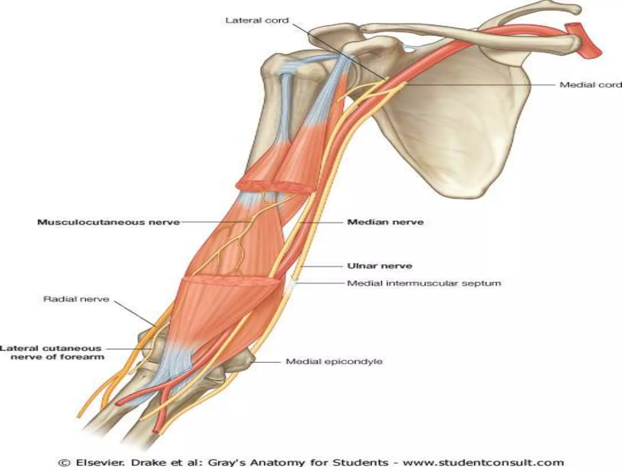
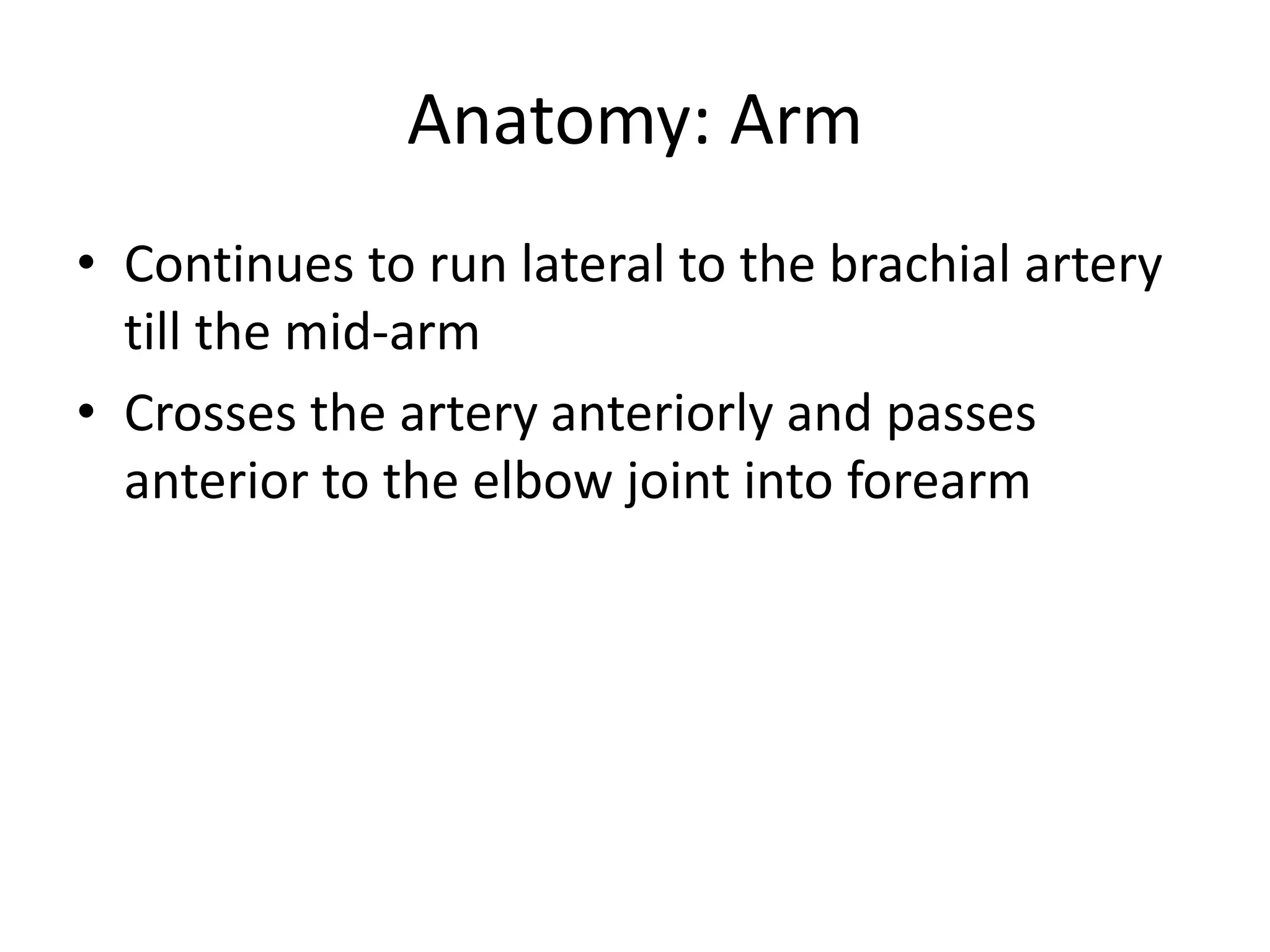
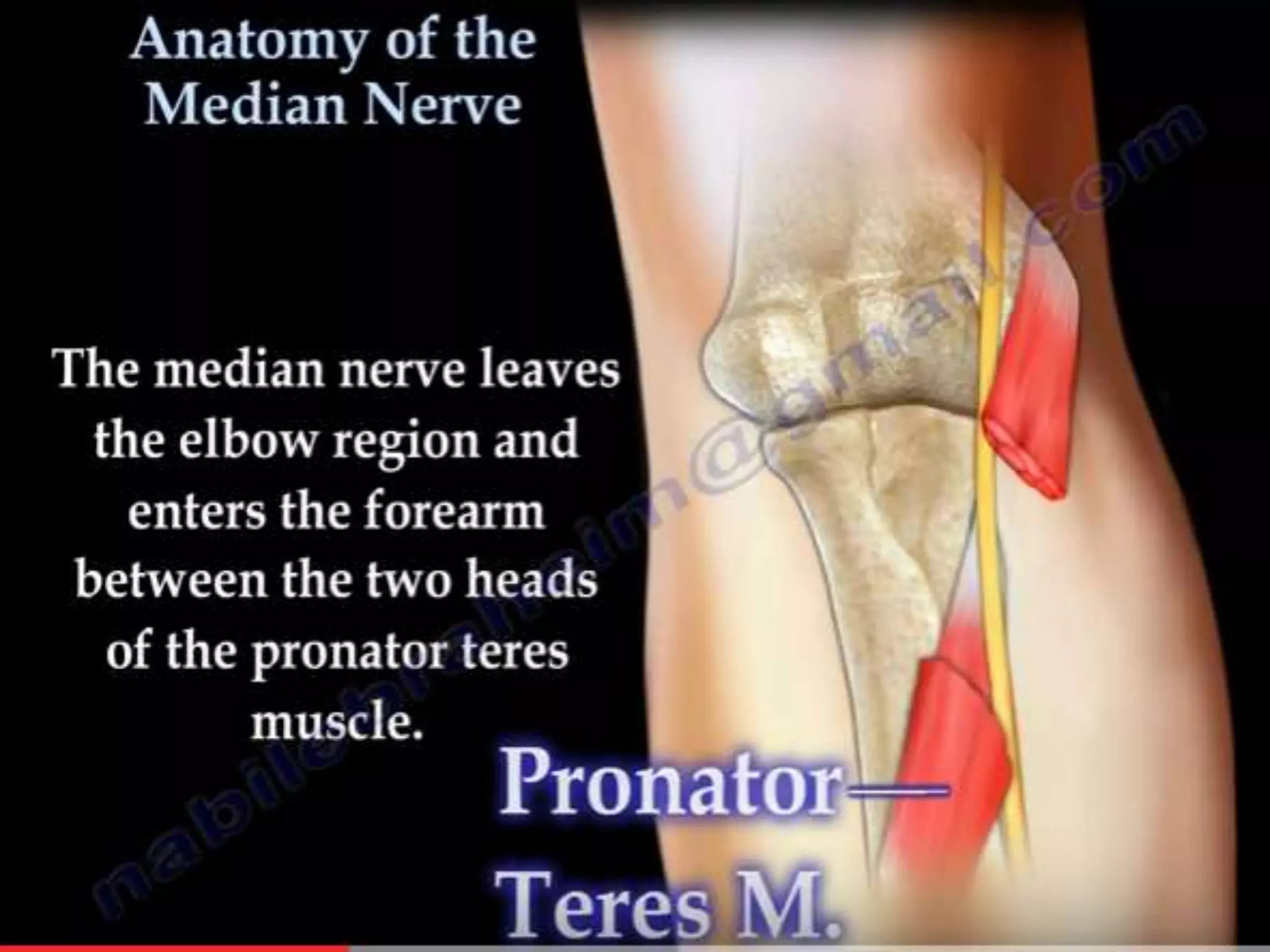
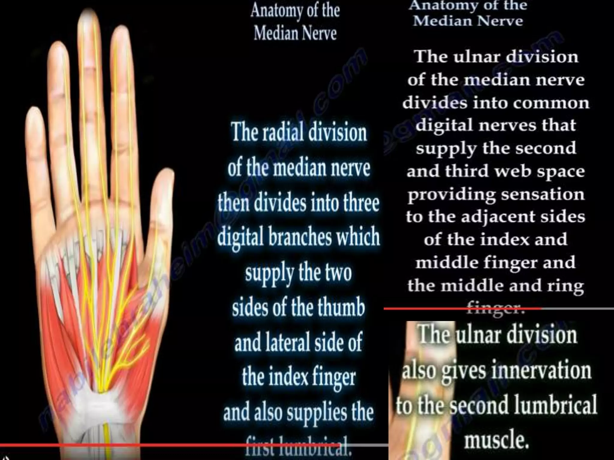
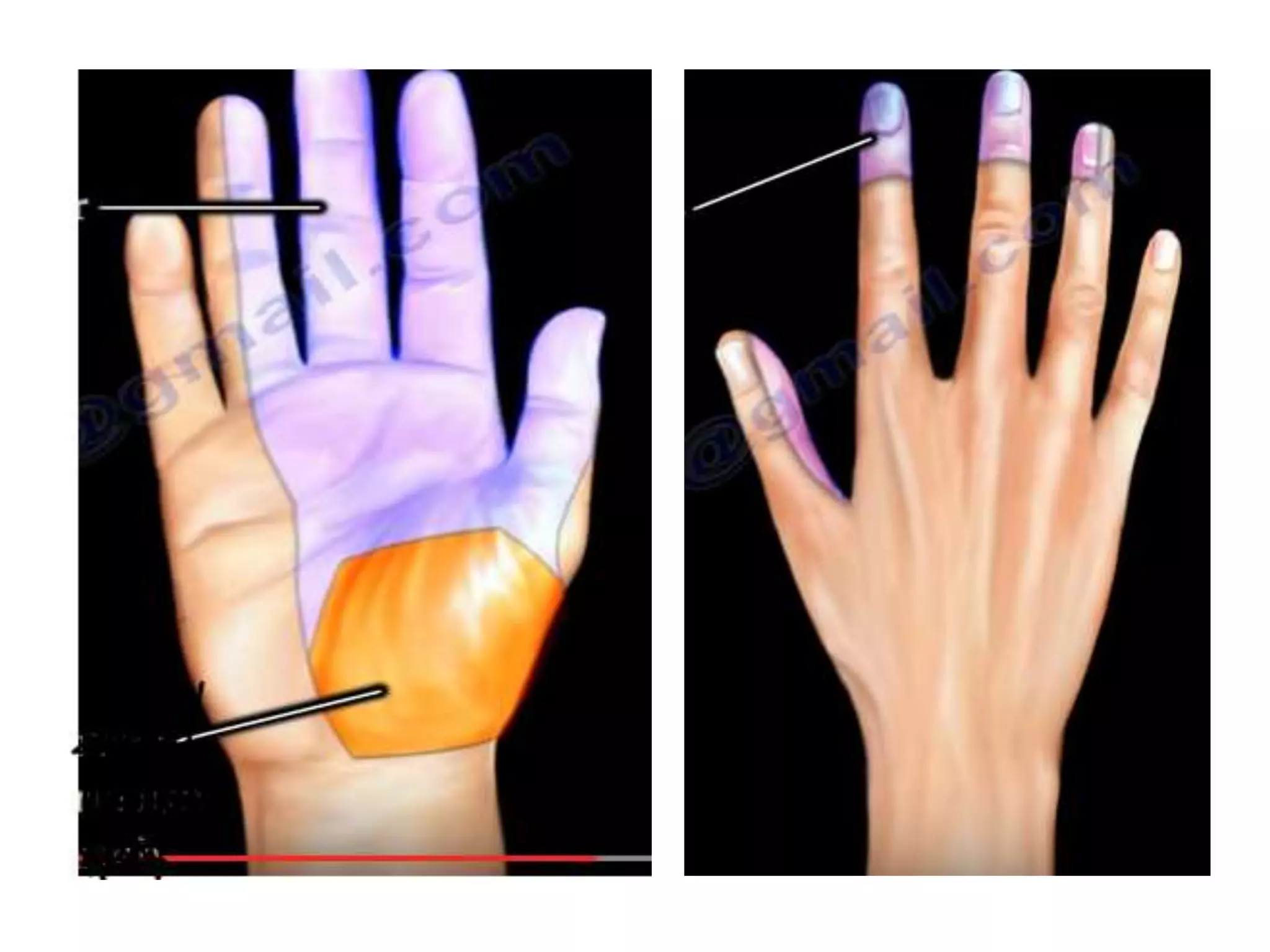
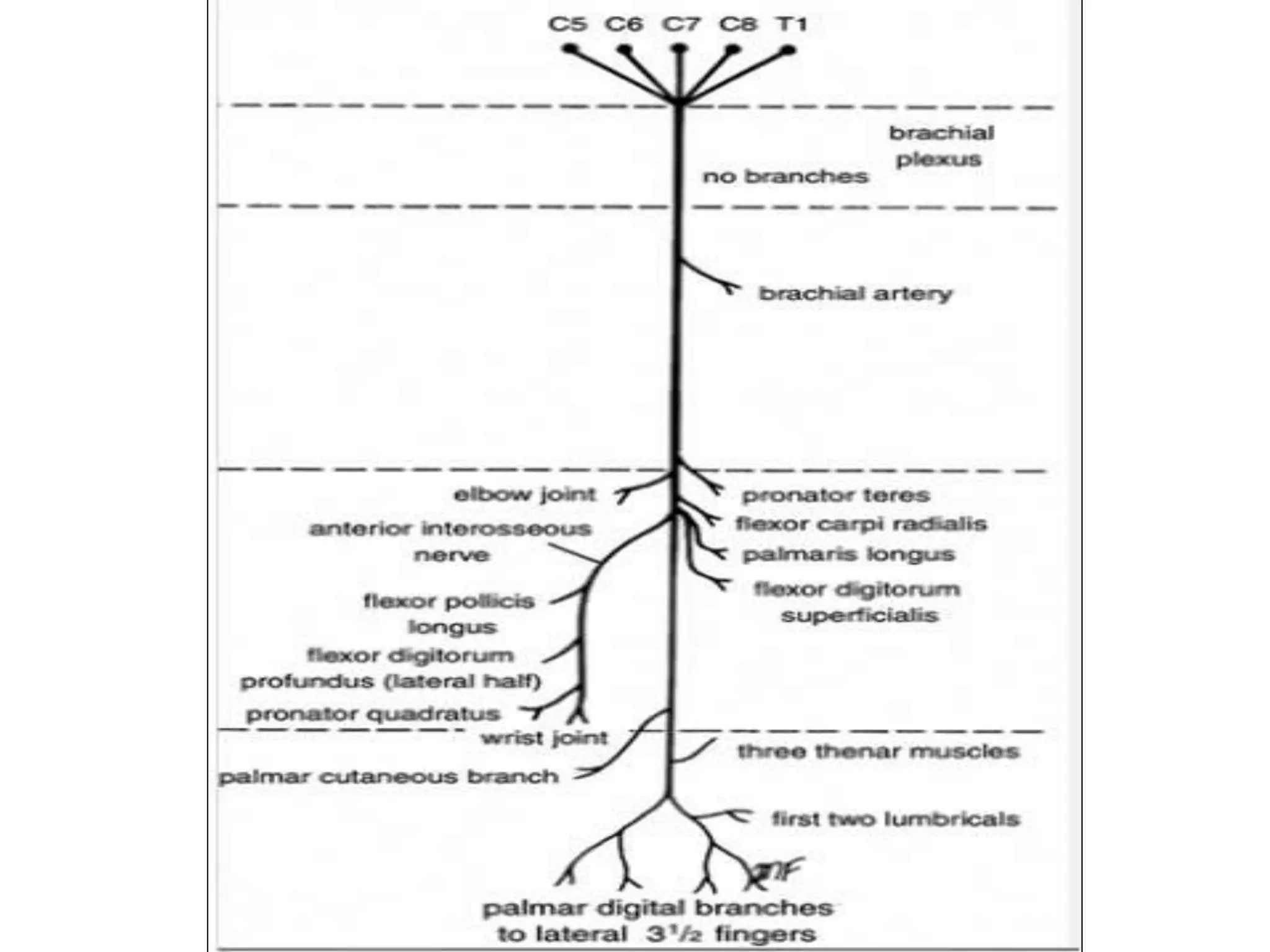
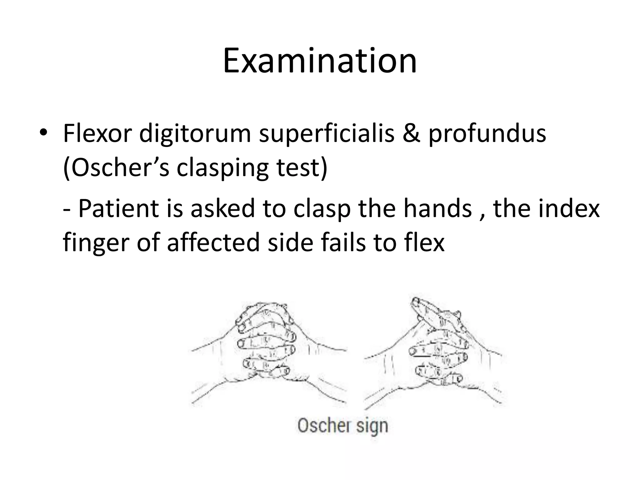
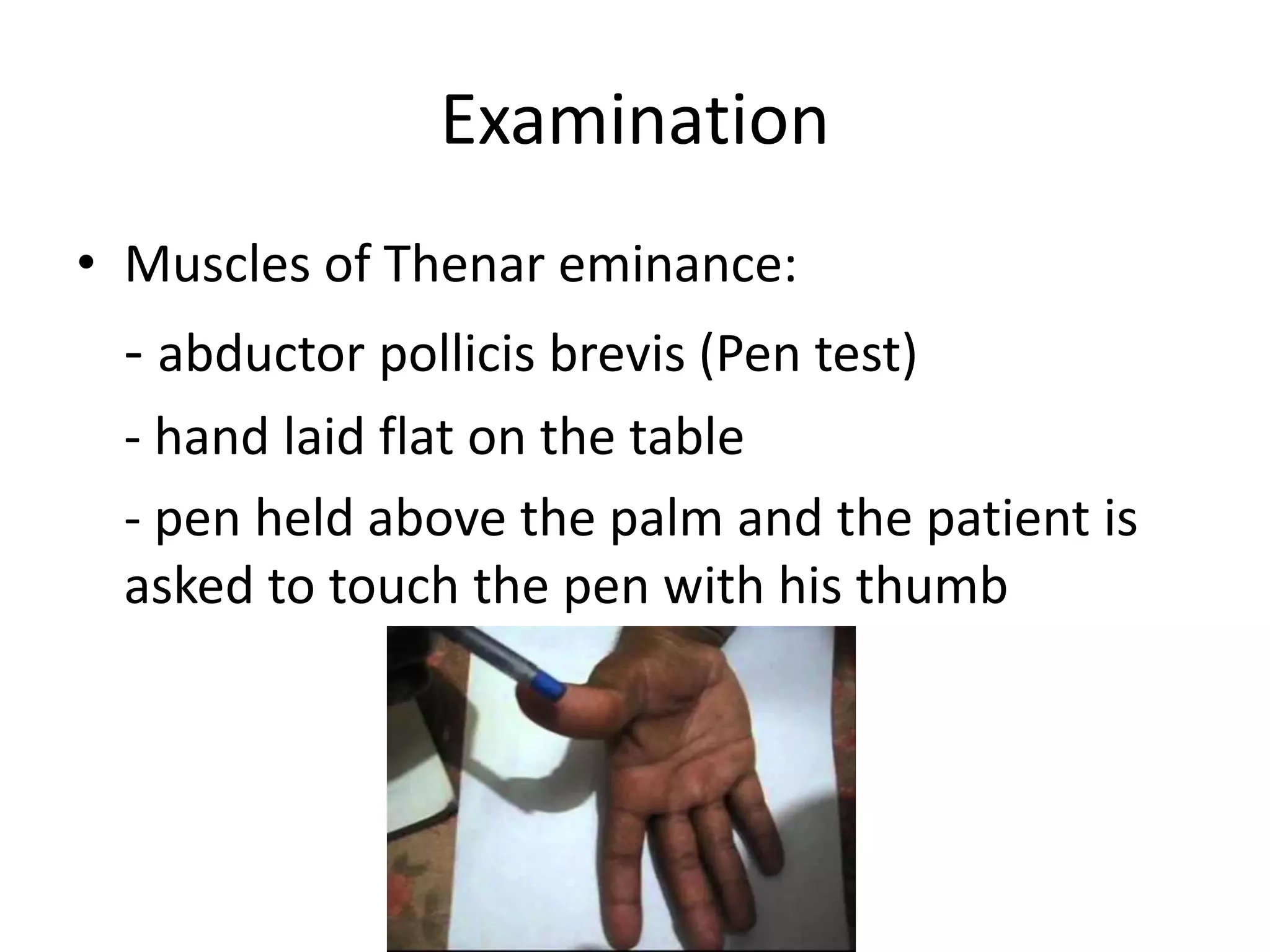
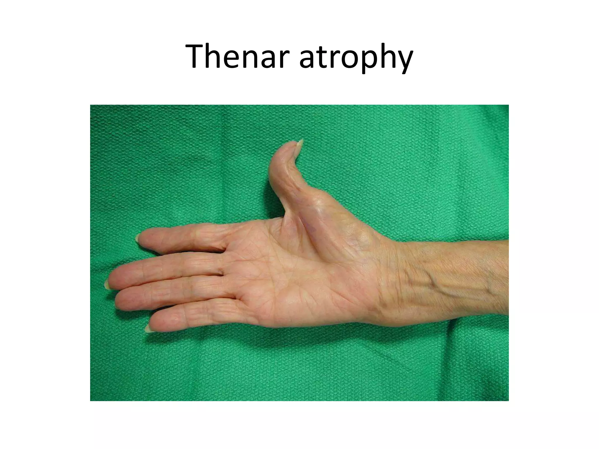
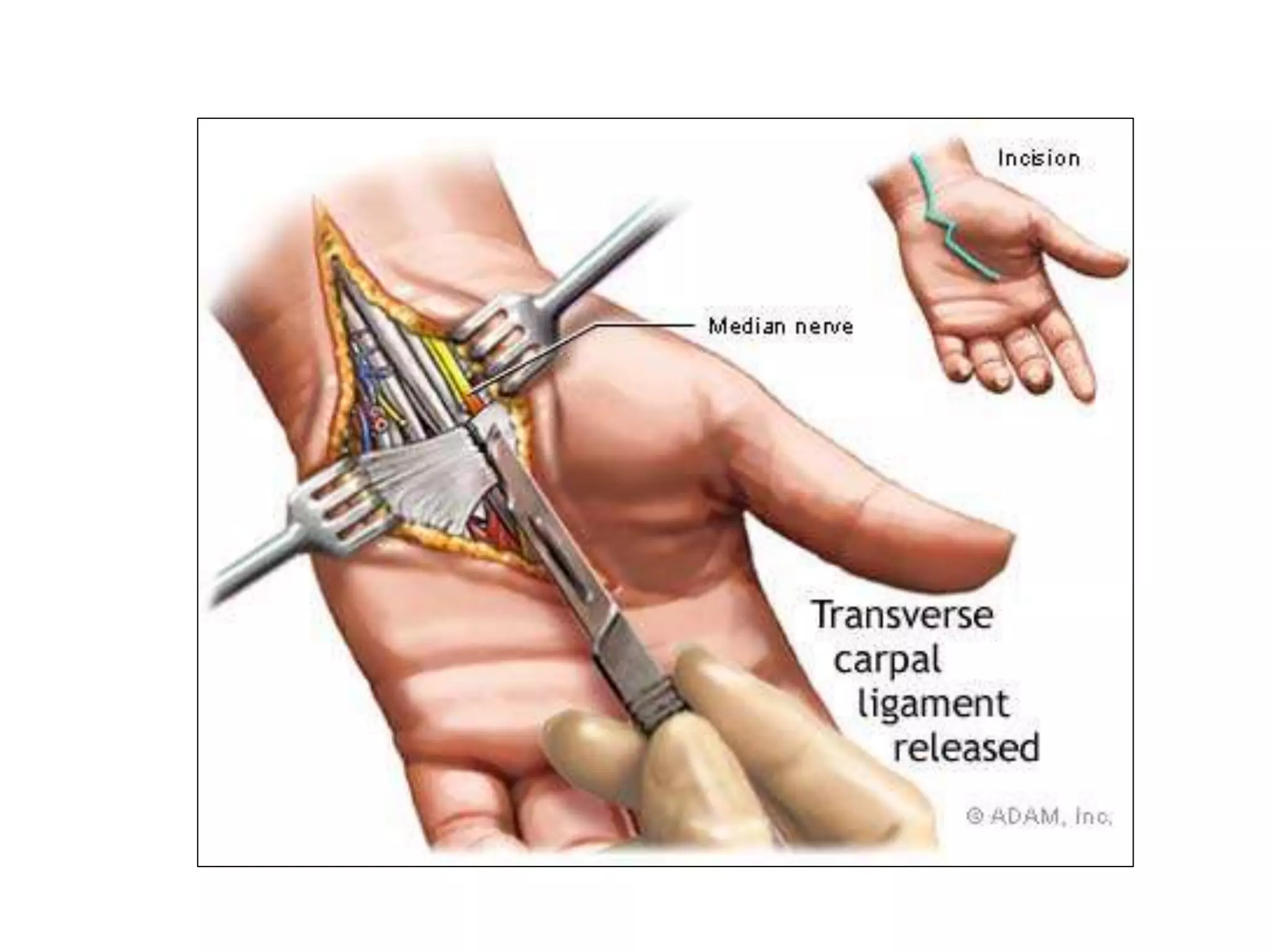
Median Nerve Injuries
Dr SD Sanyal
Anatomy
• Mixed nerve (contain motor & sensory fibers).
• Root value: C 5,6,7,8 & T1
• Runs in the median plane of the forearm , so
its called median nerve
Anatomy
• Arises in the axilla by joining:
1) Lat Cord of the brachial plexus
2) Med Cord of the brachial plexus
Anatomy: Axilla
• After being from Lateral Cord Medial Cord of
brachial plexus
Runs on the lateral aspect of Axillary artery
Anatomy: Arm
• Continues to run lateral to the brachial artery
till the mid-arm
• Crosses the artery anteriorly and passes
anterior to the elbow joint into forearm
Anatomy: Forearm
• Enters to the forearm b/w two heads of pronator teres
.
• Runs deep to the fibrous arch of FDS , in proximal 1/3rd
• Mid forearm it descends b/w FDS and FDP
• About 5 cm above wrist , it comes to lie on the lateral
side of the FDS , becomes superficial just above wrist
Anatomy: Hand
• Passes deep to the flexor retinaculum and
enters the Hand
• Muscular braches supply muscles of Thenar
eminence:
- abductor pollicis brevis
- opponens pollicis
- flexor pollicis brevis
Anatomy: Hand
• Divides into 4 to 5 palmar digital branches
supplying lateral three and half digit and their
nail beds
• Motor braches to the first and second
lumbrical muscles
Other branches
• Articular branches: supply the proximal radio-
ulnar joint
• Palmar cutaneous branch: supplies skin over
thenar eminence
Injuries
• High
• Low
High Median Nerve injuries
• Injury proximal to the elbow
• Due to forearm fractures or elbow dislocation
• Stab injuries and GSW’s
• Paralysis of all the muscles supplied by the
median nerve in the forearm and hand
Low Median Nerve Injuries
• Injury in the distal third of the forearm
• Sparing of the forearm muscles
• Muscles of the hand paralysed
• Anaesthesia over the median nerve distribution in the hand
• Thenar eminence is wasted and thumb abduction and
opposition are weak
• Sensation lost over the radial three and half digits and
trophic changes may seen
Examination
• Flexor pollicis longus : Tested by holding
thumb at its base and patient asked to flex the
terminal phalanx
Examination
• Flexor digitorum superficialis & profundus
(Oscher’s clasping test)
- Patient is asked to clasp the hands , the index
finger of affected side fails to flex
Examination
• Flexor Carpi radialis : Hand deviates to the
ulnar side when flexed against resistance
Examination
• Muscles of Thenar eminance:
- abductor pollicis brevis (Pen test)
- hand laid flat on the table
- pen held above the palm and the patient is
asked to touch the pen with his thumb
Examination
• opponens pollicis : brings the tip of the
thumb towards the tips of other fingers
Opponens pollices
Benedict Sign
Klien Nioh/ OK Sign
Ape Thumb
Principles of Surgical Management
1. Direct Injury: Nerve repair
2. Compression neuropathies: Decompression
3. Long standing cases: Tendon transfers
a) Low Median Nerve:
- Re-routing of ring/ middle finger superficial flexor
around FCU to APB to aid thumb opposition
b) High Median Nerve:
- Suturing of profundus tendons to ring and small finger
tendons for restoration of IP jt movts
- ECU re-routing and attachment to dorsal radius/
Transfer of biceps insertion from medial to lat radius for
weak forearm pronation
Median nerve Compression
Syndromes
• Carpal Tunnel
• Pronator
• Interosseous
Carpal Tunnel Syndrome
• Compressive neuropathy as the nerve passes through the
Carpal Tunnel
• Causes:
- Idiopathic : Most common
- Inflammatory : Rheumatoid Arthritis
: Wrist osteoarthritis
- Post traumatic : Bone thickening
- Endocrine : Myxoedema
: Acromegaly
- Pregnancy
- Gout
- Repetitive wrist movts: Typists & Computer users
Carpal Tunnel
Symptoms
• Hand and wrist Pain
• Paraesthesia
• Hypoaesthsia
• Sparing of Palmar cutaneous branch supply
• Patient wakes at night with burning or aching
pain and shakes the hand to obtain relief and
restore sensation
• Aggravated by elevation of hand
• Thenar atrophy and weakness of thumb
opposition and abduction may develop late
Diagnosis
• History
• Clinical examination:
- Thenar wasting
- Phalen’s sign
- Tinel’s sign
- Carpal compression test
• Electro Diagnostic Studies:
- Very reliable for evaluation
- Atypical cases may be present
Thenar atrophy
Tinel’s Sign
Carpal Compression test/ Durkan’s test
Management
• Splinting – prevents wrist flexion
• Corticosteroid/anesthetic injection
• Surgical decompression:Division of the
transverse carpal ligament
- Open
- Endoscopic
Complications
• Injury to palmar cutaneous/recurrent motor
branch of the median nerve
• Hypertrophic scarring
• Hematoma/Arterial injury
• Pillar pain
Pronator teres syndrome
• High Compression neuropathy
• It is rare compared to CTS and AIS
• Misnomer  Proximal forearm median nerve
compression
Symptoms & signs
• Symptoms are similar to those of carpal tunnel syndrome
• Sensory disturbances
- Thumb & Index > Middle finger
• Night pain is unusual and forearm pain is more common
• Hand numbness on gripping
• Phalen’s test negative
• Double crush phenomena
• Symptoms provoked by resisted elbow flexion with forearm
supinated ( tightening of bicipital aponeurosis )
• By resisted forearm pronation with the elbow extended
( pronator tension )
Management
• No relief with steroids
• Surgical decompression
Anterior Interosseous Syndrome
• Damage to the Anterior Interosseous Nerve
• Pain in the forearm
• Weakness of the gripping movement of the
thumb and index finger( unable to make ok
sign )
• Causes:
- Injury to elbow
- Injury during open/closed reduction
Management
• Corticosteroids
• Surgery:
- Resection/detachment of deep head of PT
Thank You

Median nerve injuries and mangement

  • 1.
  • 2.
    Anatomy • Mixed nerve(contain motor & sensory fibers). • Root value: C 5,6,7,8 & T1 • Runs in the median plane of the forearm , so its called median nerve
  • 4.
    Anatomy • Arises inthe axilla by joining: 1) Lat Cord of the brachial plexus 2) Med Cord of the brachial plexus
  • 6.
    Anatomy: Axilla • Afterbeing from Lateral Cord Medial Cord of brachial plexus Runs on the lateral aspect of Axillary artery
  • 8.
    Anatomy: Arm • Continuesto run lateral to the brachial artery till the mid-arm • Crosses the artery anteriorly and passes anterior to the elbow joint into forearm
  • 13.
    Anatomy: Forearm • Entersto the forearm b/w two heads of pronator teres . • Runs deep to the fibrous arch of FDS , in proximal 1/3rd • Mid forearm it descends b/w FDS and FDP • About 5 cm above wrist , it comes to lie on the lateral side of the FDS , becomes superficial just above wrist
  • 21.
    Anatomy: Hand • Passesdeep to the flexor retinaculum and enters the Hand • Muscular braches supply muscles of Thenar eminence: - abductor pollicis brevis - opponens pollicis - flexor pollicis brevis
  • 23.
    Anatomy: Hand • Dividesinto 4 to 5 palmar digital branches supplying lateral three and half digit and their nail beds • Motor braches to the first and second lumbrical muscles
  • 28.
    Other branches • Articularbranches: supply the proximal radio- ulnar joint • Palmar cutaneous branch: supplies skin over thenar eminence
  • 30.
  • 31.
    High Median Nerveinjuries • Injury proximal to the elbow • Due to forearm fractures or elbow dislocation • Stab injuries and GSW’s • Paralysis of all the muscles supplied by the median nerve in the forearm and hand
  • 32.
    Low Median NerveInjuries • Injury in the distal third of the forearm • Sparing of the forearm muscles • Muscles of the hand paralysed • Anaesthesia over the median nerve distribution in the hand • Thenar eminence is wasted and thumb abduction and opposition are weak • Sensation lost over the radial three and half digits and trophic changes may seen
  • 33.
    Examination • Flexor pollicislongus : Tested by holding thumb at its base and patient asked to flex the terminal phalanx
  • 34.
    Examination • Flexor digitorumsuperficialis & profundus (Oscher’s clasping test) - Patient is asked to clasp the hands , the index finger of affected side fails to flex
  • 35.
    Examination • Flexor Carpiradialis : Hand deviates to the ulnar side when flexed against resistance
  • 36.
    Examination • Muscles ofThenar eminance: - abductor pollicis brevis (Pen test) - hand laid flat on the table - pen held above the palm and the patient is asked to touch the pen with his thumb
  • 37.
    Examination • opponens pollicis: brings the tip of the thumb towards the tips of other fingers
  • 38.
  • 39.
  • 40.
  • 41.
  • 42.
    Principles of SurgicalManagement 1. Direct Injury: Nerve repair 2. Compression neuropathies: Decompression 3. Long standing cases: Tendon transfers a) Low Median Nerve: - Re-routing of ring/ middle finger superficial flexor around FCU to APB to aid thumb opposition b) High Median Nerve: - Suturing of profundus tendons to ring and small finger tendons for restoration of IP jt movts - ECU re-routing and attachment to dorsal radius/ Transfer of biceps insertion from medial to lat radius for weak forearm pronation
  • 43.
    Median nerve Compression Syndromes •Carpal Tunnel • Pronator • Interosseous
  • 44.
    Carpal Tunnel Syndrome •Compressive neuropathy as the nerve passes through the Carpal Tunnel • Causes: - Idiopathic : Most common - Inflammatory : Rheumatoid Arthritis : Wrist osteoarthritis - Post traumatic : Bone thickening - Endocrine : Myxoedema : Acromegaly - Pregnancy - Gout - Repetitive wrist movts: Typists & Computer users
  • 45.
  • 46.
    Symptoms • Hand andwrist Pain • Paraesthesia • Hypoaesthsia • Sparing of Palmar cutaneous branch supply • Patient wakes at night with burning or aching pain and shakes the hand to obtain relief and restore sensation • Aggravated by elevation of hand • Thenar atrophy and weakness of thumb opposition and abduction may develop late
  • 47.
    Diagnosis • History • Clinicalexamination: - Thenar wasting - Phalen’s sign - Tinel’s sign - Carpal compression test • Electro Diagnostic Studies: - Very reliable for evaluation - Atypical cases may be present
  • 48.
  • 50.
  • 51.
    Carpal Compression test/Durkan’s test
  • 52.
    Management • Splinting –prevents wrist flexion • Corticosteroid/anesthetic injection • Surgical decompression:Division of the transverse carpal ligament - Open - Endoscopic
  • 56.
    Complications • Injury topalmar cutaneous/recurrent motor branch of the median nerve • Hypertrophic scarring • Hematoma/Arterial injury • Pillar pain
  • 57.
    Pronator teres syndrome •High Compression neuropathy • It is rare compared to CTS and AIS • Misnomer  Proximal forearm median nerve compression
  • 58.
    Symptoms & signs •Symptoms are similar to those of carpal tunnel syndrome • Sensory disturbances - Thumb & Index > Middle finger • Night pain is unusual and forearm pain is more common • Hand numbness on gripping • Phalen’s test negative • Double crush phenomena • Symptoms provoked by resisted elbow flexion with forearm supinated ( tightening of bicipital aponeurosis ) • By resisted forearm pronation with the elbow extended ( pronator tension )
  • 59.
    Management • No reliefwith steroids • Surgical decompression
  • 60.
    Anterior Interosseous Syndrome •Damage to the Anterior Interosseous Nerve • Pain in the forearm • Weakness of the gripping movement of the thumb and index finger( unable to make ok sign ) • Causes: - Injury to elbow - Injury during open/closed reduction
  • 61.
    Management • Corticosteroids • Surgery: -Resection/detachment of deep head of PT
  • 62.