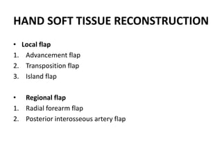
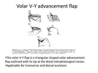
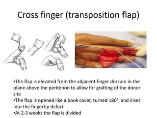
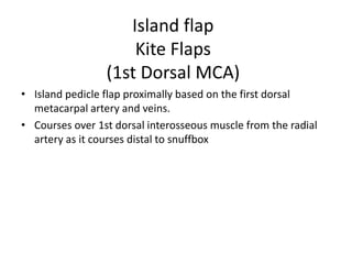
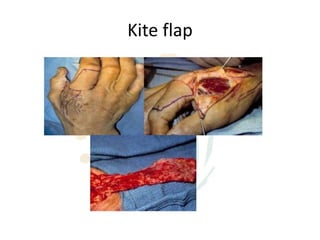
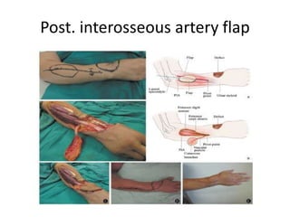
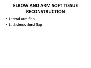
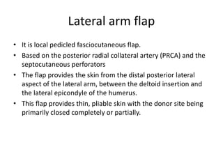
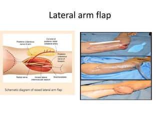
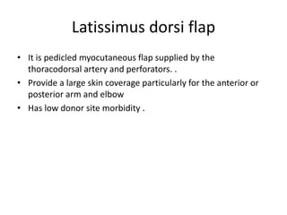
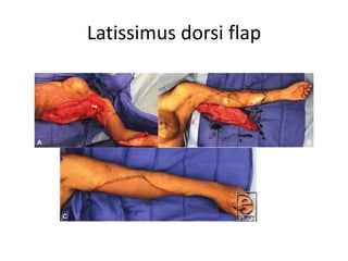
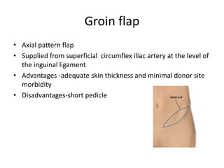
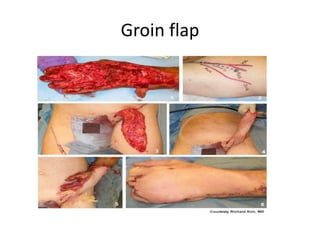
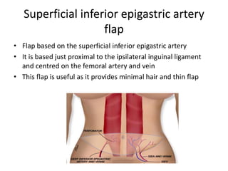
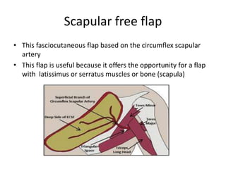
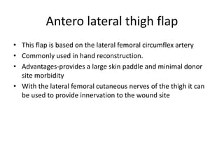
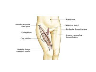
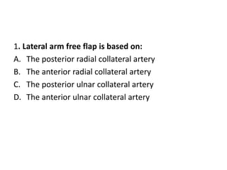
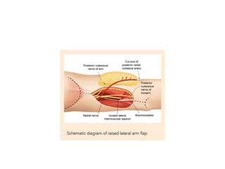
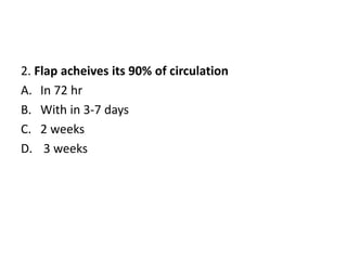
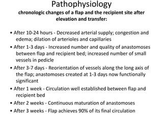
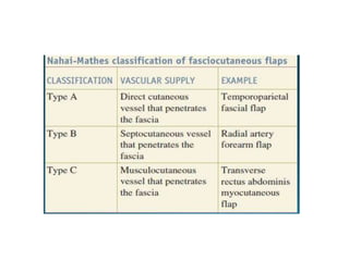
Flap Coverage in
Upper Extremities in trauma
Dr Vishal Patil
Introduction
• A flap is a vascularized block of tissue that is mobilized
from its donor site and transferred to another location,
adjacent or remote, for reconstructive purposes.
A flap is used
• To reconstruct a large primary defect
• Replace tissue loss during trauma or surgical excision.
• Provide padding over bony prominences.
• Bring in better blood supply to poorly vascularized bed.
• Improve sensation to an area
Classification of flaps
Classification of Flaps Can be based on:
1. Congruity
2. Circulation
3. Anatomical Components
Classification
CONGRUITY
• local
• Regional
• Distant
1.Pedicled
2. Free flap
• Island
CIRCULATION
• Axial pattern
• Random pattern
ANATOMICAL
COMPONENT
• Skin
• Muscle and
myocutaneous
• Fascia and
fasciocutaneous
• Free flap option is a versatile single stage procedure
facilitating simultaneous reconstruction of other critical
structures. It also allows postoperative mobilization and early
discharge and return to work
King EA, Ozer K. Free skin flap coverage of the upper extremity. Hand
Clin.2014;30:201e209. https://doi.org/10.1016/j.hcl.2014.01.003.
.
• Pedicled flaps have been the workhorse flaps for
reconstruction of the upper limb in many centers across the
world. Though the procedure includes multiple surgery,
prolonged immobilization, and joint stiffness.
Sabapathy SR. Refinements of pedicle flaps for soft tissue cover in the upper limb.
In: Venkataswami R, ed. Surgery of the Injured Hand. New Delhi (India):Jaypee
Brothers; 2009:
HAND SOFT TISSUE RECONSTRUCTION
• Local flap
1. Advancement flap
2. Transposition flap
3. Island flap
• Regional flap
1. Radial forearm flap
2. Posterior interosseous artery flap
Volar V-Y advancement flap
•The volar V-Y flap is a triangular shaped volar advancement
flap outlined with its tip at the distal interphalangeal crease.
•Applicable for transverse and dorsal avulsions
Cross finger (transposition flap)
•The flap is elevated from the adjacent finger dorsum in the
plane above the peritenon to allow for grafting of the donor
site
•The flap is opened like a book cover, turned 180°, and inset
into the fingertip defect
•At 2-3 weeks the flap is divided
Island flap
Kite Flaps
(1st Dorsal MCA)
• Island pedicle flap proximally based on the first dorsal
metacarpal artery and veins.
• Courses over 1st dorsal interosseous muscle from the radial
artery as it courses distal to snuffbox
Kite flap
Thenar flap
• A 2 cm x 4 cm thenar flap can be harvested from the MCP
crease
• Primary closure of the donor site with thumb flexion.
• Avoid injury to the neurovascular bundles and flexor pollicis
longus tendon
Regional flap
Radial forearm flap
• Most reliable and versatile flap for upper limb reconstruction
• Based on the radial artery and the accompanying
venaecomitans.
• Advantages -provision of up to 8 x 10 cm of thin, pliable and
hairless skin and ease of harvest
• Disadvantage- radial artery is sacrificed and poor donor site
morbidity with this flap
• An Allen’s test should always be performed prior
Radial forearm flap
Posterior interosseous artery flap
• A classic fasciocutaneous flap, ideal for coverage of dorsum of
hand without compromising the two main artery of hand.
• The flap is usually designed on the emergence of the artery in
the posterior compartment of the proximal forearm.
• The limits of the flap are distally 3-4 cm above the wrist
crease, medially not to extend subcutaneous border of the
ulnar and radially not extend 3-4 cm beyond the epicondylar-
ulnar line
Post. interosseous artery flap
ELBOW AND ARM SOFT TISSUE
RECONSTRUCTION
• Lateral arm flap
• Latissimus dorsi flap
Lateral arm flap
• It is local pedicled fasciocutaneous flap.
• Based on the posterior radial collateral artery (PRCA) and the
septocutaneous perforators
• The flap provides the skin from the distal posterior lateral
aspect of the lateral arm, between the deltoid insertion and
the lateral epicondyle of the humerus.
• This flap provides thin, pliable skin with the donor site being
primarily closed completely or partially.
Lateral arm flap
Latissimus dorsi flap
• It is pedicled myocutaneous flap supplied by the
thoracodorsal artery and perforators. .
• Provide a large skin coverage particularly for the anterior or
posterior arm and elbow
• Has low donor site morbidity .
Latissimus dorsi flap
Free flap for the upper extremity
Groin flap
• Axial pattern flap
• Supplied from superficial circumflex iliac artery at the level of
the inguinal ligament
• Advantages -adequate skin thickness and minimal donor site
morbidity
• Disadvantages-short pedicle
Groin flap
Superficial inferior epigastric artery
flap
• Flap based on the superficial inferior epigastric artery
• It is based just proximal to the ipsilateral inguinal ligament
and centred on the femoral artery and vein
• This flap is useful as it provides minimal hair and thin flap
Scapular free flap
• This fasciocutaneous flap based on the circumflex scapular
artery
• This flap is useful because it offers the opportunity for a flap
with latissimus or serratus muscles or bone (scapula)
Antero lateral thigh flap
• This flap is based on the lateral femoral circumflex artery
• Commonly used in hand reconstruction.
• Advantages-provides a large skin paddle and minimal donor
site morbidity
• With the lateral femoral cutaneous nerves of the thigh it can
be used to provide innervation to the wound site
Criteria of flap selection
• Patient age and health
• Condition of skin defects
• Location and size of defects
• Damage of deep structure
• Cosmetic demands
• Surgeon’s technical skill
Postoperative flap monitoring:
• The gold standard of postoperative flap monitoring is clinical
observation. It includes:
1.Flap color
2.Capillary refilling time
3.Surface temperature monitoring
4.Blanching assesment
Complications:
1.seroma formation
2.hematoma formation
3.flap necrosis
4.fat necrosis
5.Donor site infection
Causes of flap failure:
• Poor anatomical knowledge when raising the flap(such that
the blood supply is deficient from the start)
• Flap inset with too much tension.
• Local sepsis .
• The dressing applied too tightly around the pedicle
THANK YOU
Clinical vignette
1. Lateral arm free flap is based on:
A. The posterior radial collateral artery
B. The anterior radial collateral artery
C. The posterior ulnar collateral artery
D. The anterior ulnar collateral artery
2. Flap acheives its 90% of circulation
A. In 72 hr
B. With in 3-7 days
C. 2 weeks
D. 3 weeks
Pathophysiology
chronologic changes of a flap and the recipient site after
elevation and transfer:
• After 10-24 hours - Decreased arterial supply; congestion and
edema; dilation of arterioles and capillaries
• After 1-3 days - Increased number and quality of anastomoses
between flap and recipient bed; increased number of small
vessels in pedicle
• After 3-7 days - Reorientation of vessels along the long axis of
the flap; anastomoses created at 1-3 days now functionally
significant
• After 1 week - Circulation well established between flap and
recipient bed
• After 2 weeks - Continuous maturation of anastomoses
• After 3 weeks - Flap achieves 90% of its final circulation
3. According to MATHES AND NAHAI classification
radial forearm flap is
A. Type A
B. Type B
C. Type C
D. Type D
4. A Z- plasty is an example of:
A. an advancement flap
B. an island pedical flap
C. a rotation flap
D. a transposition flap
• Z-plasty transposes two interdigitating triangular flaps without
tension to use lateral skin to produce a gain in length along
the direction of the common limb of the Z.
5.Which of the following statement is true regarding
groin flap-
A. It is type of island flap
B. It is based on superficial epigastric artery
C. It has short pedicle
D. It has maximal donor site morbidity
Groin flap
• Axial pattern flap
• Supplied from the superficial femoral artery at the level of the
inguinal ligament
• Advantages -adequate skin thickness and minimal donor site
morbidity
• Disadvantages-short pedicle

Flap coverage in upper extremities in trauma

  • 1.
    Flap Coverage in UpperExtremities in trauma Dr Vishal Patil
  • 2.
    Introduction • A flapis a vascularized block of tissue that is mobilized from its donor site and transferred to another location, adjacent or remote, for reconstructive purposes.
  • 3.
    A flap isused • To reconstruct a large primary defect • Replace tissue loss during trauma or surgical excision. • Provide padding over bony prominences. • Bring in better blood supply to poorly vascularized bed. • Improve sensation to an area
  • 4.
    Classification of flaps Classificationof Flaps Can be based on: 1. Congruity 2. Circulation 3. Anatomical Components
  • 5.
    Classification CONGRUITY • local • Regional •Distant 1.Pedicled 2. Free flap • Island CIRCULATION • Axial pattern • Random pattern ANATOMICAL COMPONENT • Skin • Muscle and myocutaneous • Fascia and fasciocutaneous
  • 6.
    • Free flapoption is a versatile single stage procedure facilitating simultaneous reconstruction of other critical structures. It also allows postoperative mobilization and early discharge and return to work King EA, Ozer K. Free skin flap coverage of the upper extremity. Hand Clin.2014;30:201e209. https://doi.org/10.1016/j.hcl.2014.01.003. .
  • 7.
    • Pedicled flapshave been the workhorse flaps for reconstruction of the upper limb in many centers across the world. Though the procedure includes multiple surgery, prolonged immobilization, and joint stiffness. Sabapathy SR. Refinements of pedicle flaps for soft tissue cover in the upper limb. In: Venkataswami R, ed. Surgery of the Injured Hand. New Delhi (India):Jaypee Brothers; 2009:
  • 8.
    HAND SOFT TISSUERECONSTRUCTION • Local flap 1. Advancement flap 2. Transposition flap 3. Island flap • Regional flap 1. Radial forearm flap 2. Posterior interosseous artery flap
  • 9.
    Volar V-Y advancementflap •The volar V-Y flap is a triangular shaped volar advancement flap outlined with its tip at the distal interphalangeal crease. •Applicable for transverse and dorsal avulsions
  • 10.
    Cross finger (transpositionflap) •The flap is elevated from the adjacent finger dorsum in the plane above the peritenon to allow for grafting of the donor site •The flap is opened like a book cover, turned 180°, and inset into the fingertip defect •At 2-3 weeks the flap is divided
  • 11.
    Island flap Kite Flaps (1stDorsal MCA) • Island pedicle flap proximally based on the first dorsal metacarpal artery and veins. • Courses over 1st dorsal interosseous muscle from the radial artery as it courses distal to snuffbox
  • 12.
  • 13.
    Thenar flap • A2 cm x 4 cm thenar flap can be harvested from the MCP crease • Primary closure of the donor site with thumb flexion. • Avoid injury to the neurovascular bundles and flexor pollicis longus tendon
  • 14.
  • 15.
    Radial forearm flap •Most reliable and versatile flap for upper limb reconstruction • Based on the radial artery and the accompanying venaecomitans. • Advantages -provision of up to 8 x 10 cm of thin, pliable and hairless skin and ease of harvest • Disadvantage- radial artery is sacrificed and poor donor site morbidity with this flap • An Allen’s test should always be performed prior
  • 16.
  • 17.
    Posterior interosseous arteryflap • A classic fasciocutaneous flap, ideal for coverage of dorsum of hand without compromising the two main artery of hand. • The flap is usually designed on the emergence of the artery in the posterior compartment of the proximal forearm. • The limits of the flap are distally 3-4 cm above the wrist crease, medially not to extend subcutaneous border of the ulnar and radially not extend 3-4 cm beyond the epicondylar- ulnar line
  • 18.
  • 19.
    ELBOW AND ARMSOFT TISSUE RECONSTRUCTION • Lateral arm flap • Latissimus dorsi flap
  • 20.
    Lateral arm flap •It is local pedicled fasciocutaneous flap. • Based on the posterior radial collateral artery (PRCA) and the septocutaneous perforators • The flap provides the skin from the distal posterior lateral aspect of the lateral arm, between the deltoid insertion and the lateral epicondyle of the humerus. • This flap provides thin, pliable skin with the donor site being primarily closed completely or partially.
  • 21.
  • 22.
    Latissimus dorsi flap •It is pedicled myocutaneous flap supplied by the thoracodorsal artery and perforators. . • Provide a large skin coverage particularly for the anterior or posterior arm and elbow • Has low donor site morbidity .
  • 23.
  • 24.
    Free flap forthe upper extremity
  • 25.
    Groin flap • Axialpattern flap • Supplied from superficial circumflex iliac artery at the level of the inguinal ligament • Advantages -adequate skin thickness and minimal donor site morbidity • Disadvantages-short pedicle
  • 26.
  • 27.
    Superficial inferior epigastricartery flap • Flap based on the superficial inferior epigastric artery • It is based just proximal to the ipsilateral inguinal ligament and centred on the femoral artery and vein • This flap is useful as it provides minimal hair and thin flap
  • 28.
    Scapular free flap •This fasciocutaneous flap based on the circumflex scapular artery • This flap is useful because it offers the opportunity for a flap with latissimus or serratus muscles or bone (scapula)
  • 29.
    Antero lateral thighflap • This flap is based on the lateral femoral circumflex artery • Commonly used in hand reconstruction. • Advantages-provides a large skin paddle and minimal donor site morbidity • With the lateral femoral cutaneous nerves of the thigh it can be used to provide innervation to the wound site
  • 31.
    Criteria of flapselection • Patient age and health • Condition of skin defects • Location and size of defects • Damage of deep structure • Cosmetic demands • Surgeon’s technical skill
  • 32.
    Postoperative flap monitoring: •The gold standard of postoperative flap monitoring is clinical observation. It includes: 1.Flap color 2.Capillary refilling time 3.Surface temperature monitoring 4.Blanching assesment
  • 33.
    Complications: 1.seroma formation 2.hematoma formation 3.flapnecrosis 4.fat necrosis 5.Donor site infection
  • 34.
    Causes of flapfailure: • Poor anatomical knowledge when raising the flap(such that the blood supply is deficient from the start) • Flap inset with too much tension. • Local sepsis . • The dressing applied too tightly around the pedicle
  • 35.
  • 36.
  • 37.
    1. Lateral armfree flap is based on: A. The posterior radial collateral artery B. The anterior radial collateral artery C. The posterior ulnar collateral artery D. The anterior ulnar collateral artery
  • 39.
    2. Flap acheivesits 90% of circulation A. In 72 hr B. With in 3-7 days C. 2 weeks D. 3 weeks
  • 40.
    Pathophysiology chronologic changes ofa flap and the recipient site after elevation and transfer: • After 10-24 hours - Decreased arterial supply; congestion and edema; dilation of arterioles and capillaries • After 1-3 days - Increased number and quality of anastomoses between flap and recipient bed; increased number of small vessels in pedicle • After 3-7 days - Reorientation of vessels along the long axis of the flap; anastomoses created at 1-3 days now functionally significant • After 1 week - Circulation well established between flap and recipient bed • After 2 weeks - Continuous maturation of anastomoses • After 3 weeks - Flap achieves 90% of its final circulation
  • 41.
    3. According toMATHES AND NAHAI classification radial forearm flap is A. Type A B. Type B C. Type C D. Type D
  • 43.
    4. A Z-plasty is an example of: A. an advancement flap B. an island pedical flap C. a rotation flap D. a transposition flap
  • 44.
    • Z-plasty transposestwo interdigitating triangular flaps without tension to use lateral skin to produce a gain in length along the direction of the common limb of the Z.
  • 45.
    5.Which of thefollowing statement is true regarding groin flap- A. It is type of island flap B. It is based on superficial epigastric artery C. It has short pedicle D. It has maximal donor site morbidity
  • 46.
    Groin flap • Axialpattern flap • Supplied from the superficial femoral artery at the level of the inguinal ligament • Advantages -adequate skin thickness and minimal donor site morbidity • Disadvantages-short pedicle