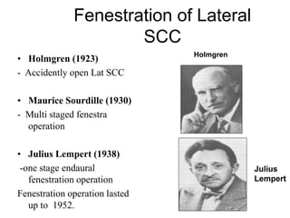
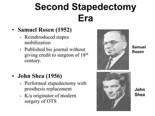
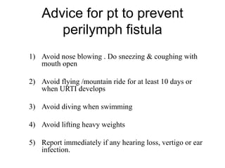
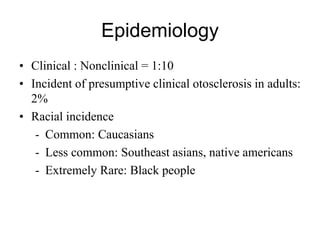
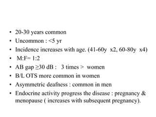
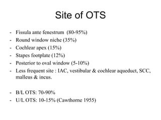
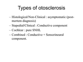
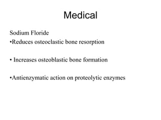
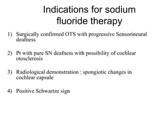
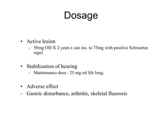
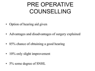
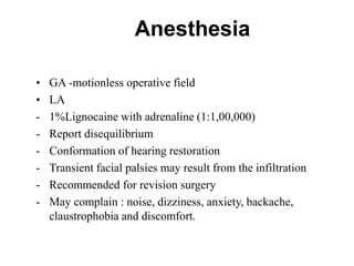
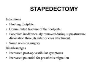
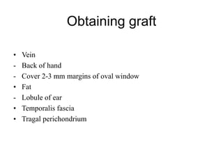
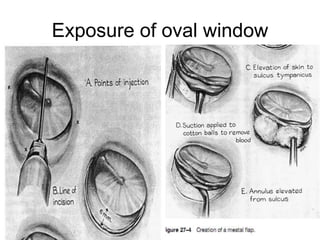
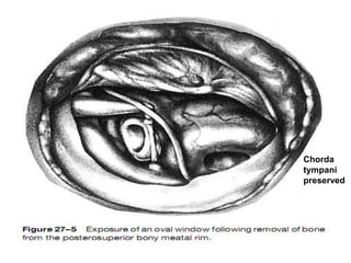
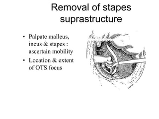
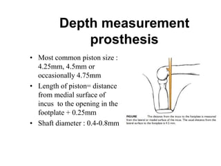
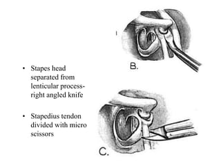
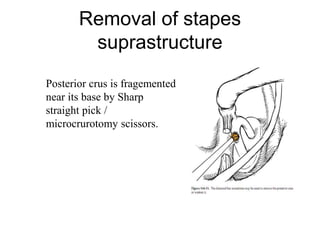
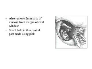
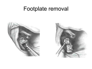
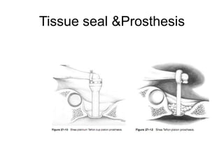
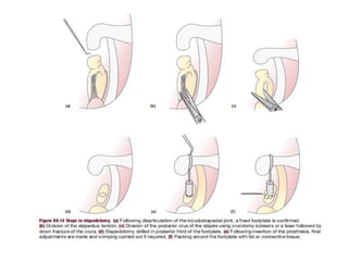
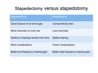
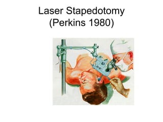
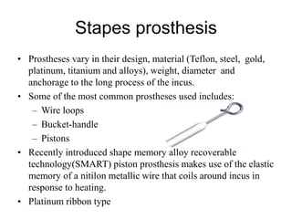
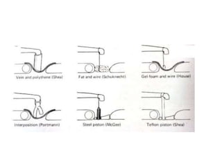
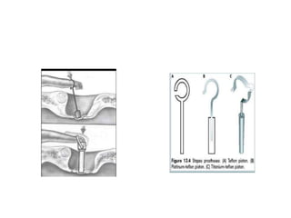
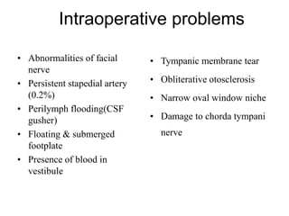
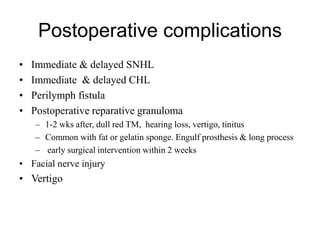
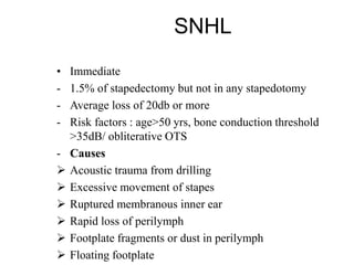
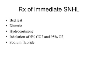
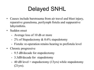
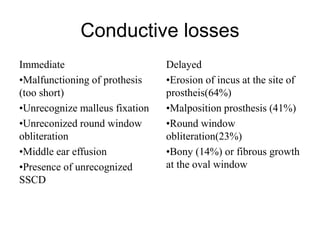
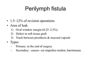
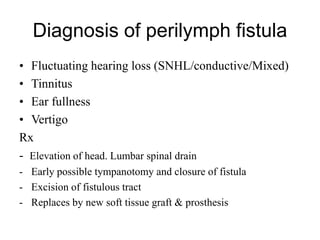
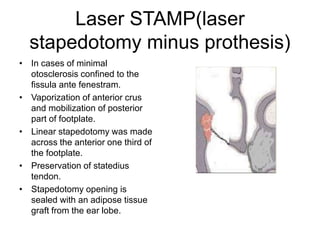
OTOSCLEROSIS
Dr. Sandeep Shrestha
3rd year resident
ENT
NMCTH
DEVELOPMENT OF THE OTIC
CAPSULE
• Thickening of surface ectoderm, otic placode -Otic placode invaginates below the
surface ectoderm - Sides closes in to form otic pit - Loses connection with the
surface to form otocyst (otic or auditory vesicles)
• The mesenchyme enclosing the otocyst becomes chondrified to form the otic
capsule.
• Ossification occurs from 14 OC – fuses to form a bony box without a single
suture line.
• 3 layers: an outer periosteal, inner endosteal and middle
endochondral layer.
• The endochondral layer is composed of bone intimately
mixed with calcified cartilagenous cells called globuli
interossei, or globuli ossei.
• Calcification is usually complete by the age of one and
there after there is very little remodeling.
• The adult otic capsule shows almost no osteoblastic or
osteoclastic activity.
• The normal otic capsule has an extremely low remodeling
rate (2%); compared to 10% annual turn over rate in other
bones.
Definition
• Localized hereditary disorder of bone metabolism of
otic capsule enchondral bone that is characterized by
disordered resorption and deposition of bone.
– Mature lamellar bone →osteoclasis→woven bone
(↑thickness, cellularity & vascularity)
– Results : new bone deposit with a different fibrillar and
cellular pattern - laid down at certain sites in temporal bone.
History
• Antonio Valsalva
– First described ankylosis of
stapes in 1741
• Joseph Toynbee (1861)
– Noted 39 stapes footplate
ankylosis out of 1959
temporal bone dissections
• Katz (1890)
– Microscopic evidence of
otosclerosis
Antonio Valsalva
Adam politzer Friedrich Siebenmann
Coined term Otosclerosis in 1894 Coined term Otospongiosis in 1912
Aetiology
THEORIES
•Metabolic
•Immune disorders
•Vascular disease
•Infection (measles virus)
•Trauma
•Anatomical & histological anomalies of
temporal bone
Genetic factors
• Autosomal dominant transmission with incomplete
penetrance. (Larsson’s 1962)
• Nearly 100% concordance in monozygotic twins.
• OTSC1 (chromosome 15q25-26)
• OTSC2 (chromosome 7q34-36)
• OTSC3 (chromosome 6p21.3-22.3) monogentic
• OTSC4 (chromosome 16q21-23.2) otosclerotic
• OTSC5 (chromosome 3q22-24) loci
• OTSC7 (chromosome 6q13-16.1)
• Other :SERPINF1 gene, COL1A1, TGFB1 and RELN.
VIRAL INFECTION
• Possible relationship between prior infection with the measles
virus and otosclerosis
- Immunohistochemical study : measles like structures + antigen
- Measles RNA were found - otosclerotic footplates.
- Perilymph study of OTS : elevated level of measles antibodies.
- Culture of measles virus have not been successful from
otosclerotic footplates.
- More recently, two studies have shown an increased expression
of specific measles virus receptor CD46 isoforms in
otosclerotic footplates.
AUTOIMMUNE DISEASE
• It has been suggested that otosclerosis represents a form of
autoimmune disease with humoral autoimmunity to type II
collagen or a closely related antigen that is abundantly present
in the regions of predilection.
• Elevated circulating antibodies against type II collagen and
type IX collagen in the blood of patients with otosclerosis have
been reported.
CYTOKINES
• Several genes encoding for cytokines have been
associated with otosclerosis.
• These cytokines are transforming growth factor β1 (TGF-
β1) and bone morphogenetic protein (BMP).
• Various isoforms, such as BMP2, BMP4, BMP5 and
BMP7 have been detected in fresh frozen footplates with
active otosclerotic foci.
• Osteoprotegerin(OPG), Receptor activator for nuclear
kappa B(RANK) and RANK L plays a role in the system
that directly controls bone turnover.
HORMONAL FACTORS
• Since angiotensin II stimulates the secretion of TNF-α,
the renin–angiotensin-aldosterone system (RAAS) may
play a role in the regulation of bone remodelling.
• There is conflicting evidence regarding the association
between genes encoding RAAS and otosclerosis.
• To date, active expression of members of the RAAS
family has not been confirmed in otosclerotic stapes
footplates.
Epidemiology
• Clinical : Nonclinical = 1:10
• Incident of presumptive clinical otosclerosis in adults:
2%
• Racial incidence
- Common: Caucasians
- Less common: Southeast asians, native americans
- Extremely Rare: Black people
• 20-30 years common
• Uncommon : <5 yr
• Incidence increases with age. (41-60y x2, 60-80y x4)
• M:F= 1:2
• AB gap ≥30 dB : 3 times > women
• B/L OTS more common in women
• Asymmetric deafness : common in men
• Endocrine activity progress the disease : pregnancy &
menopause ( increases with subsequent pregnancy).
Site of OTS
- Fissula ante fenestrum (80-95%)
- Round window niche (35%)
- Cochlear apex (15%)
- Stapes footplate (12%)
- Posterior to oval window (5-10%)
- Less frequent site : IAC, vestibular & cochlear aqueduct, SCC,
malleus & incus.
- B/L OTS: 70-90%
- U/L OTS: 10-15% (Cawthorne 1955)
Types of otosclerosis
– Histological/Non-Clinical : asymptomatic (post-
mortem diagnosis)
– Stapedial/Clinical : Conductive component
– Cochlear : pure SNHL
– Combined : Conductive + Sensorineural
component.
• Clinical otosclerosis refers to lesions that
affect the stapes, stapediovestibular joint or
round window membrane and thus cause
conductive hearing loss. In cases of mixed
hearing loss it is assumed that there are
lesions affecting the cochlear endosteum as
well.
• Cochlear otosclerosis refers to lesions
involving the cochlear endosteum without
affecting the stapes or the stapediovestibular
joint, thus causing pure sensorineural
hearing loss, with no conductive element. It
is considered very rare.
• Histologic otosclerosis refers to
histopathological lesions that do not affect
the stapes, stapediovestibular joint or
cochlear endosteum, and thus remain
asymptomatic during life.
Histopathology
• Otospongiosis(active stage)
– Characteristic feature : presence of
vascular spaces containing highly cellular
fibrous tissue.
– Mononuclear histiocytes together with
osteocytes and osteoclast are prominence.
– Blue mantles (blue bone )
• Otosclerosis (inactive stage)
– Consisting of highly mineralized bone
with a mosaic appearance
– Osteoclast dissappered,osteocytes and
osteoblasts may still be seen in the
peripheral areas.
– Vascular spaces are narrowed/obliterated
by new bone formation
– Lamellar bone - thicker and more cellular
Types of Stapedial OTS
• Anterior focus (commonest)
• Posterior focus
• Circumferential
• Biscuit or rice grain footplate with delineated margins
• Obliterative OTS
Sensorineural deafness in
otosclerosis
Possible causes of the cochlear degeneration
– Bony invasion of the scala tympani of the cochlea
– Circulatory changes in the cochlea as a result of
abnormal bony foci
– Damage to the cochlea by toxic metabolites from
abnormal bony foci
Clinical features
• B/L gradually increasing hearing loss
- Loss almost equal in both ear but often greater loss in one ear
• Betwn 3rd & 5th decade
• Noticeable after 25-30 dB hearing loss
• Paracusis Willisii
• Quiet voice with good tone
• Pure CHL later progress to SNHL
• Tinnitus
• Vertigo: usually transient nature, possibly the result of the
action of toxic enzymes which are liberated by the lesion on
the vestibule / vestibular hydrops
DIAGNOSIS
Examination
• Otoscopy :Flamingo
flush or Schwartz sign
- Indicateactive otosclerotic focus
- Red blush of TM over promontory
- Uncommon (10% cases)
- Vascular bone on promontory
- Prominent blood vessel on submucosal
layer
Tuning Fork Tests
Rinne Test: Negative (BC>AC)
Weber Test : lateralized to the ear with greatest
conductive loss
Schwabach's test : Lengthen
Pure tone audiometry
• Presence of air bone gap
• Low frequency CHL with Carhart notch
• Mixed hearing loss
• Pure sensorineural hearing loss
Air conduction threshold : deteriorate 2dB/year
AB gap & bone conduction : 1 dB/year
Carhart notch(35%)
– Decrease in bone conduction
thresholds
• 5 dB at 500 Hz
• 10 dB at 1000 Hz
• 20 dB at 2000 Hz
• 5 dB at 4000 Hz
– Proposed theory
• Stapes fixation disrupts the
normal ossicular resonance
(2000 Hz)
• Normal compressional mode of
bone conduction is disturbed
because of relative perilymph
immobility
– Reverses with stapes mobilization
Tympanometry
Acoustic reflex
How to suspect OTS in pure Sensorineural
deafness?
• Shambaugh 6 reasons (1966)
1) Positive Schwartze sign ( one or both ear)
2) Positive family history (surgically confirmed)
3) Symmetrical SNHL in both ear , one has stapes fixation.
4) Flat , Rising or cookie-bite air conduction curve with
good speech discrimination
5) Pure SNHL with insidious onset in early or adult life &
progressing with no apparent cause
6) Demonstration of stapes fixation with previous SNHL
with no apparent cause.
Radiology
• High-resolution Computed tomography (HRCT) of the
temporal bone
• Pre-op
– Characterize the extent of otosclerosis
– Severe or profound mixed hearing loss
– Evaluate for enlarge cochlear aqueduct
– Diagnosis doubt -early-age onset or associated
vertigo
• Post-op
– Recurrent CHL
» Re-obliteration/ prosthesis dislocation
» Vertigo
– Able to detect abnormal bone
densities within the otic capsule
– Shows active otosclerosis as
hypodense or lucent areas within
the otic capsule, typically
anterior to the oval window
– Stapes footplate may be seen to
be thickened.
– Other typical findings are
narrowing of the oval and round
window niches and a classic
double ring sign around the
cochlea.
– During the inactive sclerotic
phase of disease, bone densities
increase towards normal and CT
diagnosis may be more difficult
Management
• Medical
• Amplification
• Surgery
• Combinations
Medical
Sodium Floride
•Reduces osteoclastic bone resorption
• Increases osteoblastic bone formation
•Antienzymatic action on proteolytic enzymes
Indications for sodium
fluoride therapy
1) Surgically confirmed OTS with progressive Sensorineural
deafness
2) Pt with pure SN deafness with possibility of cochlear
otosclerosis
3) Radiological demonstration : spongiotic changes in
cochlear capsule
4) Positive Schwartze sign
Dosage
• Active lesion
- 50mg OD X 2 years ( can inc. to 75mg with positive Schwartze
sign)
• Stabilization of hearing
- Maintenance dose : 25 mg od life long.
• Adverse effect
- Gastric disturbance, arthritis, skeletal fluorosis
C/I of fluoride therapy
• Chronic nephritis
• Chronic rheumatoid arthritis
• Pregnant or lactating pt
• Children
• Allergic rxn
• Skeletal fluorosis ( rare condition)
BIPHOSPHONATES
• Reduce bone remodelling
• Early studies showed little benefit however a recent study
using new third generation biphosponates showed a reduction
in rate of progressive sensory hearing loss in patients with
cochlear otosclerosis.
Hearing aids
• Conventional or BAHA
• Trial should be at least discussed before surgery
– Post stapedectomy in severe OTS
– Combined & cochlear OTS
– Pt refusing & unfit for surgery
• Direct acoustic cochlear stimulation (DACS)
– Use in patients with mixed hearing loss where a successful stapedotomy alone
would not allow the patient to manage without a hearing aid
• COCHLEAR IMPLANTATION
– Patients with FAO or failed stapedotomy may be candidates for
cochlear implantation
History of stapes surgery
• Kessel (1878) – mobilized Stapes
• Boucheron (1888) : performed stapes
mobilization
• Jack of Boston (1893) performed first
successful stapedectomy
• 1900 AD – Siebenmann & Politzer
condemn Kessel
– They told the surgery useless &
dangerous
– After that surgery was not
performed for next 50 years for
OTS
Johannes Kessel
Fenestration of Lateral
SCC
• Holmgren (1923)
- Accidently open Lat SCC
• Maurice Sourdille (1930)
- Multi staged fenestra
operation
• Julius Lempert (1938)
-one stage endaural
fenestration operation
Fenestration operation lasted
up to 1952.
Holmgren
Julius
Lempert
Second Stapedectomy
Era
• Samuel Rosen (1952)
- Reindtroduced stapes
mobilization
- Published his journal without
giving credit to surgeon of 18th
century.
• John Shea (1956)
- Performed stapedectomy with
prosthesis replacement
- K/a originator of modern
surgery of OTS
Samuel
Rosen
John
Shea
Indications for surgery
1) Bone conduction level 0-25db(speech range) & air conduction
level 45-65dB : suitable candidate
2) Air bone gap atleast 15dB
3) Speech discrimination score >60%(good hearing improvement)
4) Last group : no alternative method available
Contraindications to surgery
(Morrison 1979 listed 16 C/I)
• Poor general condition
• Old age
• Children
• Conductive hearing loss from
other diseases
• Otitis externa , perforation
• Early fixation with small degree
of hearing loss
• U/L OTS
• Only one hearing ear
• Poor air bone gap
• Presence of vertigo
• Revision stapedectomy
• Second ear stapedectomy
• Young pt with otospongiosis
• Pregnancy
• Occupation : sport, airmen,
diver
• Ear with poor eustachian
tube function.
PRE OPERATIVE
COUNSELLING
• Option of hearing aid given
• Advantages and disadvantages of surgery explained
• 85% chance of obtaining a good hearing
• 10% only slight improvement
• 5% some degree of SNHL
Anesthesia
• GA -motionless operative field
• LA
- 1%Lignocaine with adrenaline (1:1,00,000)
- Report disequilibrium
- Conformation of hearing restoration
- Transient facial palsies may result from the infiltration
- Recommended for revision surgery
- May complain : noise, dizziness, anxiety, backache,
claustrophobia and discomfort.
STAPEDECTOMY
Indications
• Floating footplate
• Comminuted fracture of the footplate
• Footplate inadvertentaly removed during suprastructure
dislocation through anterior crus attachment
• Some revision surgery
Disadvantages
• Increased post-op vestibular symptoms
• Increased potential for prosthesis migration
Obtaining graft
• Vein
- Back of hand
- Cover 2-3 mm margins of oval window
• Fat
- Lobule of ear
• Temporalis fascia
• Tragal perichondrium
Exposure of oval window
Chorda
tympani
preserved
Removal of stapes
suprastructure
• Palpate malleus,
incus & stapes :
ascertain mobility
• Location & extent
of OTS focus
Depth measurement
prosthesis
• Most common piston size :
4.25mm, 4.5mm or
occasionally 4.75mm
• Length of piston= distance
from medial surface of
incus to the opening in the
footplate + 0.25mm
• Shaft diameter : 0.4-0.8mm
• Stapes head
separated from
lenticular process-
right angled knife
• Stapedius tendon
divided with micro
scissors
Removal of stapes
suprastructure
Posterior crus is fragemented
near its base by Sharp
straight pick /
microcrurotomy scissors.
• Also remove 2mm strip of
mucosa from margin of oval
window
• Small hole in thin central
part made using pick
Footplate removal
Tissue seal &Prosthesis
STAPEDOTOMY
Stapedectomy versus stapedotomy
Stapedectomy Stapedotomy
Good closure of air bone gap Comparatively less
More traumatic to inner ear Less traumatic
Decline in hearing results over time Stable hearing
More complications Fewer complications
Better low-frequency hearing gain Better high-frequency hearing gain
Laser Stapedotomy
(Perkins 1980)
Stapes prosthesis
• Prostheses vary in their design, material (Teflon, steel, gold,
platinum, titanium and alloys), weight, diameter and
anchorage to the long process of the incus.
• Some of the most common prostheses used includes:
– Wire loops
– Bucket-handle
– Pistons
• Recently introduced shape memory alloy recoverable
technology(SMART) piston prosthesis makes use of the elastic
memory of a nitilon metallic wire that coils around incus in
response to heating.
• Platinum ribbon type
Intraoperative problems
• Abnormalities of facial
nerve
• Persistent stapedial artery
(0.2%)
• Perilymph flooding(CSF
gusher)
• Floating & submerged
footplate
• Presence of blood in
vestibule
• Tympanic membrane tear
• Obliterative otosclerosis
• Narrow oval window niche
• Damage to chorda tympani
nerve
Postoperative complications
• Immediate & delayed SNHL
• Immediate & delayed CHL
• Perilymph fistula
• Postoperative reparative granuloma
– 1-2 wks after, dull red TM, hearing loss, vertigo, tinitus
– Common with fat or gelatin sponge. Engulf prosthesis & long process
– early surgical intervention within 2 weeks
• Facial nerve injury
• Vertigo
SNHL
• Immediate
- 1.5% of stapedectomy but not in any stapedotomy
- Average loss of 20db or more
- Risk factors : age>50 yrs, bone conduction threshold
>35dB/ obliterative OTS
- Causes
 Acoustic trauma from drilling
 Excessive movement of stapes
 Ruptured membranous inner ear
 Rapid loss of perilymph
 Footplate fragments or dust in perilymph
 Floating footplate
Rx of immediate SNHL
• Bed rest
• Diuretic
• Hydrocortisone
• Inhalation of 5% CO2 and 95% O2
• Sodium fluoride
Delayed SNHL
• Causes include barotrauma from air travel and blast injury,
reparative granuloma, perilymph fistula and suppurative
labyrinthitis.
• Sudden onset
- Average loss of 10 db or more
- 2% of Stapedectomy & 0.6% stapedotomy
- Fistula: re-operation retains hearing to prefistula level
• Chronic progressive
- 9.5 dB/decade for stapedectomy
- 3.3dB/decade for stapedotomy
- 40 dB level > stapedectomy (13yrs) while stapedotomy
(21yrs).
Conductive losses
Immediate
•Malfunctioning of prothesis
(too short)
•Unrecognize malleus fixation
•Unreconized round window
obliteration
•Middle ear effusion
•Presence of unrecognized
SSCD
Delayed
•Erosion of incus at the site of
prostheis(64%)
•Malposition prosthesis (41%)
•Round window
obliteration(23%)
•Bony (14%) or fibrous growth
at the oval window
Perilymph fistula
• 1.5–12% of revision operations
• Area of leak
1) Oval window margin (0.25–2.5%)
2) Defect in soft tissue graft
3) Track between prosthesis & mucosal capsule
• Types
- Primary: at the end of surgery
- Secondary : causes- cut stapedius tendon, barotrauma
Diagnosis of perilymph fistula
• Fluctuating hearing loss (SNHL/conductive/Mixed)
• Tinnitus
• Ear fullness
• Vertigo
Rx
- Elevation of head. Lumbar spinal drain
- Early possible tympanotomy and closure of fistula
- Excision of fistulous tract
- Replaces by new soft tissue graft & prosthesis
Advice for pt to prevent
perilymph fistula
1) Avoid nose blowing . Do sneezing & coughing with
mouth open
2) Avoid flying /mountain ride for at least 10 days or
when URTI develops
3) Avoid diving when swimming
4) Avoid lifting heavy weights
5) Report immediately if any hearing loss, vertigo or ear
infection.
Laser STAMP(laser
stapedotomy minus prothesis)
• In cases of minimal
otosclerosis confined to the
fissula ante fenestram.
• Vaporization of anterior crus
and mobilization of posterior
part of footplate.
• Linear stapedotomy was made
across the anterior one third of
the footplate.
• Preservation of statedius
tendon.
• Stapedotomy opening is
sealed with an adipose tissue
graft from the ear lobe.
THANK YOU

Otosclerosis

  • 1.
  • 2.
    DEVELOPMENT OF THEOTIC CAPSULE • Thickening of surface ectoderm, otic placode -Otic placode invaginates below the surface ectoderm - Sides closes in to form otic pit - Loses connection with the surface to form otocyst (otic or auditory vesicles) • The mesenchyme enclosing the otocyst becomes chondrified to form the otic capsule. • Ossification occurs from 14 OC – fuses to form a bony box without a single suture line.
  • 3.
    • 3 layers:an outer periosteal, inner endosteal and middle endochondral layer. • The endochondral layer is composed of bone intimately mixed with calcified cartilagenous cells called globuli interossei, or globuli ossei. • Calcification is usually complete by the age of one and there after there is very little remodeling. • The adult otic capsule shows almost no osteoblastic or osteoclastic activity. • The normal otic capsule has an extremely low remodeling rate (2%); compared to 10% annual turn over rate in other bones.
  • 4.
    Definition • Localized hereditarydisorder of bone metabolism of otic capsule enchondral bone that is characterized by disordered resorption and deposition of bone. – Mature lamellar bone →osteoclasis→woven bone (↑thickness, cellularity & vascularity) – Results : new bone deposit with a different fibrillar and cellular pattern - laid down at certain sites in temporal bone.
  • 5.
    History • Antonio Valsalva –First described ankylosis of stapes in 1741 • Joseph Toynbee (1861) – Noted 39 stapes footplate ankylosis out of 1959 temporal bone dissections • Katz (1890) – Microscopic evidence of otosclerosis Antonio Valsalva
  • 6.
    Adam politzer FriedrichSiebenmann Coined term Otosclerosis in 1894 Coined term Otospongiosis in 1912
  • 7.
    Aetiology THEORIES •Metabolic •Immune disorders •Vascular disease •Infection(measles virus) •Trauma •Anatomical & histological anomalies of temporal bone
  • 8.
    Genetic factors • Autosomaldominant transmission with incomplete penetrance. (Larsson’s 1962) • Nearly 100% concordance in monozygotic twins. • OTSC1 (chromosome 15q25-26) • OTSC2 (chromosome 7q34-36) • OTSC3 (chromosome 6p21.3-22.3) monogentic • OTSC4 (chromosome 16q21-23.2) otosclerotic • OTSC5 (chromosome 3q22-24) loci • OTSC7 (chromosome 6q13-16.1) • Other :SERPINF1 gene, COL1A1, TGFB1 and RELN.
  • 9.
    VIRAL INFECTION • Possiblerelationship between prior infection with the measles virus and otosclerosis - Immunohistochemical study : measles like structures + antigen - Measles RNA were found - otosclerotic footplates. - Perilymph study of OTS : elevated level of measles antibodies. - Culture of measles virus have not been successful from otosclerotic footplates. - More recently, two studies have shown an increased expression of specific measles virus receptor CD46 isoforms in otosclerotic footplates.
  • 10.
    AUTOIMMUNE DISEASE • Ithas been suggested that otosclerosis represents a form of autoimmune disease with humoral autoimmunity to type II collagen or a closely related antigen that is abundantly present in the regions of predilection. • Elevated circulating antibodies against type II collagen and type IX collagen in the blood of patients with otosclerosis have been reported.
  • 11.
    CYTOKINES • Several genesencoding for cytokines have been associated with otosclerosis. • These cytokines are transforming growth factor β1 (TGF- β1) and bone morphogenetic protein (BMP). • Various isoforms, such as BMP2, BMP4, BMP5 and BMP7 have been detected in fresh frozen footplates with active otosclerotic foci. • Osteoprotegerin(OPG), Receptor activator for nuclear kappa B(RANK) and RANK L plays a role in the system that directly controls bone turnover.
  • 12.
    HORMONAL FACTORS • Sinceangiotensin II stimulates the secretion of TNF-α, the renin–angiotensin-aldosterone system (RAAS) may play a role in the regulation of bone remodelling. • There is conflicting evidence regarding the association between genes encoding RAAS and otosclerosis. • To date, active expression of members of the RAAS family has not been confirmed in otosclerotic stapes footplates.
  • 13.
    Epidemiology • Clinical :Nonclinical = 1:10 • Incident of presumptive clinical otosclerosis in adults: 2% • Racial incidence - Common: Caucasians - Less common: Southeast asians, native americans - Extremely Rare: Black people
  • 14.
    • 20-30 yearscommon • Uncommon : <5 yr • Incidence increases with age. (41-60y x2, 60-80y x4) • M:F= 1:2 • AB gap ≥30 dB : 3 times > women • B/L OTS more common in women • Asymmetric deafness : common in men • Endocrine activity progress the disease : pregnancy & menopause ( increases with subsequent pregnancy).
  • 15.
    Site of OTS -Fissula ante fenestrum (80-95%) - Round window niche (35%) - Cochlear apex (15%) - Stapes footplate (12%) - Posterior to oval window (5-10%) - Less frequent site : IAC, vestibular & cochlear aqueduct, SCC, malleus & incus. - B/L OTS: 70-90% - U/L OTS: 10-15% (Cawthorne 1955)
  • 16.
    Types of otosclerosis –Histological/Non-Clinical : asymptomatic (post- mortem diagnosis) – Stapedial/Clinical : Conductive component – Cochlear : pure SNHL – Combined : Conductive + Sensorineural component.
  • 17.
    • Clinical otosclerosisrefers to lesions that affect the stapes, stapediovestibular joint or round window membrane and thus cause conductive hearing loss. In cases of mixed hearing loss it is assumed that there are lesions affecting the cochlear endosteum as well. • Cochlear otosclerosis refers to lesions involving the cochlear endosteum without affecting the stapes or the stapediovestibular joint, thus causing pure sensorineural hearing loss, with no conductive element. It is considered very rare. • Histologic otosclerosis refers to histopathological lesions that do not affect the stapes, stapediovestibular joint or cochlear endosteum, and thus remain asymptomatic during life.
  • 18.
    Histopathology • Otospongiosis(active stage) –Characteristic feature : presence of vascular spaces containing highly cellular fibrous tissue. – Mononuclear histiocytes together with osteocytes and osteoclast are prominence. – Blue mantles (blue bone ) • Otosclerosis (inactive stage) – Consisting of highly mineralized bone with a mosaic appearance – Osteoclast dissappered,osteocytes and osteoblasts may still be seen in the peripheral areas. – Vascular spaces are narrowed/obliterated by new bone formation – Lamellar bone - thicker and more cellular
  • 19.
    Types of StapedialOTS • Anterior focus (commonest) • Posterior focus • Circumferential • Biscuit or rice grain footplate with delineated margins • Obliterative OTS
  • 21.
    Sensorineural deafness in otosclerosis Possiblecauses of the cochlear degeneration – Bony invasion of the scala tympani of the cochlea – Circulatory changes in the cochlea as a result of abnormal bony foci – Damage to the cochlea by toxic metabolites from abnormal bony foci
  • 22.
    Clinical features • B/Lgradually increasing hearing loss - Loss almost equal in both ear but often greater loss in one ear • Betwn 3rd & 5th decade • Noticeable after 25-30 dB hearing loss • Paracusis Willisii • Quiet voice with good tone • Pure CHL later progress to SNHL • Tinnitus • Vertigo: usually transient nature, possibly the result of the action of toxic enzymes which are liberated by the lesion on the vestibule / vestibular hydrops
  • 23.
  • 24.
    Examination • Otoscopy :Flamingo flushor Schwartz sign - Indicateactive otosclerotic focus - Red blush of TM over promontory - Uncommon (10% cases) - Vascular bone on promontory - Prominent blood vessel on submucosal layer
  • 25.
    Tuning Fork Tests RinneTest: Negative (BC>AC) Weber Test : lateralized to the ear with greatest conductive loss Schwabach's test : Lengthen
  • 26.
    Pure tone audiometry •Presence of air bone gap • Low frequency CHL with Carhart notch • Mixed hearing loss • Pure sensorineural hearing loss Air conduction threshold : deteriorate 2dB/year AB gap & bone conduction : 1 dB/year
  • 27.
    Carhart notch(35%) – Decreasein bone conduction thresholds • 5 dB at 500 Hz • 10 dB at 1000 Hz • 20 dB at 2000 Hz • 5 dB at 4000 Hz – Proposed theory • Stapes fixation disrupts the normal ossicular resonance (2000 Hz) • Normal compressional mode of bone conduction is disturbed because of relative perilymph immobility – Reverses with stapes mobilization
  • 28.
  • 29.
  • 30.
    How to suspectOTS in pure Sensorineural deafness? • Shambaugh 6 reasons (1966) 1) Positive Schwartze sign ( one or both ear) 2) Positive family history (surgically confirmed) 3) Symmetrical SNHL in both ear , one has stapes fixation. 4) Flat , Rising or cookie-bite air conduction curve with good speech discrimination 5) Pure SNHL with insidious onset in early or adult life & progressing with no apparent cause 6) Demonstration of stapes fixation with previous SNHL with no apparent cause.
  • 31.
    Radiology • High-resolution Computedtomography (HRCT) of the temporal bone • Pre-op – Characterize the extent of otosclerosis – Severe or profound mixed hearing loss – Evaluate for enlarge cochlear aqueduct – Diagnosis doubt -early-age onset or associated vertigo • Post-op – Recurrent CHL » Re-obliteration/ prosthesis dislocation » Vertigo
  • 32.
    – Able todetect abnormal bone densities within the otic capsule – Shows active otosclerosis as hypodense or lucent areas within the otic capsule, typically anterior to the oval window – Stapes footplate may be seen to be thickened. – Other typical findings are narrowing of the oval and round window niches and a classic double ring sign around the cochlea. – During the inactive sclerotic phase of disease, bone densities increase towards normal and CT diagnosis may be more difficult
  • 33.
  • 34.
    Medical Sodium Floride •Reduces osteoclasticbone resorption • Increases osteoblastic bone formation •Antienzymatic action on proteolytic enzymes
  • 35.
    Indications for sodium fluoridetherapy 1) Surgically confirmed OTS with progressive Sensorineural deafness 2) Pt with pure SN deafness with possibility of cochlear otosclerosis 3) Radiological demonstration : spongiotic changes in cochlear capsule 4) Positive Schwartze sign
  • 36.
    Dosage • Active lesion -50mg OD X 2 years ( can inc. to 75mg with positive Schwartze sign) • Stabilization of hearing - Maintenance dose : 25 mg od life long. • Adverse effect - Gastric disturbance, arthritis, skeletal fluorosis
  • 37.
    C/I of fluoridetherapy • Chronic nephritis • Chronic rheumatoid arthritis • Pregnant or lactating pt • Children • Allergic rxn • Skeletal fluorosis ( rare condition)
  • 38.
    BIPHOSPHONATES • Reduce boneremodelling • Early studies showed little benefit however a recent study using new third generation biphosponates showed a reduction in rate of progressive sensory hearing loss in patients with cochlear otosclerosis.
  • 39.
    Hearing aids • Conventionalor BAHA • Trial should be at least discussed before surgery – Post stapedectomy in severe OTS – Combined & cochlear OTS – Pt refusing & unfit for surgery • Direct acoustic cochlear stimulation (DACS) – Use in patients with mixed hearing loss where a successful stapedotomy alone would not allow the patient to manage without a hearing aid • COCHLEAR IMPLANTATION – Patients with FAO or failed stapedotomy may be candidates for cochlear implantation
  • 40.
    History of stapessurgery • Kessel (1878) – mobilized Stapes • Boucheron (1888) : performed stapes mobilization • Jack of Boston (1893) performed first successful stapedectomy • 1900 AD – Siebenmann & Politzer condemn Kessel – They told the surgery useless & dangerous – After that surgery was not performed for next 50 years for OTS Johannes Kessel
  • 41.
    Fenestration of Lateral SCC •Holmgren (1923) - Accidently open Lat SCC • Maurice Sourdille (1930) - Multi staged fenestra operation • Julius Lempert (1938) -one stage endaural fenestration operation Fenestration operation lasted up to 1952. Holmgren Julius Lempert
  • 42.
    Second Stapedectomy Era • SamuelRosen (1952) - Reindtroduced stapes mobilization - Published his journal without giving credit to surgeon of 18th century. • John Shea (1956) - Performed stapedectomy with prosthesis replacement - K/a originator of modern surgery of OTS Samuel Rosen John Shea
  • 43.
    Indications for surgery 1)Bone conduction level 0-25db(speech range) & air conduction level 45-65dB : suitable candidate 2) Air bone gap atleast 15dB 3) Speech discrimination score >60%(good hearing improvement) 4) Last group : no alternative method available
  • 44.
    Contraindications to surgery (Morrison1979 listed 16 C/I) • Poor general condition • Old age • Children • Conductive hearing loss from other diseases • Otitis externa , perforation • Early fixation with small degree of hearing loss • U/L OTS • Only one hearing ear • Poor air bone gap • Presence of vertigo • Revision stapedectomy • Second ear stapedectomy • Young pt with otospongiosis • Pregnancy • Occupation : sport, airmen, diver • Ear with poor eustachian tube function.
  • 45.
    PRE OPERATIVE COUNSELLING • Optionof hearing aid given • Advantages and disadvantages of surgery explained • 85% chance of obtaining a good hearing • 10% only slight improvement • 5% some degree of SNHL
  • 46.
    Anesthesia • GA -motionlessoperative field • LA - 1%Lignocaine with adrenaline (1:1,00,000) - Report disequilibrium - Conformation of hearing restoration - Transient facial palsies may result from the infiltration - Recommended for revision surgery - May complain : noise, dizziness, anxiety, backache, claustrophobia and discomfort.
  • 47.
    STAPEDECTOMY Indications • Floating footplate •Comminuted fracture of the footplate • Footplate inadvertentaly removed during suprastructure dislocation through anterior crus attachment • Some revision surgery Disadvantages • Increased post-op vestibular symptoms • Increased potential for prosthesis migration
  • 48.
    Obtaining graft • Vein -Back of hand - Cover 2-3 mm margins of oval window • Fat - Lobule of ear • Temporalis fascia • Tragal perichondrium
  • 49.
  • 50.
  • 51.
    Removal of stapes suprastructure •Palpate malleus, incus & stapes : ascertain mobility • Location & extent of OTS focus
  • 52.
    Depth measurement prosthesis • Mostcommon piston size : 4.25mm, 4.5mm or occasionally 4.75mm • Length of piston= distance from medial surface of incus to the opening in the footplate + 0.25mm • Shaft diameter : 0.4-0.8mm
  • 53.
    • Stapes head separatedfrom lenticular process- right angled knife • Stapedius tendon divided with micro scissors
  • 54.
    Removal of stapes suprastructure Posteriorcrus is fragemented near its base by Sharp straight pick / microcrurotomy scissors.
  • 55.
    • Also remove2mm strip of mucosa from margin of oval window • Small hole in thin central part made using pick
  • 56.
  • 57.
  • 58.
  • 60.
    Stapedectomy versus stapedotomy StapedectomyStapedotomy Good closure of air bone gap Comparatively less More traumatic to inner ear Less traumatic Decline in hearing results over time Stable hearing More complications Fewer complications Better low-frequency hearing gain Better high-frequency hearing gain
  • 61.
  • 62.
    Stapes prosthesis • Prosthesesvary in their design, material (Teflon, steel, gold, platinum, titanium and alloys), weight, diameter and anchorage to the long process of the incus. • Some of the most common prostheses used includes: – Wire loops – Bucket-handle – Pistons • Recently introduced shape memory alloy recoverable technology(SMART) piston prosthesis makes use of the elastic memory of a nitilon metallic wire that coils around incus in response to heating. • Platinum ribbon type
  • 65.
    Intraoperative problems • Abnormalitiesof facial nerve • Persistent stapedial artery (0.2%) • Perilymph flooding(CSF gusher) • Floating & submerged footplate • Presence of blood in vestibule • Tympanic membrane tear • Obliterative otosclerosis • Narrow oval window niche • Damage to chorda tympani nerve
  • 66.
    Postoperative complications • Immediate& delayed SNHL • Immediate & delayed CHL • Perilymph fistula • Postoperative reparative granuloma – 1-2 wks after, dull red TM, hearing loss, vertigo, tinitus – Common with fat or gelatin sponge. Engulf prosthesis & long process – early surgical intervention within 2 weeks • Facial nerve injury • Vertigo
  • 67.
    SNHL • Immediate - 1.5%of stapedectomy but not in any stapedotomy - Average loss of 20db or more - Risk factors : age>50 yrs, bone conduction threshold >35dB/ obliterative OTS - Causes  Acoustic trauma from drilling  Excessive movement of stapes  Ruptured membranous inner ear  Rapid loss of perilymph  Footplate fragments or dust in perilymph  Floating footplate
  • 68.
    Rx of immediateSNHL • Bed rest • Diuretic • Hydrocortisone • Inhalation of 5% CO2 and 95% O2 • Sodium fluoride
  • 69.
    Delayed SNHL • Causesinclude barotrauma from air travel and blast injury, reparative granuloma, perilymph fistula and suppurative labyrinthitis. • Sudden onset - Average loss of 10 db or more - 2% of Stapedectomy & 0.6% stapedotomy - Fistula: re-operation retains hearing to prefistula level • Chronic progressive - 9.5 dB/decade for stapedectomy - 3.3dB/decade for stapedotomy - 40 dB level > stapedectomy (13yrs) while stapedotomy (21yrs).
  • 70.
    Conductive losses Immediate •Malfunctioning ofprothesis (too short) •Unrecognize malleus fixation •Unreconized round window obliteration •Middle ear effusion •Presence of unrecognized SSCD Delayed •Erosion of incus at the site of prostheis(64%) •Malposition prosthesis (41%) •Round window obliteration(23%) •Bony (14%) or fibrous growth at the oval window
  • 71.
    Perilymph fistula • 1.5–12%of revision operations • Area of leak 1) Oval window margin (0.25–2.5%) 2) Defect in soft tissue graft 3) Track between prosthesis & mucosal capsule • Types - Primary: at the end of surgery - Secondary : causes- cut stapedius tendon, barotrauma
  • 72.
    Diagnosis of perilymphfistula • Fluctuating hearing loss (SNHL/conductive/Mixed) • Tinnitus • Ear fullness • Vertigo Rx - Elevation of head. Lumbar spinal drain - Early possible tympanotomy and closure of fistula - Excision of fistulous tract - Replaces by new soft tissue graft & prosthesis
  • 73.
    Advice for ptto prevent perilymph fistula 1) Avoid nose blowing . Do sneezing & coughing with mouth open 2) Avoid flying /mountain ride for at least 10 days or when URTI develops 3) Avoid diving when swimming 4) Avoid lifting heavy weights 5) Report immediately if any hearing loss, vertigo or ear infection.
  • 74.
    Laser STAMP(laser stapedotomy minusprothesis) • In cases of minimal otosclerosis confined to the fissula ante fenestram. • Vaporization of anterior crus and mobilization of posterior part of footplate. • Linear stapedotomy was made across the anterior one third of the footplate. • Preservation of statedius tendon. • Stapedotomy opening is sealed with an adipose tissue graft from the ear lobe.
  • 75.

Editor's Notes

  • #3 Within the first few days of embryonic life (i.e. at about day  22 or 23)
  • #4 In all other bones formed in cartilage the latter is resorbed by phagocytosis, but this does not occur in the otic capsule and the material remains as calcified cartilage throughout life
  • #5 removed and replaced ,greater.
  • #6 carried in post-mortem examination on the body of a patient who believed to be deaf.
  • #7 Otospongiosis - active and and vascular stage of the process
  • #8  the cause of the development of the disease process remains obscure
  • #9 Monogenic otosclerosis is relatively rare and in most patients otosclerosis occurs without a clear familial background or with only a few affected family members, suggesting the involvement of both genetic and environmental factors. Collagen, type I, alpha 1, transforming growth factor beta 1 Reelin - extracellular glycoprotein. Pigment epithelium-derived factor (PEDF) also known as serpin F1 (SERPINF1), is a multifunctional secreted protein that has anti-angiogenic, anti-tumorigenic, and neurotrophic functions.
  • #10 evidence that has emerged thus far suggestive of ... Otosclerotic foci show all the characteristics of chronic inflammation. this hypothesis futher strengthened by decrease incident of otoclerosis following introduction of measles vaccine.
  • #11 Animals immunized with type II collagen have been found to produce lytic bone lesions in the otic capsule that strongly resemble otosclerosis.Criticism of this hypothesis is based on the ubiquitous presence of type  II collagen in other sites and the concurrent production of joint lesions in type  II collagenimmunized animals, which is not a feature of otosclerosis.Also, patients with relapsing polychondritis have extremely high titres of circulating antibodies against type II collagen, with involvement of multiple organ-sites, but without evidence of otosclerosis.75
  • #12 Bone morphogenetic protein, an inflammatory cytokine that is part of the TGF-β superfamily, may be involved in pathological bone remodelling in otosclerosis. BMP(bone morphogenic proteins) play a pivotal role in bone formation as well as the healing cascade of bone. OPG produce by fibroblasts of the spiral ligament. high in perilymph. it inhibit RANK_L thus bone remodelling. RANK L expressed by osteoblast. activation of RANK on osteoclast promotes differentiation,activation of osteoclasts.
  • #13 TNF- alpha -is a cell signaling protein involved in systemic inflammation.
  • #14 clinical otosclerosis-0.3 - 0.5 % ,histological otosclerosis -8.3-12% in caucasians and 1% in black(shambaugh) histological : 8% however in another study 3.4%
  • #15 Symptoms High progesterone levels in pregnancy have an immunological role: it let lymphocytes release PIBF (progesterone induced blocking factor, that impairs NK function and that helps to maintain pregnancy) and Th2 cytokines (there's a Th1 and Th2 shift) Progesterone binding to the alpha1-subunit of the Na/K-ATPase on the cell surface: insights from computational modeling.
  • #18  Although the prevalence of histologic otosclerosis has been estimated as high as 8.3%, an unselected series of temporal bones has found a prevalence of 2.5%. , cochlea : but prevalence is difficult to judge as some authors include cases with mixed hearing loss.
  • #19 there may be finger like processes which are spaces produced by resorption of perivascular bone.these spaces become filled with projection of remodelled bone along and around blood vessels - blue mantles. stains more basophlic. One of the earliest manifestations of otosclerosis is the “blue mantle” within the otic capsule. Blue mantles are basophilic staining regions that are seen in the otic capsule near regions of otosclerosis in temporal bones that have been stained with hematoxylin and eosin. Blue mantles (of Manasse) are nonspecific histologic changes characterized by plexus-like projections. They are formed by resorptive spaces in the otic capsule surrounding vascular spaces which stain markedly with the blue of hematoxylin and have a mantle-like appearance, hence the name. They may be the earliest histologic evidence of otosclerosis and may occur in isolation. These regions probably represent bone that has been remodeled recently. This basophilic bone may be new bone that has been deposited in a Howship lacuna after osteoclastic resorption in the vicinity of a blood vessel or merely a change in the staining pattern of bone adjacent to the bone lining cells of vascular spaces.
  • #21 obliterative otoclerosis : oval window is filled with mass of unusually dense bone and footplate cannot be identified and the crura are often enveloped.
  • #23 secondary to capsular otosclerosis. degeneration of Scarpa’s ganglion, coexistence of meniere's disease,
  • #25 of the mucous membrane of the promontory. vascular shunt
  • #27 rising audiogram or stiffness tilt
  • #28  Mechanical artifact
  • #29 lower compliance
  • #30 The first sign of early otosclerosis (even before any conductive hearing loss is detected) is a diphasic reflex pattern (ie increase compliance at the onset and cessation of the sound stimulus= “ON-OFF effect”). Probably it results from inherent elasticity in the otosclerotic anterior footplate and crura allowing the non-affected posterior footplate to move with stapedius contraction and relaxation. As stapes fixation progresses, the acoustic reflex amplitudes are reduced, followed by elevation of ipsilateral, then contralateral thresholds, and finally, disappearance of the reflexes altogether.
  • #32 imaging is not routine part of the evaluation confirming and excluding other pathology sensitivity 85-87%
  • #35 After Fluoride Therapy Fading of Schwartz sign Stabilization of SNHL Reduction of tinnitus Improved mild vestibular symptoms X-ray : recalcified focus
  • #36 Trace element : 0.1- 16 parts /million in ground water 1 part/million in drinking water prevents dental caries Osteoporosis 4 times more common in low fluoride area Stapedial fixation 4 times high in low area (Daniel 1969)
  • #37 improvement sign: stabilization of hearing , fading of schwartze sign, radiological sign of recalcification of foci
  • #38 Skeletalfluorosis?
  • #39 These drugs require further investigation but may reduce the deterioration of sensorineural hearing loss over time. risedronate, pamidronate, ibandronate, alendronate, and zoledronate.
  • #40 direct acoustic cochlear stimulation (DACS) device is of use in patients with mixed hearing loss where a successful stapedotomy alone would not allow the patient to manage without a hearing aid.Indications were a minimum average bone-conduction of over 30dB and with an additional air–bone gap of over 30dB COCHLEAR IMPLANTATION Patients with FAO or failed stapedotomy may be candidates for cochlear implantation patient with otosclerosis with average air-conduction of over 85dB and boneconduction unmeasurable due to limits of audiometric
  • #44 considered in patient with clinical otosclerosis when there are clinical indicator of stapes fixation and reasonable expectation that surgery will results in perceptile benefits to the pt. SNHL ------- air conduction bad bone conduction bad ab gap absent SDS- percentage of phonetically balanced word correctly ident at 40db above SRH Cond-- -----air conduction- bad bone conduction good ab gap present Mixed-
  • #45 Pregnancy ma delayed for 12 months after parturition. Occupation – increase risk of perilymph fistula After 70yrs- 40% chance of discrimination becoming worse, fistula formation greater Revision ma – fine adhesions between footplate and saccule or cochlear duct Technical difficulties in one ear and abnormalities in caloric response- - C/I to surgery Until activity is controlled by na fluoride Demand physical strain which may cause perilymph fistula 2nd ear stapedectomy :risk of immediate and delayed SNHL
  • #47 Hypotensive anaesthesia is useful to reduce bleeding.
  • #48 The most common causes for failure of primary stapedectomy are displaced prostheses (53%), incus erosion (26%), and bony regrowth (14%). The rate of postoperative SNHL is 7.7%, and the rate of profound loss is 1.4%. contraindication only hearing ear,in the presence of URTI/otitis externa,poor general condition. instrument : Aural specula,zollner sucker ,rosen sucker ,lempert incision knife, cawthorne semi sharp elevator, rosen curved blunt elevator, rosen curved needle,straight needle, angled needle,fine crocodile forceps , mcgee type crimping froceps,microscissors.
  • #49 Adventitia side down & intima side up
  • #50 Beginning 1mm & extending 6mm lateral to drum, 2-4 mm bony canal rim removed with angled middle ear curet, not to injure chorda tympani nerve. Bone removed until facial nerce & pyramidal eminence visualized.
  • #53 by using measuring rod Incus to footplate : 4.5mm If a 0.4mm prosthesis is being used, it is usual to fashion a perforation of approximately 0.5–0.6mm. 0.4mm prothesis can introduced to a depth of 0.5mm into the vestibule. 0.6 mm piston produced mean hearing results 4–10 dB better than a 0.4 mm piston, while a 0.8 mm piston was better than the 0.6 mm piston by 5–7 dB.
  • #55 Using a microcrurotomy burr in a microdrill Argon or CO2 laser
  • #56 transected transversly with sharp angled and straight pick into 2/3 pieces.
  • #57 Total footplate removal, post half removal or creating fenestra. Pronounced ant ots focus partial footplate removal is done.
  • #60 The stapedius tendon is now divided with a laser, scissors or microhook.The stapes superstructure may now be removed by fracturing the crura down, away from the facial nerve, but it is better to first divide the posterior crus with a laser or crurotomy scissors to reduce the risk of mobilizing the footplate. The stapedotomy should be made at the junction  of the middle and inferior thirds of the central portion of  the  footplate where the distance between the underside of the footplate and membranous labyrinth is greatest. It may be made with a laser, electrical microdrill or handheld perforator.The perforation should be slightly wider than the prosthesis. If a 0.4mm prosthesis is being used, it is usual to fashion a perforation of approximately 0.5–0.6mm. 0.7mm diameter fenestra created
  • #61 Lower incidence of: • perilymphatic fistula • sensorineural hearing impairment • lateralization of graft / prosthesis migration • post-operative vertigo • revision surgery More stable hearing gain Less ‘labyrinthine trauma’
  • #62 argon laser advantages : bloodless fenestra,reduced risk of footplate subluxation with equally good or better hearing results Cut and coagulate with great precision – Less trauma to the vestibule – Less incidence of prosthesis migration – Less fixation of prosthesis by scar tissue Argon and Potassium titanyl phosphate (KTP 532) • Wave length 500 nm • Convenient not required separate aiming beam – Carbon dioxide (CO2) • 10,000 nm • Not absorbed in perilymph • Separate aiming beam • Requirement of microscope attached delivery system • Recently hand held, flexible CO2 delivery system CO2 - advantage - not being absorsbed in the perilymph- reducing the risk to the structure within the vestibule. disadvantage - need seperate aiming beam and microscope attached delivery system to the footplate
  • #63 For wire pistons the thickness of the loop material is negligible; for the Teflon loop piston this is 0.5mm and has to be taken into consideration when cutting the shaft.The prosthesis selected should be 0.25mm longer than the distance from the undersurface of the incus to the surface of the perilymph to allow sufficient penetration. This will usually result in a 4.25mm, 4.5mm or occasionally 4.75mm prosthesis being selected. ribbon makes a wider and more stable contact point with incus compared to steel wire. The SMart 360° Piston is a completely encircling piston loop containing heat-activated nitinol (nickel-45%-titanium 55% alloy). Nitinol exhibits shape memory, returning to its original shape when heated, which dramatically simplifies the crimping maneuver. SMart 360° Pistons are available in a broad array of diameters and functional lengths.
  • #64 portmann preserved stapes suprastructure and stapedius tendon and post crus placed over vein graft. prevent noise trauma and necrosis of incus
  • #65 Nitinol is a metal alloy of nickel (45%) and titanium (55%)
  • #66 1)The facial nerve is dehiscent above the oval window in approximately 10% of ears. It may rarely herniate down over the oval window.facial nerve canal may also overhang the footplate limiting surgical access. 2)may obstruct safe access and cause troublesome bleeding. There are reports of safely dividing smaller vessels and proceeding with surgery. 3)rarely encountered. Causse and Causse quoted 0.03%.abnormally wide cochlear aqueduct or an internal auditory cana. The patient should immediately be positioned head up. It is then most commonly managed by a tissue graft over the oval window with a prosthesis to anchor it. The use of a lumber drain has been advocated by a minority. There were occasions when the flow could only be stemmed by ablation of the cochlear aqueduct or packing the internal auditory canal. 4)Minimal stapes fixation or a previous mobilization procedure predispose to a floating footplate. serious complication of stapedectomy may result dead ear,if attempt are made to extract footplate. preliminary drill hole in the foot plate before attempt to remove crural arch. 5) significantly associated with high-frequency sensorineural hearing damage. 6) radical removal of foci filling the oval window and saucerization can lead to dead hear.
  • #67 Granuloma : it is mass exuberant garnulation tissue developing in reaction to surgery, a FB(prostheis/perilymph/gelfoam) sudden deterioration of hearing 1-6weeks aftersx, sudden SNHL/mixed, nonsuppurative labyrinthitis of oval window. more conservative policy of steroids and antibiotics initially and some would consider delayed surgery if no improvement occurred. CT and MRI can be useful in assessing the extent of the granuloma which can rarely be very large and destructive. incus erosion initially manifests with a fluctuating loss intermittently improve by valsalve or changing head position. Vertigo : causes : serous labyrinthitis,reparative granuloma, perilymphatic fistula, depressed footplate, bony fragments compressing the saccule, suppurative labyrinthitis, endolymphatic hydrops and vestibular schwannoma.
  • #68 producing a hydraulic effect rapid loss of perilymph due to difficulties in removing footplace
  • #69 hydrocortisone - it protects cell membrane and has antiinflammatory,anti oedematous and antihemorrhagic sodium floride - anti enzymatic action on cochlear lesion Intravenous nicotinic acid and heparin
  • #70 speech discrimination scores deterioration 50 years or more were consistenly worse
  • #71 resorptive osteitis)
  • #72 serious complication, potentially dangerous due to risk of meningitis. it may give rise to dysequilibrium and hearing loss which will progess if fistula doesnt ger close spontaneously or by revision surgery. fistula more common with plastic prosthesis. secondary usually the results of barotrauma which break the fragile seal , months or years later.
  • #73 HL-(71–87% of cases),tinitus-28-45%,vertigo - 1/3rd of patients, positive fistula test in 2/3rd of the pt. The greatest risk factor for a fistula is repeatedly reported as having been the use of gelatin sponge to seal a stapedectomy, Revision surgery best line of treatment investigation audiometry: SNHL in the low frequency followed by a flat loss which fluctuates. vestibular test : Hallpike caloric test : paresis or hypoactive response Electronystagmography : directional fixed positional nystagmus fistula test : positive(2/3rd) radio tracer method : radioactive indium -111 DRTA is injected into the lumbar subarachnoid space-seen in nasopharyngeal secretion.
  • #74 Indication Cases for revision surgery Conductive hearing loss Dizziness Sensorineural hearing loss/perilymph fistula Far-advanced otosclerosis Distortion or vibration Previous fenestration
  • #75 reduction of hyperacusis,reduction in risk for long term postoperative inner ear injuries,no prosthesis complication, very difficult technique Revision surgery : delayed or immediate postoperative CHL of atleast 20db in speech frequency