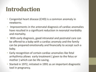
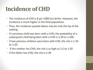
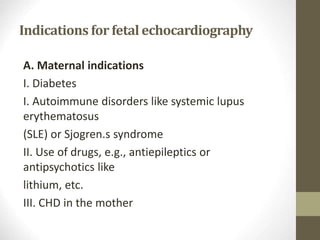
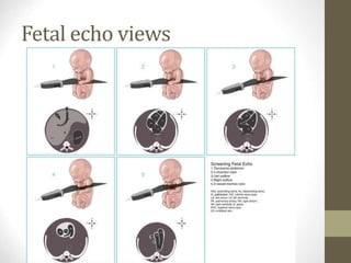
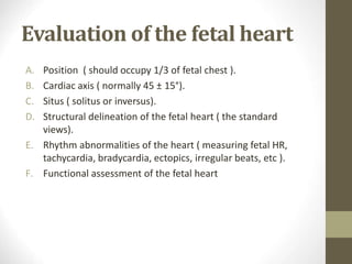
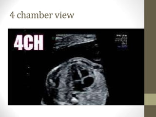
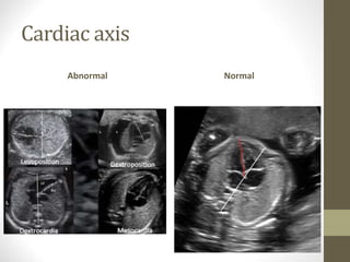
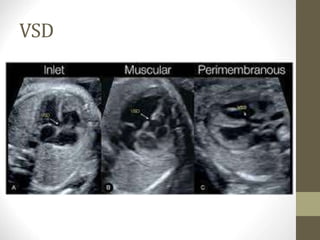
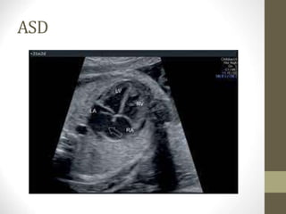
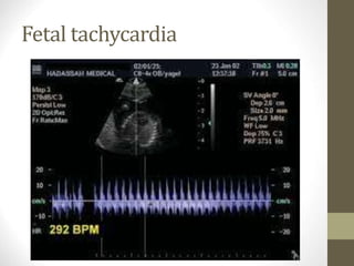
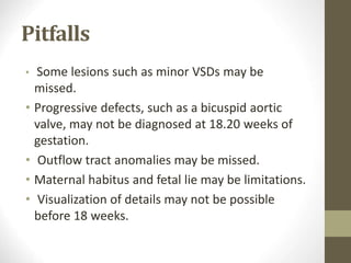
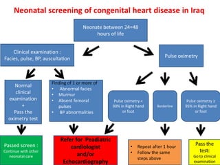
2. Introduction
• Congenital heart disease (CHD) is a common anomaly in
newborns.
• Improvements in the antenatal diagnosis of cardiac anomalies
have resulted in a significant reduction in neonatal morbidity
and mortality.
• With early diagnosis, good intranatal and postnatal care can
be offered to a baby with a cardiac anomaly and the family
can be prepared emotionally and financially to accept such a
baby.
• Early recognition of certain cardiac anomalies like fetal
arrhythmia allows early treatment ( given to the fetus or
mother ) which can be life-saving.
• Started in 1972, initiated in 1991 as an important diagnostic
tool in pregnancy.
3. Incidence of CHD
• The incidence of CHD is 8 per 1000 live births. However, the
incidence is much higher in the fetal population.
• Thus, the incidence quoted above may be only the tip of the
iceberg.
• If a previous child was born with a CHD, the probability of a
subsequent child being born with a CHD is 1:20 to 1:100.
• If two previous children were born with CHD, the risk is 1:10
to 1:20.
• If the mother has CHD, the risk is as high as 1:5 to 1:20
• If the father has CHD, the risk is 1:30
4. Indications for fetal echocardiography
A. Maternal indications
I. Diabetes
I. Autoimmune disorders like systemic lupus
erythematosus
(SLE) or Sjogren.s syndrome
II. Use of drugs, e.g., antiepileptics or
antipsychotics like
lithium, etc.
III. CHD in the mother
5. Indications for fetal echocardiography
B. Fetal indications:
I. History of a sibling with a cardiac anomaly
II. Prominent nuchal translucency or
increased nuchal fold thickness
III. Structural defect in other systems
IV. Fetal infections
V. IUGR in the mid-trimester
6. Indications for convertinga routine scan
into fetalechocardiography
I. Chamber asymmetry
II. Altered cardiac axis
III. Altered position of the fetal heart
IV. Enlarged fetal heart
V. Arrhythmia
7. Timing
• The fetal heart can be evaluated at any time during the
gestation period when USG is done.
• In the first trimester (11.14 weeks), cardiac details may
not be elicited well.
• The fetal heart can be evaluated in the third trimester,
but there are a lot of limitations due to oligoamnios and
shadowing from the fetal spine, ribs, and limbs.
• The best time to evaluate the fetal heart is between
18.22 weeks gestation ( 2nd trimester ).
10. Evaluation of the fetal heart
A. Position ( should occupy 1/3 of fetal chest ).
B. Cardiac axis ( normally 45 ± 15°).
C. Situs ( solitus or inversus).
D. Structural delineation of the fetal heart ( the standard
views).
E. Rhythm abnormalities of the heart ( measuring fetal HR,
tachycardia, bradycardia, ectopics, irregular beats, etc ).
F. Functional assessment of the fetal heart
16. Pitfalls
• Some lesions such as minor VSDs may be
missed.
• Progressive defects, such as a bicuspid aortic
valve, may not be diagnosed at 18.20 weeks of
gestation.
• Outflow tract anomalies may be missed.
• Maternal habitus and fetal lie may be limitations.
• Visualization of details may not be possible
before 18 weeks.
17. Neonatal screening of congenital heart disease in Iraq
Neonate between 24=48
hours of life
Pulse oximetry
Clinical examination :
Facies, pulse, BP, auscultation
Pulse oximetry ≥
95% in Right hand
or foot
Pulse oximetry <
90% in Right hand
or foot
Pass the
test:
Go to clinical
examination
Borderline
• Repeat after 1 hour
• Follow the same
steps above
Refer for Peadiatric
cardiologist
and/or
Echocardiography
Finding of 1 or more of
• Abnormal facies
• Murmur
• Absent femoral
pulses
• BP abnormalities
Normal
clinical
examination
+
Pass the
oximetry test
Passed screen :
Continue with other
neonatal care