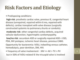
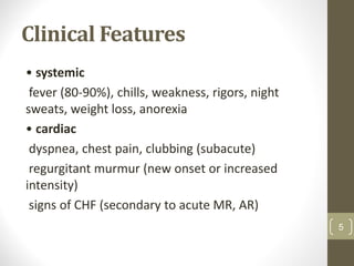
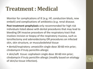
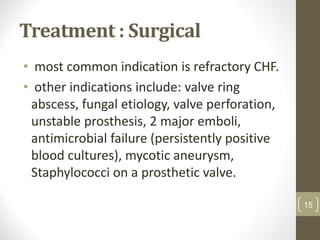
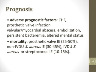
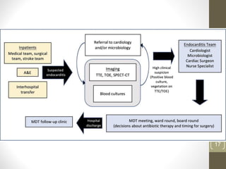
CVS lectures :
Infective
Endocarditis
Professor Ali A. Hadi Al-Saady
University of Al-Ameed / College of Medicine
Department of Medicine
Definition of Infective
Endocarditis ( IE )
• infection of cardiac endothelium, most
commonly the valves
• classifications: acute vs. subacute, native
valve vs. prosthetic valve, right sided vs. left
sided
• leaflet vegetations are made of platelet-
fibrin thrombi, WBCs, and bacteria.
2
Risk Factors and Etiology
• Predisposing conditions:
high risk: prosthetic cardiac valve, previous IE, congenital heart
disease (unrepaired, repaired within 6 mo, repaired with
defects), cardiac transplant with valve disease (surgically
constructed systemic-to pulmonary shunts or conduits).
moderate risk: other congenital cardiac defects, acquired
valvular dysfunction, hypertrophic cardiomyopathy.
low/no risk: secundum ASD or surgically repaired ASD < VSD,
PDA, MV prolapse, ischemic heart disease, previous CABG.
Opportunity for bacteremia: IVDU, indwelling venous catheter,
hemodialysis, poor dentition, DM, HIV.
• frequency of valve involvement : MV >> AV > TV > PV
but in 50% of IVDU-related IE the tricuspid valve is involved
3
4
Clinical Features
• systemic
fever (80-90%), chills, weakness, rigors, night
sweats, weight loss, anorexia
• cardiac
dyspnea, chest pain, clubbing (subacute)
regurgitant murmur (new onset or increased
intensity)
signs of CHF (secondary to acute MR, AR)
5
Clinical Features
• embolic/vascular
petechiae over legs, splinter hemorrhages (linear, reddish-
brown lesion within nail bed)
Janeway lesions (painless, 5 mm, erythematous, hemorrhagic
pustular lesions on soles/palms)
focal neurological signs (CNS emboli), headache (mycotic
aneurysm)
splenomegaly (subacute)
microscopic hematuria, flank pain (renal emboli) ± active
sediment
• immune complex
Osler’s nodes (painful, raised, red/brown, 3-15 mm on digits)
glomerulonephritis, arthritis , Roth’s spots (retinal hemorrhage
with pale centre)
6
7
Roth`s spots
8
DiagnosisofIE:ModiedDukeCriteria
Definitivediagnosisif:2major,OR1major+3minor,OR5minor
Possiblediagnosisif:1major+1minor,OR3minor
9
Investigations
• serial blood cultures: 3 sets (each containing one aerobic
and one anaerobic sample) collected from different sites
>1 h apart.
persistent bacteremia is the hallmark of endovascular
infection (such as IE).
• repeat blood cultures (at least 2 sets) after 48-72 h of
appropriate antibiotics to confirm clearance.
• blood work: CBC and differential (normochromic,
normocytic anemia), ESR (increased), RF (+), urea/Cr
• urinalysis (proteinuria, hematuria, red cell casts) and
urine C&S. 10
Investigations
ECG: prolonged PR interval may indicate perivalvular abscess
• echo findings: vegetations, regurgitation, abscess.
TTE (poor sensitivity) inadequate in 20% (obesity, COPD, chest
wall deformities).
TEE indicated if TTE is non-diagnostic in patients with at least
possible endocarditis or if suspect prosthetic valve endocarditis
or complicated endocarditis (e.g. paravalvular
abscess/perforation) (~90% sensitivity).
• TEE transesophageal echo
• TTE transthoracic echo
11
Vegetations on echocardiography
12
Treatment : Medical
• usually non-urgent and can wait for conformation of etiology
before initiating treatment unless patient is septic.
- empiric antibiotic therapy if patient is unstable; administer
ONLY after blood cultures have been taken. Generally, S. aureus,
coagulase-negative staphylococcus (CNST), and Gram-negative
coverage is important.
First line empiric treatment for native valve: vancomycin +
gentamicin OR ceftriaxone.
First line empiric treatment for prosthetic valve: vancomycin +
gentamicin + rifampin.
Targeted antibiotic therapy: antibiotic and duration (usually 4-6
wk) adjusted based on valve,organism, and susceptibilities. 13
Treatment : Medical
Monitor for complications of IE (e.g. HF, conduction block, new
emboli) and complications of antibiotics (e.g. renal disease.
Post treatment prophylaxis only recommended for high risk
individuals listed above with dental procedures that may lead to
bleeding OR invasive procedure of the respiratory tract that
involves incision or biopsy of the respiratory mucosa, such as
tonsillectomy and adenoidectomy OR procedures on infected
skin, skin structure, or musculoskeletal tissue
• dental/respiratory: amoxicillin single dose 30-60 min prior;
clindamycin if truly penicillin-allergic
• skin/soft tissue: cephalexin single dose 30-60 min prior;
clindamycin if truly penicillin-allergic (modify based on etiology
of skin/so tissue infection).
14
Treatment : Surgical
• most common indication is refractory CHF.
• other indications include: valve ring
abscess, fungal etiology, valve perforation,
unstable prosthesis, 2 major emboli,
antimicrobial failure (persistently positive
blood cultures), mycotic aneurysm,
Staphylococci on a prosthetic valve.
15
Prognosis
• adverse prognostic factors: CHF,
prosthetic valve infection,
valvular/myocardial abscess, embolization,
persistent bacteremia, altered mental status
• mortality: prosthetic valve IE (25-50%),
non-IVDU S. aureus IE (30-45%), IVDU S.
aureus or streptococcal IE (10-15%).
16
17
Infective Endocarditis.pdf

Infective Endocarditis.pdf

  • 1.
    CVS lectures : Infective Endocarditis ProfessorAli A. Hadi Al-Saady University of Al-Ameed / College of Medicine Department of Medicine
  • 2.
    Definition of Infective Endocarditis( IE ) • infection of cardiac endothelium, most commonly the valves • classifications: acute vs. subacute, native valve vs. prosthetic valve, right sided vs. left sided • leaflet vegetations are made of platelet- fibrin thrombi, WBCs, and bacteria. 2
  • 3.
    Risk Factors andEtiology • Predisposing conditions: high risk: prosthetic cardiac valve, previous IE, congenital heart disease (unrepaired, repaired within 6 mo, repaired with defects), cardiac transplant with valve disease (surgically constructed systemic-to pulmonary shunts or conduits). moderate risk: other congenital cardiac defects, acquired valvular dysfunction, hypertrophic cardiomyopathy. low/no risk: secundum ASD or surgically repaired ASD < VSD, PDA, MV prolapse, ischemic heart disease, previous CABG. Opportunity for bacteremia: IVDU, indwelling venous catheter, hemodialysis, poor dentition, DM, HIV. • frequency of valve involvement : MV >> AV > TV > PV but in 50% of IVDU-related IE the tricuspid valve is involved 3
  • 4.
  • 5.
    Clinical Features • systemic fever(80-90%), chills, weakness, rigors, night sweats, weight loss, anorexia • cardiac dyspnea, chest pain, clubbing (subacute) regurgitant murmur (new onset or increased intensity) signs of CHF (secondary to acute MR, AR) 5
  • 6.
    Clinical Features • embolic/vascular petechiaeover legs, splinter hemorrhages (linear, reddish- brown lesion within nail bed) Janeway lesions (painless, 5 mm, erythematous, hemorrhagic pustular lesions on soles/palms) focal neurological signs (CNS emboli), headache (mycotic aneurysm) splenomegaly (subacute) microscopic hematuria, flank pain (renal emboli) ± active sediment • immune complex Osler’s nodes (painful, raised, red/brown, 3-15 mm on digits) glomerulonephritis, arthritis , Roth’s spots (retinal hemorrhage with pale centre) 6
  • 7.
  • 8.
  • 9.
  • 10.
    Investigations • serial bloodcultures: 3 sets (each containing one aerobic and one anaerobic sample) collected from different sites >1 h apart. persistent bacteremia is the hallmark of endovascular infection (such as IE). • repeat blood cultures (at least 2 sets) after 48-72 h of appropriate antibiotics to confirm clearance. • blood work: CBC and differential (normochromic, normocytic anemia), ESR (increased), RF (+), urea/Cr • urinalysis (proteinuria, hematuria, red cell casts) and urine C&S. 10
  • 11.
    Investigations ECG: prolonged PRinterval may indicate perivalvular abscess • echo findings: vegetations, regurgitation, abscess. TTE (poor sensitivity) inadequate in 20% (obesity, COPD, chest wall deformities). TEE indicated if TTE is non-diagnostic in patients with at least possible endocarditis or if suspect prosthetic valve endocarditis or complicated endocarditis (e.g. paravalvular abscess/perforation) (~90% sensitivity). • TEE transesophageal echo • TTE transthoracic echo 11
  • 12.
  • 13.
    Treatment : Medical •usually non-urgent and can wait for conformation of etiology before initiating treatment unless patient is septic. - empiric antibiotic therapy if patient is unstable; administer ONLY after blood cultures have been taken. Generally, S. aureus, coagulase-negative staphylococcus (CNST), and Gram-negative coverage is important. First line empiric treatment for native valve: vancomycin + gentamicin OR ceftriaxone. First line empiric treatment for prosthetic valve: vancomycin + gentamicin + rifampin. Targeted antibiotic therapy: antibiotic and duration (usually 4-6 wk) adjusted based on valve,organism, and susceptibilities. 13
  • 14.
    Treatment : Medical Monitorfor complications of IE (e.g. HF, conduction block, new emboli) and complications of antibiotics (e.g. renal disease. Post treatment prophylaxis only recommended for high risk individuals listed above with dental procedures that may lead to bleeding OR invasive procedure of the respiratory tract that involves incision or biopsy of the respiratory mucosa, such as tonsillectomy and adenoidectomy OR procedures on infected skin, skin structure, or musculoskeletal tissue • dental/respiratory: amoxicillin single dose 30-60 min prior; clindamycin if truly penicillin-allergic • skin/soft tissue: cephalexin single dose 30-60 min prior; clindamycin if truly penicillin-allergic (modify based on etiology of skin/so tissue infection). 14
  • 15.
    Treatment : Surgical •most common indication is refractory CHF. • other indications include: valve ring abscess, fungal etiology, valve perforation, unstable prosthesis, 2 major emboli, antimicrobial failure (persistently positive blood cultures), mycotic aneurysm, Staphylococci on a prosthetic valve. 15
  • 16.
    Prognosis • adverse prognosticfactors: CHF, prosthetic valve infection, valvular/myocardial abscess, embolization, persistent bacteremia, altered mental status • mortality: prosthetic valve IE (25-50%), non-IVDU S. aureus IE (30-45%), IVDU S. aureus or streptococcal IE (10-15%). 16
  • 17.