Bangalore Call Girl Whatsapp Number 100% Complete Your Sexual Needs
Lindsay impacto
1. Fetal and Maternal Medicine Review 2004; 15:4 327–341 C 2004 Cambridge University Press
DOI: 10.1017/S0965539504001330
IMPACT OF PRENATAL DIAGNOSIS ON THE PAEDIATRIC
MANAGEMENT OF HEART DEFECTS
LINDSEY ALLAN
King’s College Hospital, London
INTRODUCTION
Ultrasound has been used in cardiac diagnosis since the 1960s. The original modality
used was M-mode, which recorded the movement of heart structures relative to a
single line of sound passed through the heart and was displayed as a paper tracing.
During the 1970s, a two-dimensional image became possible, but it was a static
image, which had limited value for cardiac evaluation. However, by the end of the
1970s, advances in ultrasound equipment allowed the heart to be displayed in real-
time. This technology was applied initially in the adult and subsequently in the
child. Echocardiography proved particularly suitable for children, partly because it is
non-invasive and repeatable, but also because most heart disease in children is due
to malformation of anatomical structure, which ultrasound can ideally display. At
the same time, obstetric ultrasound was progressing rapidly and descriptions of the
appearances of malformations in most fetal systems began to be published by the
end of the 1970s. It was not until real-time equipment became generally available in
obstetrics that the fetal heart could be satisfactorily evaluated. This led to descriptions
by several authors of normal fetal cardiac anatomy as seen echocardiographically in
1980.1,2,3
The appearances of the echocardiogram in different forms of congenital
heart disease (CHD) in children were published in the late 1970’s, setting the stage for
diagnosis in fetal life. As a result, by the mid-1980’s, most major forms of CHD had
been detected prenatally.4
At this time, detailed fetal cardiac evaluation was confined to specialists in fetal
echocardiography and to those patients selected for expert analysis because they
fell into a high-risk category for CHD. These high-risk categories included mothers
with a family history of CHD, those with maternal diabetes and in fetuses where
extracardiac malformations had been identified. However, a novel idea, which had
the potential of greatly increasing the impact of fetal echocardiography, was put
forward by a French group in 1985. Building on their nationwide network of skilled
Address for correspondence: Lindsey Allen, Consultant in Fetal Cardiology, Harris Birthright Centre for
Fetal Medicine, King’s College Hospital, Denmark Hill, London SE5 9RS, United Kingdom.
2. 328 L Allan
obstetric ultrasonographers, they suggested teaching them to incorporate a single view
of the fetal heart, namely the four chamber view, into routine obstetric scanning.
Although this single view of the heart would not detect all major structural heart
malformations, theoretically, if systematically applied, it could detect about 60%
of them.5
This introduced the concept of screening the normal pregnant population
for CHD. At the time, it appeared that about 90% of CHD occurred in low-risk
pregnancies, therefore an effective screen of the heart during the obstetric scan
enormously expanded the potential for the detection of CHD. The stimulus to
detect cardiac abnormalities prenatally is partly driven by the fact that they are
still an important cause of mortality in infancy with severe cardiac abnormalities
accounting for approximately 20% of neonatal deaths.6
In a significant proportion of
these cases the cardiac defect is not recognised during life, but found on post mortem
examination.7
As the skill of the obstetric ultrasonographer and the image quality of ultrasound
equipment has improved over the years, great artery assessment has been added to
the four chamber view during routine examination, such that over 90% of major
CHD is potentially detectable in skilled hands. The reality is, however, that success
of detection of cardiac anomalies varies8,9,10
widely and tends to be linked to the
standard and intensity of more general ultrasound screening. Thus, where a routine
fetal anomaly scan, applied to all pregnancies and performed to a high specification,
is in place, detection of all anomalies, including heart defects, is high. However,
the use of ultrasound during pregnancy varies widely nationally and geographically
and even between neighbouring obstetric units, although overall detection of cardiac
anomalies of all kinds has shown steady improvement over the 25 years that fetal
echocardiography has been possible. As the four-chamber view is technically easier to
obtain and analyse, defects which alter the appearance of this view, are preferentially
detected prenatally. This was clearly shown in a UK-wide study published in 1999,11
of
all cases of CHD found over a 3 year period. In the cases detected in fetal life, diagnoses
such as the hypoplastic left heart syndrome, univentricular heart and atrioventricular
septal defect predominated, in contrast to the postnatal series where arch anomalies,
tetralogy of Fallot and ventricular septal defects predominated.
The detection of a possible cardiac abnormality during the routine scan allows
the patient to be referred to the fetal cardiologist for more detailed diagnosis and
counselling. The ideal fetal cardiologist has skill in obstetric imaging, a thorough
understanding of the fetal spectrum of CHD, coupled with up-to-date knowledge of
local and international results of the management of different types of CHD, both
immediate and long-term, in order to offer the detailed counselling which the parents
need when a cardiac malformation is found in the fetus.
The advent in the mid-1990s of screening for nuchal translucency (NT) at
12 weeks gestation was a further significant advance in the fetal echocardiographic
field.12,13
Nuchal translucency detected a significant group of pregnancies at high-
risk of CHD, which had not been identified as such before. It was shown that
if the NT measurement was above the 95th centile, there was an association not
only with chromosomal defects, which was the primary aim of nuchal translucency
3. Impact of prenatal diagnosis on the paediatric management of heart defects 329
Table 1 Incidence of CHD per 1000, with increasing NT
0
20
40
60
80
100
120
140
160
180
200
2.5-3.5 3.5-4.5 4.5-5.5 5.5-6.5 >6.5
measurement, but also with CHD. The incidence of CHD increases with increased
levels of NT. In a recent analysis of our own data, there was a rate of CHD at twice
normal if the NT was between the 95–99th centile (about 2.6–3.5 mm) increasing to
twenty-six times the normal rate if the NT was over 6.5 mm (Table 1). The group
of patients with NT > 95th centile is probably the most useful high-risk group
for selection for detailed fetal echocardiography. Interestingly, increased NT is not
associated with any particular form of CHD. Table 2 shows a recent analysis of the
type of CHD seen in 123 cases with increased NT and normal chromosomes, where a
similar scatter of CHD was seen to the spectrum which might be expected in infants.
Coarctation was slightly over-represented in the fetus but some of these cases are
likely to have progressed to the hypoplastic left heart syndrome by birth. There has
been some debate about the incidence of increased NT in those with CHD, with rates
of 17% and 56% found in different studies.14,15
The real figure is probably around
25%.
The incidence of CHD is about 8/1000 livebirths. About 4–5 of these 8 children have
ventricular septal defects, secundum atrial septal defects, mild pulmonary stenosis or
persistent arterial ducts. With the exception of moderate to large ventricular septal
defects, none of these lesions are detectable in the fetus. However, all these lesions
can be readily treated surgically or by catheter techniques, or even may not require
intervention at all, and are compatible with a normal quality of life and life-span. In
contrast, about 3/8 cases of CHD per 1000 are major forms of CHD, which nearly all
require surgical treatment, and sometimes repeated intervention, at a varying risk of
mortality and morbidity, depending on the type of defect. These forms of CHD can
4. 330 L Allan
Table 2 Types of CHD seen in a series of 123 cases with increased NT and normal chromosomes.
DIV = double inlet ventricle, HLH = hypoplastic left heart syndrome, DORV = double outlet right ventricle,
VSD = ventricular septal defect, TOF = tetralogy of Fallot, AVSD = atrioventricular septal defect.
0 5 10 15 20 25
Pulmonary atresia
Mitral atresia
Ebsteins
Isomerism
Aortic stenosis
Tricuspid atresia
DIV
HLH
DORV
Transposition
VSD
TOF
AVSD
Coarctation
severely impact the child’s life and, even if successfully treated, will nearly all impair
the quality of life and/or shorten the lifespan. It is this severe spectrum of disease that
is typically detected in fetal life.
TERMINATION OF PREGNANCY
The detection of CHD during pregnancy offers the patient the possibility of
termination of pregnancy. In the UK, prior to 24 weeks gestation, this course of
action may be chosen legally for fetal anomalies. However, in the setting of CHD,
termination is more likely to be chosen by the parents when there are associated
extracardiac anomalies and/or chromosomal anomalies, or where the complexity
of the heart malformation is such that a poor outlook is anticipated. In addition,
in general, the earlier the diagnosis is made of fetal abnormality, the more likely
termination is to be chosen. Thus, the more widespread application of nuchal
translucency screening has led to earlier diagnosis of CHD, even as early as 12 weeks
gestation, which in turn is likely to lead to a higher rate of pregnancy interruption for
CHD in the future. Termination of pregnancy after 24 weeks gestation is confined
to the most severe forms of CHD or those with multiple malformations and is
uncommon.
5. Impact of prenatal diagnosis on the paediatric management of heart defects 331
As a result of increasing detection of major CHD prenatally, the prevalence of
some major forms of heart malformation has been shown to have fallen as a result
of termination.16
In a study of all cases of pulmonary atresia with intact ventricular
septum seen in the UK between 1991 and 1995, the prevalence in postnatal life was
shown to have fallen from 5.6 to 4.1 in England because of prenatal detection and
termination of pregnancy. It is likely that this effect may increase in the future as
prenatal detection of CHD improves, reducing the number of the most complex cases
of CHD coming to the paediatric cardiologist. However, counterbalancing this is
the fact that as the prognosis for surgical repair of many heart defects continues to
improve, the counsellor is able to be more optimistic about the results and termination
may be chosen less often.
PROGRESSION OF CHD
One of the most interesting observations in fetal cardiology has been that some forms
of CHD progress in fetal life. This has been seen in both aortic and pulmonary stenosis
where the valve appeared echocardiographically normal in early pregnancy but became
stenosed later, or where the degree of stenosis has progressed from mild to severe or
even to atresia during gestation. In pulmonary stenosis, this can occur in isolated
valve disease or in more complex CHD where pulmonary stenosis is a component.
Thus, for example, in tetralogy of Fallot, transposition with ventricular septal defect
or double outlet right ventricle, the pulmonary valve obstruction can increase during
pregnancy to pulmonary atresia and thus duct dependency after birth.17
In some cases
of critical aortic stenosis, the left ventricle is usually dilated in early pregnancy but it
fails to keep pace with normal growth resulting in a small left ventricle by term, with
quite different implications for surgical management.18
In coarctation, poor growth of
the aortic arch can occur, such that an extended arch repair through a sternotomy is
necessary after birth, with a higher morbidity and mortality, rather than the simpler
surgical approach of a lateral thoractomy. Valvar incompetence can also progress in
utero, particularly in the tricuspid valve, leading to hydrops and even intrauterine
death. In addition, there are some cardiac lesions which are not usually evident early
in pregnancy but which evolve towards term. Examples include cardiac tumours and
cardiomyopathies. Thus, although a normal fetal cardiac scan in competent hands
at 20 weeks can exclude most major heart disease, there are forms of major disease
which progress from milder disease or may develop in later pregnancy. However, such
examples are rare.
FETAL THERAPY
There are some fetal conditions where prenatal treatment is appropriate. In fetal
tachycardias, the fetus can be treated by maternal anti-arrhythmic drugs which
cross the placenta. Treatment can prevent the development of cardiac failure, if the
tachycardia is detected soon after its initiation. Once the fetus is in cardiac failure
6. 332 L Allan
RV
LV
spine
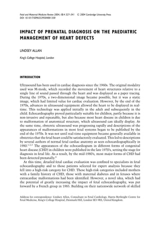
Figure 1 The left ventricle is dilated, globular in shape and poorly contracting in the moving image. This
appearance is typically seen associated with a critically obstructed aortic valve, which is amenable to prenatal
balloon valvoplasty. LV = left ventricle, RV = right ventricle.
due to a tachycardia, the fetus is at considerable risk of mortality and treatment can
be life-saving.19
Although these cases are uncommon, treatment is well-established
in this context.
Prenatal balloon valvoplasty was first suggested at the end of the 1980s for
conditions where an isolated obstructive lesion was present, such as aortic stenosis
(Figure 1). It was observed that as pregnancy advanced in a fetus with a critically
obstructed aortic valve, the left ventricle either failed to grow sufficiently or became
irreparably damaged by endocardial fibroelastosis. At this time, there was no surgical
treatment available for a hypoplastic or inadequate left ventricle, and thus these cases
were associated with a high mortality after birth. The rationale for prenatal treatment
was to relieve valvar obstruction sufficiently early in pregnancy to avoid the secondary
effects of the obstructed valve on the left ventricle and give it “recovery time” prior
to birth, while the circulation was also supported by the right ventricle. The left
ventricular apex was punctured directly using a needle through which a guide wire
7. Impact of prenatal diagnosis on the paediatric management of heart defects 333
and subsequent balloon catheter could be passed. It was then positioned and inflated
in the valve orifice. There was limited success in the early cases and some difficulties
with balloon technology,20
while at the same time, treatment strategies in the form
of the Norwood operation for the hypoplastic left heart syndrome began to achieve
better results21
and thus the technique fell out of use. Interest has been rekindled
more recently, partly because of improved technology, and partly in an attempt to
avoid one ventricle repair on either the right or left sides of the heart.22,23
It remains
to be seen whether this technique will find an established role in Fetal/Paediatric
Cardiology practise.
DELIVERY OF A FETUS WITH CHD
Depending on the type of defect, presentation in CHD varies from acute
decompensation soon after birth to gradual deterioration over the first months of
life, with all stages in between. Examples of conditions which can cause symptoms
immediately after birth include severe Ebstein’s malformation and tricuspid dysplasia,
transposition of the great arteries and total anomalous pulmonary venous drainage.
Ebstein’s malformation is an anomaly of the tricuspid valve in which the orifice
of the tricuspid valve is displaced into the right ventricle. With a mild degree of
displacement, the neonate is likely to be asymptomatic but with more severe degrees,
especially if there is severe tricuspid regurgitation, postnatal ventilation can be
difficult due to lung hypoplasia. This, in turn, is due to compression of the fetal
lung fields by cardiomegaly. If the neonate can be supported through the early period
and there is sufficient lung volume, tricuspid regurgitation tends to lessen as the
pulmonary pressure falls in the first weeks of life. However, some extreme examples
do not survive this early period despite maximum support.
In the majority of cases of transposition of the great arteries (Figure 2), the neonate
is noticed to be cyanosed in the first days of life but about 20% present more
acutely, becoming hypoxic and acidotic within even an hour of birth. This subgroup of
cases have a restrictive or intact atrial septum, preventing oxygenated blood reaching
the systemic circulation, resulting in rapid deterioration and even death before the
neonate can reach a cardiology centre. These infants can be successfully treated by
ultrasound-guided tearing of the atrial septum by a balloon catheter, if they reach help
in time.
In total anomalous pulmonary venous drainage, if the pulmonary veins drain to an
obstructed confluence, which can occur with both supra and infracardiac drainage,
the neonate can become severely cyanosed and require urgent surgical relief. In
transposition and total anomalous pulmonary venous drainage, maintaining ductal
patency is not usually of much benefit. However, there is a group of patients who are
termed “duct-dependent” and who do benefit from maintaining the duct open using
prostaglandins. In these cases, depending on the cardiac defect, either the pulmonary
or systemic circulation is dependent on the patency of the arterial duct. The arterial
duct naturally starts to close some hours or days after birth but can do so acutely, either
causing severe cyanosis (if the pulmonary blood flow is affected) or systemic collapse
8. 334 L Allan
RV Ao
LV
PA
Figure 2 The great arteries do not show their usual spiral relationship but arise in parallel orientation.
This is characteristic of transposition of the great arteries. In this condition, delivery where immediate
cardiological care can be instituted can improve the outcome. LV = left ventricle, RV = right ventricle,
Ao = aorta, PA = pulmonary artery.
(if the aortic blood flow is affected). Conditions where the pulmonary circulation is
duct dependent include severe pulmonary stenosis or atresia, which can be isolated or
be a component of different forms of intracardiac malformation such as tetralogy of
Fallot (Figure 3), or double outlet right ventricle. Conditions where the systemic
circulation is duct dependent include aortic stenosis or atresia (Figure 4), and
interruption or coarctation of the aortic arch. In contrast, many conditions, such as
an atrioventricular septal defect, for example, are associated with the gradual devel-
opment of cardiac failure over the first 2–4 months of life and do not present urgently
(Figure 5).
From the time of the earliest reports of fetal cardiac diagnosis, it has been clear
that the outcome for the fetus with CHD is less good than would be expected in
postnatal life.24,25
This is because of the bias towards detecting the most severe
CHD prenatally, due to the way patients are selected for fetal echocardiography.
9. Impact of prenatal diagnosis on the paediatric management of heart defects 335
LV
RV
<Ao
RV
PA>
<Ao
spine
Figure 3 In the left panel, there is a ventricular septal defect with the aorta overriding the crest of the
ventricular septum. The scan plane immediately above this level is shown in the right hand panel, where the
pulmonary artery is seen crossing over the aortic origin in a normal fashion. However, the pulmonary artery
is smaller than the aorta, which is the reverse of normal. This finding in association with aortic override,
indicates a diagnosis of tetralogy of Fallot. Some cases of tetralogy of Fallot are duct dependent and should
benefit from prenatal diagnosis.
Those fetuses with gross cardiac abnormalities detected on screening and those
with extracardiac anomalies or hydrops are preferentially referred. There is a higher
incidence of chromosomal anomalies in fetal series than in live-births.25
There is
a small but fairly consistent incidence of spontaneous intrauterine death of between
5–10% in fetal series, partly due to the presence of associated chromosomal anomalies
but not entirely. Some fetuses with CHD die in utero for no apparent reason. The
outcome after the diagnosis of CHD in a recent series of 956 cases is shown in Table 3.
A high rate of termination reflects our high incidence of chromosomal anomalies, as
many of our cases of CHD come from the NT screening programme, but there is also
an incidence of spontaneous intrauterine death and neonatal loss despite maximal
treatment.
In a series of cases of the hypoplastic left heart syndrome diagnosed in fetal life, the
mortality in the fetally diagnosed cases was almost twice that of those submitted to
first stage Norwood surgery in the same institution.26
This could be accounted for by
chromosomal anomalies, other extracadiac malformations or extreme prematurity in
the fetal “intention-to-treat” group, all of which were less common in the postnatal
group, or such postnatal patients were not submitted for surgery and therefore not
counted in the surgical series. In addition, there were some cases of a restrictive
atrial septum in the fetal series who decompensated so acutely after birth, that they
would have died before reaching the surgical centre, had they not been prenatally
diagnosed.
There is no evidence that early or operative delivery in CHD will improve
outcome, although both might be considered in the rare cases where cardiac function
deteriorates in late gestation. Early delivery might be considered in the setting of
10. 336 L Allan
RA
RV
LA
Figure 4 In this four chamber view, the mitral valve is atretic and the left ventricle hypoplastic. This
was a case of the hypoplastic left heart syndrome. In such a fetus the systemic perfusion is dependent on
the patency of the arterial duct which naturally closes within hours or days of birth, sometimes acutely. If
the neonate is not treated urgently with prostaglandins, irreparable damage can occur to the kidneys and
liver. Thus, this cardiac condition would be expected to benefit from prenatal detection and delivery in a
specialised centre.
increasing hydrops in CHD or in a fetal tachycardia resistant to therapy but the
disadvantages of prematurity, and lung immaturity in particular, must be weighed
against any possible advantage. However, it seems intuitively obvious that those cases
where early or urgent cardiological treatment is required must benefit from prenatal
diagnosis, by being delivered at, or close to, the site of cardiological care. This is
particularly relevant in settings where the specialised cardiac centre is geographically
remote, as intrauterine transfer is much more stable than the transfer of a sick
11. Impact of prenatal diagnosis on the paediatric management of heart defects 337
RV
LV
<
<
Figure 5 This four-chamber view shows a defect at the crux of the heart, an atrioventricular septal defect.
Such a malformation will not cause immediate symptoms in the newborn, so can be safely delivered locally.
neonate. Although many authors have tried to prove the benefit of planned delivery
after prenatal diagnosis, it has been difficult to show for various reasons. In many
cases, this is because the numbers of prenatally diagnosed cases are not sufficient
for statistical analysis.27,28
Some authors have looked at mixed diagnostic groups,
with the more severe lesions predominating in the fetal series.29
Also, in general,
this question cannot be addressed by a tertiary referral centre, as they do not usually
have a method to ascertain cases who have died in the periphery prior to referral.
Had they been included, such cases would have had the effect of increasing the
mortality in the postnatally diagnosed cases, but they are not usually recognised.
Deaths from unrecognised CHD occurred in up to 15% of cases of the hypoplastic
left heart syndrome in one reported series.7
Subsets of cases of the hypoplastic left
heart syndrome which are at especially high risk of early death, such as those with
a restrictive atrial septum are over-represented in the fetal series, supporting the
concept that these cases would not have reached the referral centre had they not been
detected prenatally. The only series which was sufficiently large to analyse and was
population based and therefore ascertained all cases in a geographical area, whether
12. 338 L Allan
Table 3 Outcome of 956 cases of fetal CHD evaluated between 1998 and 2004. TOP = termination of
pregnancy, IUD = intrauterine death, NND = neonatal death, InfD = infant death, uk = unknown or presently
continuing pregnancies
0
100
200
300
400
500
600
TOP IUD Alive NND InfD uk
they reached the referral centre or not, was that of Bonnet et al.30
This was a ten
year study of a single diagnostic category, transposition of the great arteries, and
was based in the Paris area of France. This showed a clear advantage for the cases
prenatally diagnosed with no mortality in this group, as compared to an 8% mortality
prior to surgery, plus an 8% mortality after surgery in the postnatally diagnosed
group. More recently, similar results were reported in a study of neonates with
coarctation of the aorta, demonstrating that the risk of postnatal death was decreased
in the prenatally diagnosed group and that they were more haemodynamically stable
preoperatively.31
Clearly, mortality is rather a crude outcome measure and long-term morbidity in
infants with CHD should be explored in more detail. In all the studies which have
looked at prenatal versus postnatal diagnosis in terms of outcome, the prenatally
diagnosed cases have been haemodynamically more stable in the immediate postnatal
period than those postnatally diagnosed.32
This would be expected to translate into a
better neurological result in the long-term for those prenatally diagnosed. A number
of studies have shown that after staged surgical reconstruction for hypoplastic left
heart there is a major risk of long-term neurodisability.33,34,35
This risk appears to be
improving with advancing era36
but such data still gives cause for concern. There
is clear evidence to show a correlation between length of stay in the intensive
care unit and IQ.37
In a significant number of neonates requiring intervention,
prenatal diagnosis reduces the time in the intensive care unit and this in turn
may influence long-term morbidity and neurological outcome. Only large studies,
evaluating neurological development in children 5–15 years after treatment for CHD
will prove this point and these are notoriously hard to perform.
13. Impact of prenatal diagnosis on the paediatric management of heart defects 339
SUMMARY
Prenatal diagnosis of CHD disease has influenced the practise of Paediatric Cardiology
in a variety of ways and is likely to have an increasing effect in the future. Fetal
echocardiography now constitutes a significant proportion of the work of a specialised
Paediatric Cardiology unit. Early diagnosis and termination of pregnancy may reduce
the prevalence of complex disease in postnatal series. At the present time, at least
a third of neonates presenting to most units will have been identified in fetal life and
this figure is likely to increase as routine screening continues to improve, and nuchal
translucency screening becomes more widely adopted. Planning the delivery where
skilled and experienced cardiac support can be initiated immediately, and intervention
performed where appropriate, will potentially salvage those with urgently presenting
or duct dependent lesions. Long-term morbidity associated with the repair of CHD in
infancy is likely to improve as a result of ideal perinatal management.
REFERENCES
1 Lange LW, Sahn DJ, Allen HD, Goldberg SJ, Anderson C, Giles H. Qualitative real-time cross-sectional
echocardiographic imaging of the human fetus during the second half of pregnancy. Circulation 1980;
62: 799–806.
2 Kleinman CS, Hobbins JC, Jaffe CC, Lynch DC, Talner NS. Echocardiographic studies of the human
fetus: prenatal diagnosis of congenital heart disease and cardiac dysrhythmias. Pediatrics 1980; 65:
1059–67.
3 Allan LD, Tynan MJ, Campbell S, Wilkinson J, Anderson RH. Echocardiographic and anatomical
correlates in the fetus. Br Heart J 1980; 44: 444–51.
4 Allan LD, Crawford DC, Anderson RH, Tynan MJ. Echocardiographic and anatomical correlations in
fetal congenital heart disease. Br Heart J 1984; 52: 542–48.
5 Allan LD, Crawford DC, Chita SK, Tynan MJ. Prenatal screening for congenital heart disease. Br Med
J 1986; 292: 1717–719.
6 Young ID, Clarke M. Lethal malformations and perinatal mortality: A ten year review with comparison
of ethnic differences. Br Med J 1987; 295: 88–91.
7 Abu-Harb M, Hey E, Wren C. Death in infancy from unrecognised congenital heart disease. Arch Dis
Child 1994; 71: 3–7.
8 Sharland GK, Allan LD. Screening for congenital heart disease prenatally. Results of a 2 1/2-year study
in the South East Thames Region. Br J Obstet Gynaecol 1992; 99: 220–25.
9 Tegnander E, Eik-Nes SH, Linker DT. Incorporating the four-chamber view of the fetal heart into the
second-trimester routine fetal examination. Ultrasound Obstet Gynecol 1994; 4: 24–28.
10 Garne E, Stoll C, Clementi M. Euroscan Group. Evaluation of prenatal diagnosis of congenital heart
diseases by ultrasound: experience from 20 European registries. Ultrasound Obstet Gynecol 2001; 17:
386–91.
11 Bull C. On behalf of the British Paediatric Cardiac Association. Current and potential impact of fetal
diagnosis on the prevalence and spectrum of serious congenital heart disease at term. Lancet 1999;
354: 1242–247.
12 Snijders RJ, Johnson S, Sebire NJ, Noble PL, Nicolaides KH. First-trimester ultrasound screening for
chromosomal defects. Ultrasound Obstet Gynecol 1996; 7: 216–26.
13 Hyett JA, Perdu M, Sharland GK, Snijders RS, Nicolaides KH. Increased nuchal translucency at
10–14 weeks of gestation as a marker for major cardiac defects. Ultrasound Obstet Gynecol 1997;
10: 242–46.
14. 340 L Allan
14 Mavrides E, Cobian-Sanchez F, Tekay A, Moscoso G, Campbell S, Thilaganathan B, Carvalho JS.
Limitations of using first-trimester nuchal translucency measurement in routine screening for major
congenital heart defects. Ultrasound Obstet Gynecol 2001; 17: 106–10.
15 Hyett J, Perdu M, Sharland G, Snijders R, Nicolaides KH. Using fetal nuchal translucency to screen for
major congenital cardiac defects at 10–14 weeks of gestation: population based cohort study. Br Med J
1999; 318: 81–85.
16 Daubeney PE, Sharland GK, Cook AC, Keeton BR, Anderson RH, Webber SA. Pulmonary atresia
with intact ventricular septum: impact of fetal echocardiography on incidence at birth and postnatal
outcome. UK and Eire collaborative study of pulmonary atresia with intact ventricular septum.
Circulation 1998; 98: 562–66.
17 Allan LD, Sharland GS, Tynan M. Natural history of hypoplastic left heart syndrome. Int J Cardiol
1989; 25: 341–43.
18 Hornberger LK, Sanders SP, Sahn DJ, Rice MJ, Spevak PJ, Benacerraf BR et al. In utero pulmonary artery
and aortic growth and potential for progression of pulmonary outflow tract obstruction in tetralogy of
Fallot. J Am Coll Cardiol 1995; 25: 739–45.
19 Maxwell DJ, Crawford DC, Curry PVM, Tynan MJ, Allan LD. Obstetric importance, diagnosis and
management of fetal tachycardias. Br Med J 1988; 297: 107–10.
20 Maxwell DJ, Allan LD, Tynan MJ. Balloon dilatation of the aortic valve in the fetus. Br Heart J 1991;
65: 256–58.
21 Bu’Lock FA, Stumper O, Jagtap R, Silove ED, DeGiovanni JW, Wright JG et al. Surgery for infants with
a hypoplastic systemic ventricle and severe outflow tract obstruction: early results form a modified
Norwood procedure. Br Heart J 1995; 73: 456–61.
22 Tulzer G, Arzt W, Franklin RCG, Loughna PV, Mair R, Gardiner HM. Fetal pulmonary valvuloplasty
for critical pulmonary stenosis or atresia with intact septum. Lancet 2002; 360: 1567–568.
23 Tworetzky W, Marshall AC. Balloon valvuloplasty for congenital heart disease in the fetus. Clin
Perinatol 2003; 30: 541–50.
24 Paladini D, Russo M, Teodoro A, Pacileo G, Capozzi G, Martinelli P et al. Prenatal diagnosis of
congenital heart disease in the Naples area during the years 1994–1999 – the experience of a joint
fetal-pediatric cardiology unit. Prenat Diagn 2002; 22: 545–52.
25 Allan LD, Sharland GK, Milburn A, Lockhart SM, Groves AM, Anderson RH et al. Prospective
diagnosis of 1006 consecutive cases of congenital heart disease in the fetus. J Am Coll Cardiol 1994;
23: 1452–458.
26 Allan LD, Apfel HD, Printz BF. Outcome after prenatal diagnosis of hypoplastic left heart syndrome.
Heart 1998; 79: 371–73.
27 Chang AC, Huhta JC, Yoon GC, Wood DG, Tulzer G, Cohen et al. Diagnosis, transport and outcome
in fetuses with left ventricular outflow tract obstruction. J Thoac Cardovasc Surg 1991; 102: 841–48.
28 Kumar RK, Newburger JW, Gauvreau K, Kamenir SA, Hamberger LK. Comparison of outcome when
hypoplastic left heart syndrome and transposition of the great arteries are diagnosed prenatally versus
when diagnosis of these two conditions is only made postnatally. Am J Cardiol 1999; 83: 1649–653.
29 Copel JA, Tan AS, Kleinmann CS. Does prenatal diagnosis of congenital heart disease alter short term
outcome?Ultrasound Obstet Gynaecol 1997; 10: 237–41.
30 Bonnet D, Coltri A, Butera G, Fermont L, Bidois J, Kachaner J, Sidi D. Detection of transposition of
the great arteries in fetuses reduces neonatal morbidity and mortality. Circulation 1999; 99: 916–18.
31 Franklin O, Burch M, Manning N, Sleeman K, Gould S, Archer N. Prenatal diagnosis of coarctation of
the aorta improves survival and reduces morbidity. Heart 2002; 87: 67–69.
32 Verheijen PM, Lisowski LA, Stoutenbeek P, Hitchcock JF, Bennink GB, Meijboom EJ. Lactacidosis
in the neonate is minimized by prenatal detection of congenital heart disease. Ultrasound Obstet
Gynecol 2002; 19: 552–55.
33 Rogers BT, Msall ME, Buck GM, Lyon NR, Norris MK, Roland JM et al. Neurodevelopmental outcome
in infants with hypoplastic left heart syndrome. J Pediatr 1995; 126: 496–98.
34 Kern JH, Hayes CJ, Michler RE, Gersony WM, Quaegebeur JM. Survival and risk factor analysis for
the Norwood procedure for the hypoplastic left heart syndrome. Am J Cardiol 1997; 80: 170–74.
15. Impact of prenatal diagnosis on the paediatric management of heart defects 341
35 Mahle WT, Clancy RR, McGaurn SP, Goin JE, Clark BJ. Impact of prenatal diagnosis on survival and
early neurological morbidity in neonates with the hypoplastic left heart syndrome. Pediatrics 2001;
107: 1277–282.
36 Goldberg CS, Schwartz EM, Brunberg JA, Mosca RS, Bove EL, Schork MA et al. Neurodevelopmental
outcome of patients after the Fontan operation: a comparison between children with hypoplastic left
heart syndrome and other single functional ventricle lesions. J Pediatrics 2000; 137: 646–52.
37 Newburger JW, Wypij D, Bellinger DC, du Plessi AJ, Kuban KC, Rappaport LA et al. Length of stay
after infant heart surgery is related to cognitive outcome at age 8 years. J Pediatr 2003; 143: 67–73.