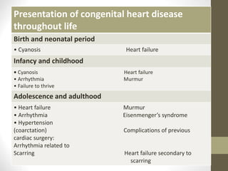
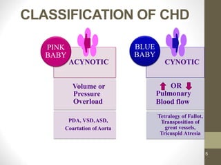
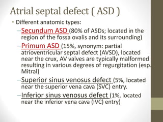
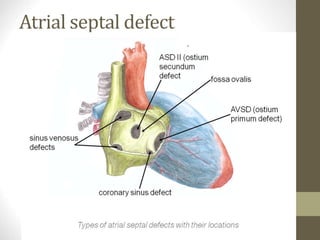
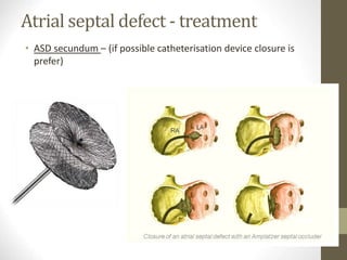
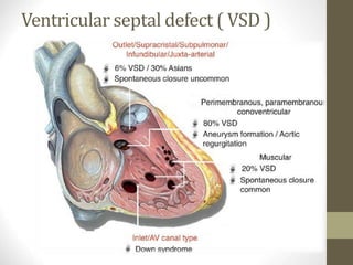
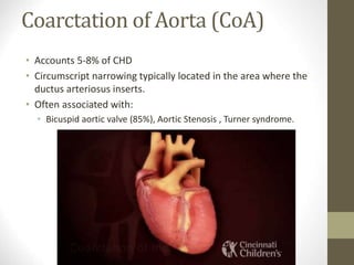
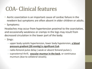
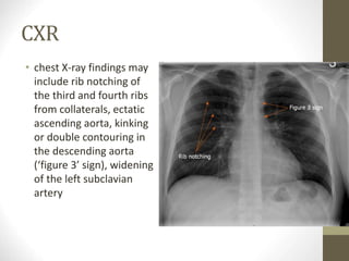
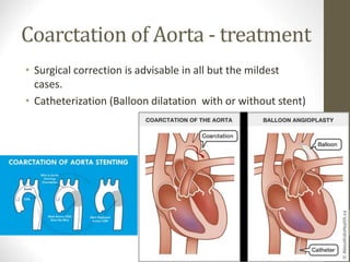
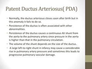
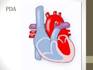
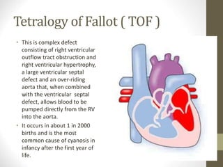
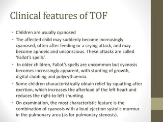
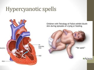
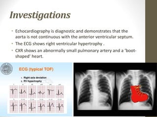
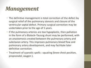
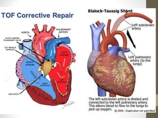
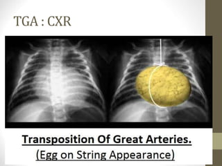
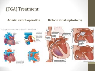
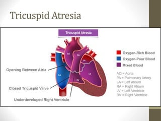
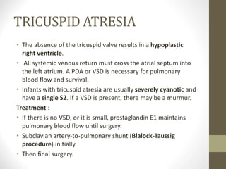
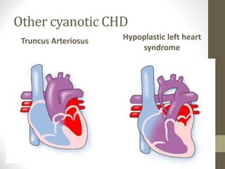
This document provides information on congenital heart disease (CHD), including epidemiology, classification, diagnosis, and treatment. It discusses several specific types of CHD such as atrial septal defect (ASD), ventricular septal defect (VSD), patent ductus arteriosus (PDA), coarctation of the aorta, tetralogy of Fallot, transposition of the great arteries, tricuspid atresia, truncus arteriosus, and hypoplastic left heart syndrome. It describes the clinical presentation, investigations, and management approaches for each condition.
This document provides an overview of basics of ECG, including:
- A brief history of ECG development from 1842 to present day.
- An explanation of what an ECG measures and how it can be used to identify arrhythmias, ischemia, infarction and other cardiac conditions.
- A breakdown of the components of a normal ECG waveform including the P wave, PR interval, QRS complex, ST segment, and T wave.
- Descriptions of the 12-lead ECG system and how each lead views electrical activity from different angles in the heart.
- Explanations of how to analyze an ECG, including determining heart rate and cardiac axis. Bradyarrhythm
Congenital heart diseases are structural abnormalities present at birth that affect the normal formation and function of the heart. The most common types are ventricular septal defect (VSD), atrial septal defect (ASD), patent ductus arteriosus (PDA), tetralogy of Fallot, pulmonary stenosis, coarctation of the aorta, and transposition of the great arteries (TGA). Symptoms range from none to signs of heart failure. Treatment options include medical management, catheter-based procedures such as angioplasty or device closure, and open heart surgery to repair or palliate the defects. Prognosis depends on the specific type of congenital heart disease and whether complete repair is possible.
1. The document discusses different types of arrhythmias including tachycardia and bradycardia. It describes various causes of arrhythmias including functional, organic, hemodynamic, hormonal and congenital factors.
2. Types of arrhythmias are classified based on location in the heart as atrial, junctional, or ventricular. Common atrial arrhythmias include premature atrial contractions and atrial fibrillation.
3. Supraventricular tachycardia is a junctional arrhythmia and the most common type is atrioventricular nodal reentrant tachycardia. Symptoms can include a pounding heart, shortness of breath, chest pain and d
The document discusses congenital heart diseases, which occur in approximately 1% of live births. It describes several types of congenital heart defects including atrial septal defect (ASD), ventricular septal defect (VSD), atrioventricular canal defect, and patent ductus arteriosus - all of which involve increased pulmonary blood flow. It also discusses obstructive defects like aortic stenosis and pulmonary stenosis. The document provides details on the pathophysiology, clinical manifestations, diagnosis, and treatment of these various congenital heart conditions.
This document discusses aortic valve stenosis, including its anatomy, function, causes, pathophysiology, classification, signs and symptoms, diagnostic evaluation, and treatment options. Key points include:
- Aortic stenosis is caused by calcification and stiffening of the aortic valve leaflets, restricting their opening and increasing the pressure gradient between the left ventricle and aorta.
- It can be congenital due to a bicuspid aortic valve or acquired from degeneration, rheumatic fever, or radiation exposure.
- Symptoms include angina, syncope, and dyspnea as the left ventricle hypertrophies and diastolic function declines in response to the increased
This document provides guidelines for managing adult congenital heart disease (ACHD). It discusses how ACHD involves a range of cardiac abnormalities present at birth, excluding inherited disorders. When ACHD involves acquired disease, the focus is on differences from that disease due to anatomy, other lesions, or patient age. Survival of severe CHD to adulthood is now over 90%, but ACHD patients are heterogeneous in anatomy, physiology, and surgical history. The document presents a classification system to comprehensively assess ACHD severity based on anatomy and physiology.
This document discusses atrioventricular septal defects (AVSDs), including their embryogenesis, classification, clinical features, imaging, and management. It describes the spectrum of AVSDs from partial to complete. Partial AVSDs involve a primum atrial septal defect with a cleft in the mitral valve. Complete AVSDs have a large ventricular septal defect with a common atrioventricular valve. Imaging like echocardiography is important for evaluating the anatomy and determining appropriate treatment, which ranges from observation to surgical repair.
Ventricular tachycardia (VT) is an arrhythmia originating in the ventricles with a heart rate over 100 beats per minute and wide QRS complexes of at least 120 ms. VT can be either idiopathic or structural, sustained or non-sustained, and monomorphic or polymorphic. The ECG can diagnose VT based on the wide QRS complexes. VT has subtypes including bundle branch reentry VT and idiopathic monomorphic VT. Treatment options include medical therapies like amiodarone, implantable cardioverter defibrillators based on major trials, and catheter ablation.
Total anomalous pulmonary venous connections seminar ppt.Pawan Ola
This document provides information on total anomalous pulmonary venous connection (TAPVC). It defines TAPVC as a condition where the pulmonary veins drain anomalously into the right atrium or systemic veins rather than the left atrium. The document discusses the history, classification, embryology, clinical features, investigations and management of TAPVC. It describes the different types of TAPVC based on the site of drainage and presence of obstruction. Echocardiography is highlighted as the main diagnostic tool to identify the anomalous connections and assess for obstruction. The clinical presentation and hemodynamics vary depending on the type and presence of obstruction.
This document discusses three types of dextrocardia:
1. Situs inversus totalis, where the organs are mirrored and the cardiac apex is located on the right side.
2. Dextroversion with situs solitus, where the ventricles can rotate to the right side and defects like VSD and pulmonary stenosis are common.
3. Dextroposition, where external factors like lung hypoplasia cause the heart to be displaced to the right side, and defects causing left-to-right shunts often occur.
Diagnosis involves inspection of the precordium, auscultation, chest X-ray, and echocardiogram. Situs
The document discusses various types of congenital heart defects including cyanotic and acyanotic defects, summarizing key features such as symptoms, physical exam findings, treatments, and anesthetic considerations for septal defects, atrial septal defects, patent ductus arteriosus, interrupted aortic arch, tetralogy of Fallot, and single ventricle physiology. It provides an overview of important congenital heart conditions from an anesthesiology perspective.
Congenital cyanotic heart disease approachVarsha Shah
This document discusses cyanotic congenital heart disease (CCHD). It notes that the incidence of moderate to severe CHD is 6-8 per 1,000 live births. The top five diagnoses presenting in the first week of life include transposition of the great arteries, hypoplastic left ventricle, tetralogy of Fallot, coarctation of the aorta, and ventricular septal defect. Clinical findings, ECG patterns, and prognosis after surgery are reviewed for various CCHDs, including tetralogy of Fallot, transposition of the great arteries, tricuspid atresia, Ebstein's anomaly, and hypoplastic left heart syndrome. Medical management including prostagland
This document discusses congenital cardiovascular anomalies, specifically atrial septal defects and ventricular septal defects. It provides information on the incidence, causes, classifications, clinical presentations, investigations, and management of these conditions. The key points are that atrial septal defects and ventricular septal defects are the most common congenital heart diseases, accounting for around 30-35% of cases. They typically present with no cyanosis but can cause heart failure if left untreated. Echocardiography is the main investigation used to diagnose them. Small atrial septal defects may close spontaneously while larger defects usually require surgical closure.
Ebstein’s anomaly is a rare congenital heart disorder occurring in ≈1 per 200 000 live births and accounting for <1% of all cases of congenital heart disease. This anomaly was described by Wilhelm Ebstein in 1866 in a report titled, “Concerning a very rare case of insufficiency of the tricuspid valve caused by a congenital malformation.” The patient was a 19-year-old cyanotic man with dyspnea, palpitations, jugular venous distension, and cardiomegaly. At autopsy, Ebstein described an enlarged and fenestrated anterior leaflet of the tricuspid valve. The posterior and septal leaflets were hypoplastic, thickened, and adherent to the right ventricle. There was also a thinned and dilated atrialized portion of the right ventricle, an enlarged right atrium, and a patent foramen ovale. By 1950, only 3 cases of this anomaly had been published.
Based on the size of the defect, perimembranous VSDs between 4-18 mm in diameter would be suitable for closure with the Amplatzer VSD occluder. The device size would need to be selected based on the actual defect size as assessed by echocardiography. Adequate rims around the defect are required but specifics on rim measurements are not provided in this document. Other factors such as indications for closure and no contraindications to the percutaneous approach would also need to be evaluated for a particular patient.
This document provides information on Ebstein's anomaly, including its anatomy, embryology, clinical presentation, diagnosis, and natural history. Some key points:
- Ebstein's anomaly is a congenital defect involving downward displacement of the tricuspid valve into the right ventricle. This can cause dilation of the right atrium and dysfunction of the right ventricle.
- Clinical presentation varies from neonatal congestive heart failure to later cyanosis, arrhythmias, and right heart failure in adults. Associated defects are common.
- Diagnosis is made through echocardiogram demonstrating displacement of the tricuspid valve leaflets. Other tests like ECG, chest x-ray, and
An aortic dissection occurs when blood tears the inner layer of the aorta, separating it from the middle layer. It is classified by location and timing of symptoms. Risk factors include hypertension, connective tissue disorders, and family history. Treatment depends on location but may include surgery, endovascular stent grafting, or medical management of blood pressure. Prognosis depends on type and treatment, with mortality rates declining with advances in surgical and endovascular techniques.
1) Cardiac arrhythmias are common in the ICU and represent a major source of morbidity and potential increased mortality. Arrhythmias may be the primary reason for admission or develop during critical illness.
2) Factors that increase the risk of life-threatening arrhythmias in ICU patients include their underlying critical illnesses, drugs, electrolyte imbalances, hypoxia, sepsis and other metabolic disturbances, and fluctuations in intravascular volume.
3) Arrhythmias can be life-threatening if the heart rate is too fast or slow resulting in hemodynamic instability, if it degenerates to ventricular fibrillation, or if associated with severe hypokalemia/hypomagnesemia or underlying
This document discusses coarctation of the aorta, including its embryology, nomenclature, pathophysiology, natural history, and clinical features. Some key points include:
- Coarctation of the aorta is a congenital narrowing of the aorta near the ductus arteriosus. Left untreated, 50% of patients will die within 10 years primarily due to heart failure.
- Associated anomalies include ventricular septal defects (40% of cases) and bicuspid aortic valves (46% of cases).
- Long-term complications include hypertension, aneurysm formation, dissection, and rupture.
- Natural history studies show mortality rates increase significantly from 25% at age
The document discusses atrial septal defect (ASD), including its embryology, types, pathophysiology, natural history, evaluation, and management. ASD is a congenital heart defect characterized by an opening in the interatrial septum that causes blood to flow from the left atrium to the right atrium. The size and location of the defect determines symptoms and treatment, which may include medical management, interventional closure, or surgical repair.
Transposition of the great arteries is a serious but rare heart defect present at birth (congenital), in which the two main arteries leaving the heart are reversed (transposed). The condition is also called dextro-transposition of the great arteries.
Surgical management of tetralogy of fallotrahul arora
This document discusses the diagnosis and management of Tetralogy of Fallot. It begins with describing the clinical examination findings and various investigations used. Echocardiography, ECG, chest x-ray, cardiac catheterization, CT, and MRI are discussed. Palliative treatments like Blalock-Taussig shunt are explained. Factors deciding definitive repair are covered, along with the surgical techniques and risks of early and late complications. Post-operative care and follow up are briefly mentioned.
A congenital heart defect is a problem with the structure of the heart. It is present at birth. Congenital heart defects are the most common type of birth defect. The defects can involve the walls of the heart, the valves of the heart, and the arteries and veins near the heart. They can disrupt the normal flow of blood through the heart. The blood flow can slow down, go in the wrong direction or to the wrong place, or be blocked completely.
Doctors use a physical exam and special heart tests to diagnose congenital heart defects. They often find severe defects during pregnancy or soon after birth. Signs and symptoms of severe defects in newborns include
Rapid breathing
Cyanosis - a bluish tint to the skin, lips, and fingernails
Fatigue
Poor blood circulation
Many congenital heart defects cause few or no signs and symptoms. They are often not diagnosed until children are older.
Many children with congenital heart defects don't need treatment, but others do. Treatment can include medicines, catheter procedures, surgery, and heart transplants. The treatment depends on the type of the defect, how severe it is, and a child's age, size, and general health.
The causes listed are all potential causes for syncope in children. The most common causes are neurocardiogenic syncope, breath-holding spells, and seizures. Other important considerations include cardiac causes like long QT syndrome, hypertrophic cardiomyopathy, and aortic stenosis.
This document discusses acute coronary syndrome (ACS) and non-ST-segment elevation acute coronary syndrome (NSTE-ACS). It defines unstable angina (UA) and non-ST elevation myocardial infarction (NSTEMI) and covers their clinical presentation, diagnostic criteria, laboratory investigation, and management. The key goals of diagnosis and treatment for NSTE-ACS patients are to recognize or exclude myocardial infarction, detect resting ischemia, and identify coronary artery obstruction. Treatment involves anti-ischemic, antithrombotic medications and consideration of coronary revascularization.
Clinical approach to congenital heart diseaseHariz Jaafar
1. Congenital heart disease affects 8 in 1000 live births and includes defects like ventricular septal defects, atrial septal defects, patent ductus arteriosus, aortic stenosis, pulmonary stenosis, and coarctation of the aorta.
2. Acyanotic heart defects involve left-to-right shunting of blood and obstructive lesions, and may cause symptoms like fatigue, poor growth, and murmurs during infancy or childhood.
3. Ventricular septal defects are the most common congenital cardiac lesion and cause symptoms like fast heart rate, failure to gain weight, and congestive heart failure if large.
1. The document provides an outline and overview of congenital heart disease for anesthesia students.
2. It discusses the prevalence, etiology, evaluation, and management of various congenital heart lesions including acyanotic lesions like ASD, VSD, PDA, and obstructive lesions as well as cyanotic lesions with decreased and increased pulmonary blood flow.
3. The document describes the pathophysiology, clinical manifestations, diagnosis, and treatment of each lesion in detail.
This document discusses congenital heart disease in adults. It notes that 1 million adults in the US have congenital heart disease, with 20,000 more reaching adulthood each year due to increased survival of children with CHD. Common adult presentations of CHD include effort dyspnea, atrial fibrillation, and right heart failure. The document reviews the pathophysiology, clinical features, diagnostic evaluation, and management of various CHD lesions that may present in adulthood, such as atrial septal defects, ventricular septal defects, patent ductus arteriosus, tetralogy of Fallot, Ebstein's anomaly, and coarctation of the aorta. Surgical and percutaneous interventions are discussed
Congenital Heart Disorders (TOF, TGV, COA) Kishore Rajan
This document discusses several congenital heart defects including coarctation of the aorta, tetralogy of Fallot, and transposition of the great arteries. Coarctation is a narrowing of the aorta near the ductus arteriosus. Tetralogy of Fallot consists of four defects that result in deoxygenated blood mixing with oxygenated blood. Transposition of the great arteries is a condition where the pulmonary artery and aorta are connected to the wrong ventricles, preventing proper oxygenation of blood. The document provides details on the pathophysiology, clinical presentation, diagnostic workup and management of each of these conditions.
This document summarizes various acyanotic congenital heart diseases. It describes the pathophysiology, clinical features, diagnosis, and treatment of conditions including atrial septal defect, ventricular septal defect, patent ductus arteriosus, patent foramen ovale, pulmonary valve stenosis, and aortic valve stenosis. Common causes are genetic defects, maternal infections, or drug/alcohol use during pregnancy. These conditions are characterized by left-to-right shunts, which can cause pulmonary hypertension and right heart hypertrophy if significant in size.
This document provides information on congenital heart disease, including definitions, classifications, and details on specific conditions. It defines congenital heart disease and notes its prevalence of about 1% of live births. It classifies heart defects into left-to-right shunt lesions, right to left shunt lesions, and obstructive lesions. Details are given on specific conditions including atrial septal defect, ventricular septal defect, and patent ductus arteriosus. For each, the document describes hemodynamics, clinical findings, diagnosis, and treatment. Echocardiography is highlighted as the primary diagnostic tool for congenital heart disease.
Approach to cyanotic congenital heart disease in new bornJigar Patel
This document provides an overview of the approach to cyanotic congenital heart disease in newborns. It discusses the importance of early recognition and stabilization of these patients before transporting them to a cardiac care center. Physical exam findings, tests like chest x-ray and echocardiogram can help make the clinical diagnosis which is then confirmed by echocardiography. Specific cyanotic lesions discussed in detail include transposition of the great arteries, total anomalous pulmonary venous connection, truncus arteriosus, tetralogy of Fallot, hypoplastic left heart syndrome, and tricuspid atresia.
This document discusses various congenital heart diseases including Tetralogy of Fallot, Ventricular Septal Defect, and their clinical presentations, investigations, management, and treatment. It provides an overview of Tetralogy of Fallot describing the four anatomical malformations, typical symptoms like cyanosis and "Tet spells", management of spells, and surgical procedures. It also covers Ventricular Septal Defect, describing the types, pathophysiology, clinical features, investigations, complications, indications for intervention, and surgical correction techniques.
This document discusses Tetralogy of Fallot (TOF), a common cyanotic congenital heart defect characterized by four anatomical abnormalities: pulmonary stenosis, overriding aorta, ventricular septal defect, and right ventricular hypertrophy. Patients typically present with cyanosis and "tet spells" involving dyspnea, cyanosis, and loss of consciousness. Investigations include echocardiogram, ECG, and CXR showing the classic "boot-shaped" heart. Management involves medical procedures like shunts as well as complete repair of the defects. Complications include anoxic spells, endocarditis, and hypoxic damage if left unrepaired.
Adult Congenital Heart Disease can affect over 1 million adults in the US. Common conditions include Atrial Septal Defects, Ventricular Septal Defects, Patent Ductus Arteriosus, Bicuspid Aortic Valve, Coarctation of the Aorta, Tetralogy of Fallot, and Transposition of the Great Arteries. Clinical presentation and treatment depends on the specific condition and degree of severity. Long term monitoring is important for complications. Pregnancy can also pose additional risks for some congenital heart conditions.
Congenital Heart Disease is present in approximately 0.8% of children. It is commonly divided into cyanotic and noncyanotic categories based on the direction of blood flow. Tetralogy of Fallot is the most common cyanotic congenital heart defect, characterized by four anatomical abnormalities that result in deoxygenated blood mixing with oxygenated blood. Clinical features include progressive cyanosis after infancy and hypoxic spells in young children. Surgical repair is usually required. Atrial septal defect is a common noncyanotic defect where a hole exists between the upper chambers of the heart. It often causes no symptoms and may close on its own, but larger defects require closure to prevent lung and heart damage.
The document provides information on congenital heart defects, including their causes, types, signs and symptoms, diagnosis, and treatment. It discusses several specific defects in detail, including aortic stenosis, coarctation of the aorta, pulmonary stenosis, and patent ductus arteriosus. The key points are:
1. Congenital heart defects can involve the heart's chambers, valves, or vessels and have various causes including genetic syndromes.
2. Specific defects like aortic stenosis and pulmonary stenosis can cause obstruction to blood flow while others like patent ductus arteriosus allow extra blood flow to the lungs.
3. Symptoms depend on the severity of the defect but may include heart failure,
This document discusses coarctation of the aorta, a congenital heart defect where the aorta is narrowed or constricted. It accounts for 5-8% of congenital heart defects. Coarctation can occur isolated or with other defects like bicuspid aortic valve or ventricular septal defect. Clinical presentation depends on age - infants may present with heart failure while older children and adults often present with hypertension. Imaging like chest x-ray and echocardiogram are used for diagnosis. Treatment involves surgery to repair the coarctation.
This document provides information on various congenital heart diseases including their incidence, etiology, classification, clinical presentation and treatment. It discusses conditions such as atrial septal defect (ASD), ventricular septal defect (VSD), patent ductus arteriosus (PDA), tetralogy of Fallot (TOF) and transposition of the great arteries (TGA). Key information includes their definitions, types, signs and symptoms, diagnostic evaluation through imaging studies, and management approaches including medical therapy or surgical intervention.
The document discusses cardiovascular disorders in children, including congenital heart diseases like ventricular septal defect (VSD). It provides details on the anatomy and physiology of the cardiovascular system in children, changes after birth, and diagnostic techniques for congenital heart diseases such as echocardiography and catheterization. VSD is described as a hole in the septum between the right and left ventricles allowing left-to-right shunting, which can cause congestive heart failure or pulmonary hypertension.
This document provides an overview of congenital heart disease, including common lesions and their physiology. It discusses the fetal circulation and how defects are more tolerated in utero but often cause issues after birth as pulmonary vascular resistance decreases. Common left-to-right shunt lesions like atrial septal defects, ventricular septal defects, and patent ductus arteriosus are explained. Obstructive lesions such as coarctation of the aorta and pulmonary valve stenosis are also summarized. The document provides details on presentation, diagnosis, and treatment of these various congenital heart conditions.
Dilated CMP case presentation and treatment .pptDrAliAlsaady1
This case report describes a 10-month-old male infant presenting with dyspnea, fever, poor feeding, and signs of a chest infection. Echocardiography revealed features of dilated cardiomyopathy. Treatment with diuretics, enalapril, and carnitine supplementation resulted in dramatic improvement within one month, even after discontinuing heart failure medications. Follow up over 4 years showed normal growth, development, and cardiac function with carnitine therapy, indicating carnitine deficiency was the underlying cause of the patient's dilated cardiomyopathy. Primary carnitine deficiency can cause dilated cardiomyopathy but is treatable if identified early, as complete resolution of heart disease is possible with carnitine supplementation.
Case of Kawasaki disease case report .pptDrAliAlsaady1
This document discusses a case of Kawasaki disease in a 3-year old male patient who presented with fever and developed recurrent seizures. Kawasaki disease is a multisystem vasculitis that commonly affects coronary arteries in children. The patient was diagnosed based on meeting the diagnostic criteria and treated with intravenous immunoglobulin and aspirin. He showed improvement in his seizures, consciousness, and coronary artery abnormalities after treatment. The case highlights that central nervous system complications can rarely occur in Kawasaki disease.
This document discusses fetal echocardiography, which is an important diagnostic tool used to detect congenital heart disease (CHD) in fetuses. It provides indications for fetal echocardiography including maternal factors like diabetes or autoimmune disorders, and fetal factors like a family history of CHD or abnormalities found on other scans. The best time for a fetal echocardiogram is between 18-22 weeks gestation. The exam evaluates heart structure, rhythm, positioning and function. It can detect various defects but may miss some minor issues. The document also discusses limitations and neonatal screening for CHD in Iraq.
Fetal arrhythmia is a condition where the fetus has an abnormal heart rhythm. This document discusses the management of fetal arrhythmia written by Professor Ali A. Hadi Alsaady from Al-Zahrawi Unicollege. The document likely provides guidance for doctors on diagnosing and treating abnormal heart rhythms in fetuses.
This document discusses fever, including its definition, causes, types, stages, treatment and management. It defines fever as a body temperature above the normal range of 36.6-37.2°C due to a raised hypothalamic temperature set-point in response to pyrogens. Causes can be infectious, inflammatory or toxic. Treatment focuses on hydration, antipyretics and cooling measures, with fever itself not usually requiring treatment below 40.6°C.
This document provides information on the global epidemiology and transmission of COVID-19. It discusses trends in cases and deaths globally and in the US. It reviews proposed routes of transmission, including via aerosols, droplets, fomites, and the environment. The viability of SARS-CoV-2 on different surfaces is summarized. Prevention strategies like hand washing, social distancing and face coverings are also covered.
1. Heart valve disease occurs when the heart valves do not function properly, either becoming narrowed (stenosis) or allowing backward leakage (regurgitation).
2. The four heart valves (mitral, aortic, tricuspid, pulmonary) ensure one-way blood flow through the heart and prevent backflow.
3. Common causes of valve disease include rheumatic fever, congenital defects, infections, and aging. Symptoms depend on the severity and valve affected but can include fatigue, shortness of breath, chest pain, and heart failure.
1) The document discusses different types of myocardial diseases including myocarditis, dilated cardiomyopathy, hypertrophic cardiomyopathy, and restrictive cardiomyopathy.
2) Myocarditis is defined as an inflammatory process of the myocardium that can be caused by infections, toxins, autoimmune disorders, and other systemic diseases. Common signs and symptoms include chest pain, arrhythmias, and heart failure.
3) Dilated cardiomyopathy is characterized by dilation and impaired contraction of the left and right ventricles and can result from alcohol use, inherited factors, viral infections, and other causes. Treatment focuses on managing heart failure and preventing arrhythmias.
This document provides an overview of infective endocarditis. It defines infective endocarditis as a cardiac valve infection caused by platelet-fibrin vegetations containing bacteria and white blood cells. Risk factors include pre-existing heart conditions and procedures that cause bacteremia. Symptoms include fever, heart murmurs, heart failure, embolic events, and immunological findings. Diagnosis is based on modified Duke criteria using blood cultures, echocardiography, and clinical features. Treatment involves antibiotics targeted to the identified organism with consideration of surgery for complications or refractory infection. Prognosis depends on patient and infection factors with higher mortality for prosthetic valves and complications.
This document discusses arrhythmias and their classification, pathogenesis, clinical presentation, diagnosis and management. It covers topics such as:
1. Arrhythmias are abnormal heart rhythms that can cause symptoms and health risks if left untreated. They are classified as bradyarrhythmias or tachyarrhythmias.
2. Causes of arrhythmias include reentry mechanisms, enhanced automaticity of ectopic foci, and multiple enhanced foci.
3. Management depends on the specific arrhythmia but may involve rate control with medications, cardioversion, ablation procedures, or pacemaker implantation.
The facial nerve, also known as cranial nerve VII, is one of the 12 cranial nerves originating from the brain. It's a mixed nerve, meaning it contains both sensory and motor fibres, and it plays a crucial role in controlling various facial muscles, as well as conveying sensory information from the taste buds on the anterior two-thirds of the tongue.
We are one of the top Massage Spa Ajman Our highly skilled, experienced, and certified massage therapists from different corners of the world are committed to serving you with a soothing and relaxing experience. Luxuriate yourself at our spas in Sharjah and Ajman, which are indeed enriched with an ambiance of relaxation and tranquility. We could confidently claim that we are one of the most affordable Spa Ajman and Sharjah as well, where you can book the massage session of your choice for just 99 AED at any time as we are open 24 hours a day, 7 days a week.
Visit : https://massagespaajman.com/
Call : 052 987 1315
Unlocking the Secrets to Safe Patient Handling.pdfLift Ability
Furthermore, the time constraints and workload in healthcare settings can make it challenging for caregivers to prioritise safe patient handling Australia practices, leading to shortcuts and increased risks.
Can Allopathy and Homeopathy Be Used Together in India.pdfDharma Homoeopathy
This article explores the potential for combining allopathy and homeopathy in India, examining the benefits, challenges, and the emerging field of integrative medicine.
Let's Talk About It: Breast Cancer (What is Mindset and Does it Really Matter?)bkling
Your mindset is the way you make sense of the world around you. This lens influences the way you think, the way you feel, and how you might behave in certain situations. Let's talk about mindset myths that can get us into trouble and ways to cultivate a mindset to support your cancer survivorship in authentic ways. Let’s Talk About It!
2024 HIPAA Compliance Training Guide to the Compliance OfficersConference Panel
Join us for a comprehensive 90-minute lesson designed specifically for Compliance Officers and Practice/Business Managers. This 2024 HIPAA Training session will guide you through the critical steps needed to ensure your practice is fully prepared for upcoming audits. Key updates and significant changes under the Omnibus Rule will be covered, along with the latest applicable updates for 2024.
Key Areas Covered:
Texting and Email Communication: Understand the compliance requirements for electronic communication.
Encryption Standards: Learn what is necessary and what is overhyped.
Medical Messaging and Voice Data: Ensure secure handling of sensitive information.
IT Risk Factors: Identify and mitigate risks related to your IT infrastructure.
Why Attend:
Expert Instructor: Brian Tuttle, with over 20 years in Health IT and Compliance Consulting, brings invaluable experience and knowledge, including insights from over 1000 risk assessments and direct dealings with Office of Civil Rights HIPAA auditors.
Actionable Insights: Receive practical advice on preparing for audits and avoiding common mistakes.
Clarity on Compliance: Clear up misconceptions and understand the reality of HIPAA regulations.
Ensure your compliance strategy is up-to-date and effective. Enroll now and be prepared for the 2024 HIPAA audits.
Enroll Now to secure your spot in this crucial training session and ensure your HIPAA compliance is robust and audit-ready.
https://conferencepanel.com/conference/hipaa-training-for-the-compliance-officer-2024-updates
The best massage spa Ajman is Chandrima Spa Ajman, which was founded in 2023 and is exclusively for men 24 hours a day. As of right now, our parent firm has been providing massage services to over 50,000+ clients in Ajman for the past 10 years. It has about 8+ branches. This demonstrates that Chandrima Spa Ajman is among the most reasonably priced spas in Ajman and the ideal place to unwind and rejuvenate. We provide a wide range of Spa massage treatments, including Indian, Pakistani, Kerala, Malayali, and body-to-body massages. Numerous massage techniques are available, including deep tissue, Swedish, Thai, Russian, and hot stone massages. Our massage therapists produce genuinely unique treatments that generate a revitalized sense of inner serenely by fusing modern techniques, the cleanest natural substances, and traditional holistic therapists.
Letter to MREC - application to conduct studyAzreen Aj
Application to conduct study on research title 'Awareness and knowledge of oral cancer and precancer among dental outpatient in Klinik Pergigian Merlimau, Melaka'
Exploring the Benefits of Binaural Hearing: Why Two Hearing Aids Are Better T...Ear Solutions (ESPL)
Binaural hearing using two hearing aids instead of one offers numerous advantages, including improved sound localization, enhanced sound quality, better speech understanding in noise, reduced listening effort, and greater overall satisfaction. By leveraging the brain’s natural ability to process sound from both ears, binaural hearing aids provide a more balanced, clear, and comfortable hearing experience. If you or a loved one is considering hearing aids, consult with a hearing care professional at Ear Solutions hearing aid clinic in Mumbai to explore the benefits of binaural hearing and determine the best solution for your hearing needs. Embracing binaural hearing can lead to a richer, more engaging auditory experience and significantly improve your quality of life.
Gemma Wean- Nutritional solution for Artemiasmuskaan0008
GEMMA Wean is a high end larval co-feeding and weaning diet aimed at Artemia optimisation and is fortified with a high level of proteins and phospholipids. GEMMA Wean provides the early weaned juveniles with dedicated fish nutrition and is an ideal follow on from GEMMA Micro or Artemia.
GEMMA Wean has an optimised nutritional balance and physical quality so that it flows more freely and spreads readily on the water surface. The balance of phospholipid classes to- gether with the production technology based on a low temperature extrusion process improve the physical aspect of the pellets while still retaining the high phospholipid content.
GEMMA Wean is available in 0.1mm, 0.2mm and 0.3mm. There is also a 0.5mm micro-pellet, GEMMA Wean Diamond, which covers the early nursery stage from post-weaning to pre-growing.
Healthy Eating Habits:
Understanding Nutrition Labels: Teaches how to read and interpret food labels, focusing on serving sizes, calorie intake, and nutrients to limit or include.
Tips for Healthy Eating: Offers practical advice such as incorporating a variety of foods, practicing moderation, staying hydrated, and eating mindfully.
Benefits of Regular Exercise:
Physical Benefits: Discusses how exercise aids in weight management, muscle and bone health, cardiovascular health, and flexibility.
Mental Benefits: Explains the psychological advantages, including stress reduction, improved mood, and better sleep.
Tips for Staying Active:
Encourages consistency, variety in exercises, setting realistic goals, and finding enjoyable activities to maintain motivation.
Maintaining a Balanced Lifestyle:
Integrating Nutrition and Exercise: Suggests meal planning and incorporating physical activity into daily routines.
Monitoring Progress: Recommends tracking food intake and exercise, regular health check-ups, and provides tips for achieving balance, such as getting sufficient sleep, managing stress, and staying socially active.
2. CHD – Epidemiology
• Congenital heart disease can be the result of defects in the
formation of the heart or great vessels or can arise because the
anatomical changes that occur during transition between the fetus
and the newborn child fail to proceed normally.
• Congenital heart disease usually presents in childhood but some
patients do not present until adult life.
• Incidence is about 0.8% of live births
• The most common CHD is Ventricular Septal Defect (VSD) , 30-40%
of all CHD .
• Bicuspid Aortic Valve – 1-2% of General Population.
• Inheritance in multifactorial ( genetic + Environmental causes like
drugs, radiation , infection, etc… ).
3. •Most of CHD diagnosed in newborn and
early childhood (VSD, AVSD, ToF, TGA,
CoA, PA, etc.).
•Some of them can be diagnosed in
adults: ASD (only smaller), CoA, ccTGA,
Ebstein disease of TV, etc. (generally less
serious forms)
CHD - Diagnosis
4. Presentation of congenital heart disease
throughout life
Birth and neonatal period
• Cyanosis Heart failure
Infancy and childhood
• Cyanosis Heart failure
• Arrhythmia Murmur
• Failure to thrive
Adolescence and adulthood
• Heart failure Murmur
• Arrhythmia Eisenmenger’s syndrome
• Hypertension
(coarctation) Complications of previous
cardiac surgery:
Arrhythmia related to
Scarring Heart failure secondary to
scarring
5. CLASSIFICATION OF CHD
ACYNOTIC
Volume or
Pressure
Overload
PDA, VSD,ASD,
Coartation ofAorta
PINK
BABY
CYNOTIC
OR
Pulmonary
Blood flow
Tetralogy of Fallot,
Transposition of
great vessels,
Tricuspid Atresia
BLUE
BABY
5
7. Atrial septal defect ( ASD )
• Different anatomic types:
–Secundum ASD (80% of ASDs; located in the
region of the fossa ovalis and its surrounding)
–Primum ASD (15%, synonym: partial
atrioventricular septal defect (AVSD), located
near the crux, AV valves are typically malformed
resulting in various degrees of regurgitation (esp.
Mitral)
–Superior sinus venosus defect (5%, located
near the superior vena cava (SVC) entry.
–Inferior sinus venosus defect (1%, located
near the inferior vena cava (IVC) entry)
10. Atrial septal defect -
pathophysiology
• Naturally L to R shunt (higher BP in LA)
• Volume overloading of RV – dilation RA+RV
• Arrhythmias (Atrial fibrillation, flutter) (5th
decade)
• Increase of transpulmonary flow – reactive
higher pulmonary vascular resistance
• severe PAH (only in 5%) and bidirectional shunt
(Eisenmenger physiology).
11. Atrial septal defect - diagnosing
• Most children are asymptomatic for many years and the
condition is often detected at routine clinical examination or
following a chest X-ray.
• Symptoms depend on size of ASD & amount of shunt,
generally may not present until 3rd decade of life.
• Symptoms – exertional shortness of breath, fatigue,
palpitation
• Signs – fixed splitting of the second heart sound, systolic
pulmonary flow murmur.
• ECG – RBBB, right axis deviation.
13. Atrial septal defect - treatment
• Surgical or trans-catheter device closure.
• Medical management is rarely indicated. If a significant shunt is
still present at around 3 years of age, closure is usually
recommended.
• Many secundum ASDs can be closed with an ASD closure device
in the catheterization laboratory.
• Primum and sinus venosus defects require surgical closure.
• Eisenmenger`s syndrome(severe PAH with high pulmonary
vascular resistance) – contraindication of closure
14. Atrial septal defect - treatment
• ASD secundum – (if possible catheterisation device closure is
prefer)
17. VSD- Pathophysiology
• Naturally L to R shunt (much higher BP in LV)
• If significant ( large ) defect:
• Volume overloading of LV – dilation
• Pressure overloading of RV – hypertrophy
• Increase of transpulmonary flow and blood
pressure in pulmonary aretry – reactive increase
pulmonary vascular resistance
• severe PAH and bidirectional shunt (Eisenmenger
physiology) is developed early (within 1st year)
• (Infants are operated in 3-5months)
18. VSD-Clinical features
• Flow from the high-pressure LV to the low-pressure RV during
systole produces a pansystolic murmur ( with thrill ), small
defects produce loudest murmurs with no other effects.
• a large defect produces a softer murmur with features of
heart failure.
• ECG : Biventricular hypertrophy.
• CXR : cardiomegaly
20. Ventricular septal defect - treatment
• Insignificant small (restrictive) defect (with loud systolic murmur
and high pressure gradient) : Conservative, only observation, IE
prophylaxis
• Significant large (unrestrictive) – (prefer) surgical closure or
trans-catheter closer according to site.
21. Coarctation of Aorta (CoA)
• Accounts 5-8% of CHD
• Circumscript narrowing typically located in the area where the
ductus arteriosus inserts.
• Often associated with:
• Bicuspid aortic valve (85%), Aortic Stenosis , Turner syndrome.
22. COA- Clinical features
• Aortic coarctation is an important cause of cardiac failure in the
newborn but symptoms are often absent in older children or adults.
• Symptoms:
Headaches may occur from hypertension proximal to the coarctation,
and occasionally weakness or cramps in the legs may result from
decreased circulation in the lower part of the body.
• Sings:
– upper body systolic hypertension, lower body hypotension, a blood
pressure gradient (20 mmHg) is significant CoA
– radio-femoral pulse delay ( weak or absent femoral pulses ).
– suprasternal thrill, vascular murmur in the back, or continuous
murmurs (due to collateral vessels).
23. CXR
• chest X-ray findings may
include rib notching of
the third and fourth ribs
from collaterals, ectatic
ascending aorta, kinking
or double contouring in
the descending aorta
(‘figure 3’ sign), widening
of the left subclavian
artery
24. Coarctation of Aorta - treatment
• Surgical correction is advisable in all but the mildest
cases.
• Catheterization (Balloon dilatation with or without stent)
26. Patent Ductus Arteriosus( PDA)
• Normally, the ductus arteriosus closes soon after birth but in
this anomaly it fails to do so.
• Persistence of the ductus is often associated with other
abnormalities.
• Persistence of the ductus causes a continuous AV shunt from
the aorta to the pulmonary artery since pressure in the aorta
is higher than that in the pulmonary circulation.
• The volume of the shunt depends on the size of the ductus.
• A large left-to-right shunt in infancy may cause a considerable
rise in pulmonary artery pressure and sometimes this leads to
progressive pulmonary vascular damage.
28. PDA- Clinical features
• Small shunts there may be no symptoms for years, but
when the ductus is large, growth and development may
be retarded.
• with large PDA : Cardiac failure may eventually ensue,
dyspnoea being the first symptom.
• A continuous ‘machinery’ murmur is heard with late
systolic accentuation, maximal in the second left
intercostal space below the clavicle.
• Pulses are increased in volume ( Bounding pulses ).
• If left untreated → the pulmonary artery pressure may
rise until it equals or exceeds aortic pressure → The
shunt through the defect may reverse →Eisenmenger’s
syndrome.
29. Investigations
• ECG : Right or left
Ventricular
hypertrophy .
• Echocardiography is
the investigation of
choice ( diagnose the
defect & measure the
size of PDA & signs of
pulmonary
hypertension ).
30. Treatment of PDA
• A prostaglandin synthetase inhibitor (Indometacin or ibuprofen)
may be used in the first week of life to induce closure.
Medical
• Closure should be undertaken in infancy if the shunt is
significant and pulmonary resistance not elevated, but this may
be delayed until later childhood in those with smaller shunts.
Trans-catheter device closure
• For anatomically difficult lesions or if PDA is associated with
other CHD
Surgical
33. Tetralogy of Fallot ( TOF )
• This is complex defect
consisting of right ventricular
outflow tract obstruction and
right ventricular hypertrophy,
a large ventricular septal
defect and an over-riding
aorta that, when combined
with the ventricular septal
defect, allows blood to be
pumped directly from the RV
into the aorta.
• It occurs in about 1 in 2000
births and is the most
common cause of cyanosis in
infancy after the first year of
life.
34. Clinical features of TOF
• Children are usually cyanosed
• The affected child may suddenly become increasingly
cyanosed, often after feeding or a crying attack, and may
become apnoeic and unconscious. These attacks are called
‘Fallot’s spells’.
• In older children, Fallot’s spells are uncommon but cyanosis
becomes increasingly apparent, with stunting of growth,
digital clubbing and polycythaemia.
• Some children characteristically obtain relief by squatting after
exertion, which increases the afterload of the left heart and
reduces the right-to-left shunting.
• On examination, the most characteristic feature is the
combination of cyanosis with a loud ejection systolic murmur
in the pulmonary area (as for pulmonary stenosis).
36. Investigations
• Echocardiography is diagnostic and demonstrates that the
aorta is not continuous with the anterior ventricular septum.
• The ECG shows right ventricular hypertrophy .
• CXR shows an abnormally small pulmonary artery and a ‘boot-
shaped’ heart.
37. Management
• The definitive management is total correction of the defect by
surgical relief of the pulmonary stenosis and closure of the
ventricular septal defect. Primary surgical correction may be
undertaken prior to the age of 5 years.
• If the pulmonary arteries are too hypoplastic, then palliation
in the form of a Blalock–Taussig shunt may be performed, with
an anastomosis created between the pulmonary artery and
subclavian artery. This improves pulmonary blood flow and
pulmonary artery development, and may facilitate later
definitive correction.
• Treatment of cyanotic spells : squating (knee-chest position,
propranalol, oxygen ).
38.
39. Transposition of the Great Arteries
• 3.8% of all congenital heart disease.
• In complete TGA, the aorta arises from the right ventricle
and the pulmonary artery from the left ventricle.
• As a result, the systemic venous flow (i.e., blood with low
oxygen content) is returned to the right ventricle and is
then pumped to the body through the aorta without
passing through the lungs for gas exchange.
• Oxygenation and survival depend on mixing between the
systemic and pulmonary circulations at the atrial,
ventricular, or PDA level ( 50% of cases have ASD, VSD,
PDA, other lesions )
41. Clinical Manifestations
• A history of cyanosis is always present, although it depends on
the amount of mixing.
• Quiet tachypnea and a single S2 are typically present.
• If the ventricular septum is intact, there may be no murmur.
• Children with transposition and a large VSD have improved
intracardiac mixing and less cyanosis. They may present with
signs of heart failure. The heart is hyperdynamic, with
palpable left and right ventricular impulses. A loud VSD
murmur is heard with single S2.
• ECG : RV Hypertrophy, Right axis deviation.
• CXR : increased pulmonary vascularity, and the cardiac
shadow is classically an egg on a string.
• Echocardiography: shows TGA & associated lesions.
43. Treatment
• Initial medical management includes prostaglandin E1 to
maintain ductal patency.
• If significant hypoxia persists on prostaglandin therapy, a
balloon atrial septostomy improves mixing between the two
circulations ( to create ASD in order to mix blood to allow
oxygenation).
• Complete surgical repair is most often an arterial switch. The
arterial switch usually is performed within the first 2 weeks of
life, when the left ventricle can still maintain systemic
pressure.
46. TRICUSPID ATRESIA
• The absence of the tricuspid valve results in a hypoplastic
right ventricle.
• All systemic venous return must cross the atrial septum into
the left atrium. A PDA or VSD is necessary for pulmonary
blood flow and survival.
• Infants with tricuspid atresia are usually severely cyanotic and
have a single S2. If a VSD is present, there may be a murmur.
Treatment :
• If there is no VSD, or it is small, prostaglandin E1 maintains
pulmonary blood flow until surgery.
• Subclavian artery-to-pulmonary shunt (Blalock-Taussig
procedure) initially.
• Then final surgery.