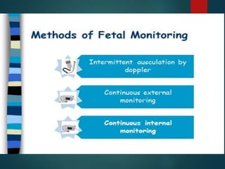
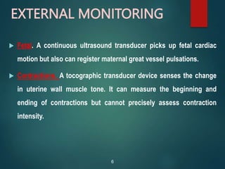
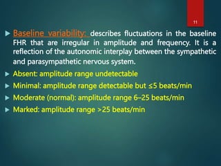
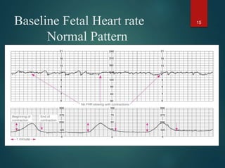
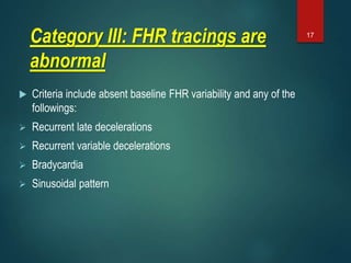
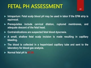
Intrapartum monitoring
DR. AHMED MOHAMED ABBAS, MD
Assistant Professor of OB-GYN
Faculty of Medicine- Assiut University
FETAL HEART RATE MONITORING
 Normal fetal heart rate (FHR) findings are highly reassuring of fetal
well-being.
 Abnormal FHR findings are poor predictors of fetal compromise.
 Wide usage of electronic FHR monitoring has not lowered the rate
of cerebral palsy (CP) because the antecedents of CP appear not to
be intrapartum events, but rather antenatal events.
 So, the false-positive rate for electronic FHR monitoring for
predicting CP is >99%.
2
3
Intermittent auscultation devices
Pinard stethoscope DeLee stethoscope
4
EXTERNAL MONITORING
 External devices (most common) are placed on the uterine
fundus.
 Advantage used before significant cervical dilation and
membrane rupture.
 Disadvantages are poor quality tracing with maternal
obesity and maternal discomfort from the device belts.
5
EXTERNAL MONITORING
 Fetal. A continuous ultrasound transducer picks up fetal cardiac
motion but also can register maternal great vessel pulsations.
 Contractions. A tocographic transducer device senses the change
in uterine wall muscle tone. It can measure the beginning and
ending of contractions but cannot precisely assess contraction
intensity.
6
Cardiotocography External Monitoring
7
INTERNAL DEVICES
 Internal devices are placed through the dilated cervix.
 Advantages include optimum signal quality, which is unaffected by
maternal obesity.
 Disadvantages include limitation to labor when cervical dilation and
membrane rupture have occurred.
 Fetal. A direct scalp electrode precisely senses each QRS complex of the
fetal cardiac cycle. Complications can include fetal scalp trauma and
infection.
 Contractions. An intrauterine pressure catheter (IUPC) placed into the
uterine cavity precisely registers intrauterine hydrostatic changes with
each contraction.
.
8
9
INTRAPARTUM FETAL HEART RATE
MONITORING
DEFINITIONS
 Baseline Fetal Heart Rate (FHR): The mean FHR rounded
to increments of 5 beats/min during a 10-minute segment.
 Normal FHR baseline is 110–160 beats/minute.
 Tachycardia: FHR baseline is >160 beats/min.
 Bradycardia : FHR baseline is <110 beats/min.
10
 Baseline variability: describes fluctuations in the baseline
FHR that are irregular in amplitude and frequency. It is a
reflection of the autonomic interplay between the sympathetic
and parasympathetic nervous system.
 Absent: amplitude range undetectable
 Minimal: amplitude range detectable but ≤5 beats/min
 Moderate (normal): amplitude range 6–25 beats/min
 Marked: amplitude range >25 beats/min
11
Variability 12
Normal/ increased variability or irregularity of a cardiac rhythm.
Absence or decreased variability, or a smooth flat baseline, is a sign
of fetal compromise
FETAL HEART RATE CATEGORIES
 A three-tiered system for the categorization of FHR patterns is
recommended.
 Categorization evaluates the fetus at that point in time; tracing
patterns can and will change.
 FHR tracing may move back and forth between the categories
depending on the clinical situation and management strategies.
13
Category I: FHR tracings are normal
Criteria include all of the following:
 Baseline rate: 110–160 beats/min
 Baseline FHR variability: moderate
 Late or variable decelerations: absent
 Early decelerations: present or absent
 Accelerations: present or absent
14
Baseline Fetal Heart rate
Normal Pattern
15
Category II: FHR tracings are
indeterminate
 These include all FHR tracings not categorized as category I
or III and may represent a great portion of those encountered
in clinical care.
16
Category III: FHR tracings are
abnormal
 Criteria include absent baseline FHR variability and any of the
followings:
 Recurrent late decelerations
 Recurrent variable decelerations
 Bradycardia
 Sinusoidal pattern
17
Poor or absent variability
18
19
INTRAUTERINE RESUSCITATION
 Decrease uterine contractions: Turn off any IV oxytocin
infusion or administer terbutaline 0.25 mg subcutaneously to
enhance intervillous placental blood flow.
 Augment IV fluid volume: Infuse 500 mL bolus of intravenous
normal saline rapidly to enhance uteroplacental infusion.
 Administer high-flow oxygen: Give the parturient 8–10 L of
oxygen by facemask to increase delivery of maternal oxygen
to the placenta.
20
 Amnioinfusion is useful for reducing the severity of variable
decelerations.
 Change position: Removing the parturient from the supine position
decreases inferior vena cava compression and enhances cardiac
return, thus cardiac output to the placenta. Turning the parturient
from one lateral position to the other may relieve any umbilical cord
compression that may be present.
 Vaginal examination: Perform a digital vaginal examination to rule
out possible prolapsed umbilical cord.
 Scalp stimulation: Perform a digital scalp stimulation observing for
accelerations, which would be reassuring of fetal condition.
21
INTRAUTERINE RESUSCITATION
FETAL PH ASSESSMENT
 Intrapartum: Fetal scalp blood pH may be used in labor if the EFM strip is
equivocal.
 Prerequisites include cervical dilation, ruptured membranes, and
adequate descent of the fetal head.
 Contraindications are suspected fetal blood dyscrasia.
 A small, shallow fetal scalp incision is made resulting in capillary
bleeding.
 The blood is collected in a heparinized capillary tube and sent to the
laboratory for blood gas analysis.
 Normal fetal pH is ≥7.20.
22
CASE SCENARIO
 A 20-year-old primigravida at 39 weeks’ gestation is in active
labor at 7 cm of cervical dilation.
 The EFM strip shows a baseline heart rate of 175 beats/min,
and variability is absent, but repetitive late decelerations are
seen after each contraction. No accelerations are noted.
23
 Examine the EFM strip carefully looking for baseline FHR, degree of
variability, and presence of periodic changes (accelerations,
decelerations).
 Confirm abnormal findings using criteria discussed (category II or
III).
 Identify nonhypoxic causes present that could explain the abnormal
findings.
 Initiate the intrauterine resuscitation measures described previously
to enhance placental perfusion and fetal oxygenation.
 Observe for normalization of the EFM tracing.
 Prepare for delivery promptly if resuscitation measures do not
normalize EFM tracing.
24
Specific interventions if immediate delivery Is indicated:
 In stage 1 of labor, the only option is emergency cesarean
section.
 In stage 2 of labor, an operative vaginal delivery (e.g.,
vacuum extractor
25
26
Thank you

IPFA.ppt

  • 1.
    Intrapartum monitoring DR. AHMEDMOHAMED ABBAS, MD Assistant Professor of OB-GYN Faculty of Medicine- Assiut University
  • 2.
    FETAL HEART RATEMONITORING  Normal fetal heart rate (FHR) findings are highly reassuring of fetal well-being.  Abnormal FHR findings are poor predictors of fetal compromise.  Wide usage of electronic FHR monitoring has not lowered the rate of cerebral palsy (CP) because the antecedents of CP appear not to be intrapartum events, but rather antenatal events.  So, the false-positive rate for electronic FHR monitoring for predicting CP is >99%. 2
  • 3.
  • 4.
    Intermittent auscultation devices Pinardstethoscope DeLee stethoscope 4
  • 5.
    EXTERNAL MONITORING  Externaldevices (most common) are placed on the uterine fundus.  Advantage used before significant cervical dilation and membrane rupture.  Disadvantages are poor quality tracing with maternal obesity and maternal discomfort from the device belts. 5
  • 6.
    EXTERNAL MONITORING  Fetal.A continuous ultrasound transducer picks up fetal cardiac motion but also can register maternal great vessel pulsations.  Contractions. A tocographic transducer device senses the change in uterine wall muscle tone. It can measure the beginning and ending of contractions but cannot precisely assess contraction intensity. 6
  • 7.
  • 8.
    INTERNAL DEVICES  Internaldevices are placed through the dilated cervix.  Advantages include optimum signal quality, which is unaffected by maternal obesity.  Disadvantages include limitation to labor when cervical dilation and membrane rupture have occurred.  Fetal. A direct scalp electrode precisely senses each QRS complex of the fetal cardiac cycle. Complications can include fetal scalp trauma and infection.  Contractions. An intrauterine pressure catheter (IUPC) placed into the uterine cavity precisely registers intrauterine hydrostatic changes with each contraction. . 8
  • 9.
  • 10.
    INTRAPARTUM FETAL HEARTRATE MONITORING DEFINITIONS  Baseline Fetal Heart Rate (FHR): The mean FHR rounded to increments of 5 beats/min during a 10-minute segment.  Normal FHR baseline is 110–160 beats/minute.  Tachycardia: FHR baseline is >160 beats/min.  Bradycardia : FHR baseline is <110 beats/min. 10
  • 11.
     Baseline variability:describes fluctuations in the baseline FHR that are irregular in amplitude and frequency. It is a reflection of the autonomic interplay between the sympathetic and parasympathetic nervous system.  Absent: amplitude range undetectable  Minimal: amplitude range detectable but ≤5 beats/min  Moderate (normal): amplitude range 6–25 beats/min  Marked: amplitude range >25 beats/min 11
  • 12.
    Variability 12 Normal/ increasedvariability or irregularity of a cardiac rhythm. Absence or decreased variability, or a smooth flat baseline, is a sign of fetal compromise
  • 13.
    FETAL HEART RATECATEGORIES  A three-tiered system for the categorization of FHR patterns is recommended.  Categorization evaluates the fetus at that point in time; tracing patterns can and will change.  FHR tracing may move back and forth between the categories depending on the clinical situation and management strategies. 13
  • 14.
    Category I: FHRtracings are normal Criteria include all of the following:  Baseline rate: 110–160 beats/min  Baseline FHR variability: moderate  Late or variable decelerations: absent  Early decelerations: present or absent  Accelerations: present or absent 14
  • 15.
    Baseline Fetal Heartrate Normal Pattern 15
  • 16.
    Category II: FHRtracings are indeterminate  These include all FHR tracings not categorized as category I or III and may represent a great portion of those encountered in clinical care. 16
  • 17.
    Category III: FHRtracings are abnormal  Criteria include absent baseline FHR variability and any of the followings:  Recurrent late decelerations  Recurrent variable decelerations  Bradycardia  Sinusoidal pattern 17
  • 18.
    Poor or absentvariability 18
  • 19.
  • 20.
    INTRAUTERINE RESUSCITATION  Decreaseuterine contractions: Turn off any IV oxytocin infusion or administer terbutaline 0.25 mg subcutaneously to enhance intervillous placental blood flow.  Augment IV fluid volume: Infuse 500 mL bolus of intravenous normal saline rapidly to enhance uteroplacental infusion.  Administer high-flow oxygen: Give the parturient 8–10 L of oxygen by facemask to increase delivery of maternal oxygen to the placenta. 20
  • 21.
     Amnioinfusion isuseful for reducing the severity of variable decelerations.  Change position: Removing the parturient from the supine position decreases inferior vena cava compression and enhances cardiac return, thus cardiac output to the placenta. Turning the parturient from one lateral position to the other may relieve any umbilical cord compression that may be present.  Vaginal examination: Perform a digital vaginal examination to rule out possible prolapsed umbilical cord.  Scalp stimulation: Perform a digital scalp stimulation observing for accelerations, which would be reassuring of fetal condition. 21 INTRAUTERINE RESUSCITATION
  • 22.
    FETAL PH ASSESSMENT Intrapartum: Fetal scalp blood pH may be used in labor if the EFM strip is equivocal.  Prerequisites include cervical dilation, ruptured membranes, and adequate descent of the fetal head.  Contraindications are suspected fetal blood dyscrasia.  A small, shallow fetal scalp incision is made resulting in capillary bleeding.  The blood is collected in a heparinized capillary tube and sent to the laboratory for blood gas analysis.  Normal fetal pH is ≥7.20. 22
  • 23.
    CASE SCENARIO  A20-year-old primigravida at 39 weeks’ gestation is in active labor at 7 cm of cervical dilation.  The EFM strip shows a baseline heart rate of 175 beats/min, and variability is absent, but repetitive late decelerations are seen after each contraction. No accelerations are noted. 23
  • 24.
     Examine theEFM strip carefully looking for baseline FHR, degree of variability, and presence of periodic changes (accelerations, decelerations).  Confirm abnormal findings using criteria discussed (category II or III).  Identify nonhypoxic causes present that could explain the abnormal findings.  Initiate the intrauterine resuscitation measures described previously to enhance placental perfusion and fetal oxygenation.  Observe for normalization of the EFM tracing.  Prepare for delivery promptly if resuscitation measures do not normalize EFM tracing. 24
  • 25.
    Specific interventions ifimmediate delivery Is indicated:  In stage 1 of labor, the only option is emergency cesarean section.  In stage 2 of labor, an operative vaginal delivery (e.g., vacuum extractor 25
  • 26.