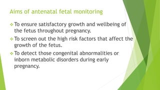
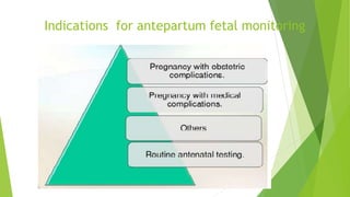
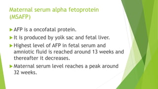
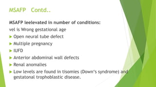
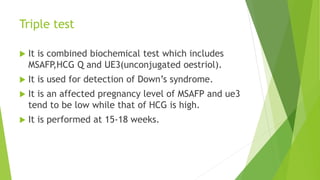
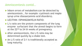
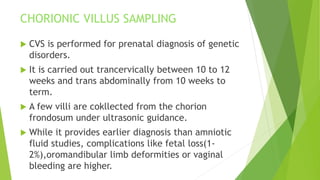
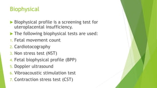
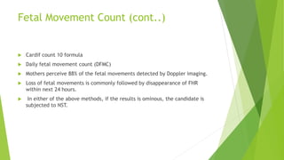
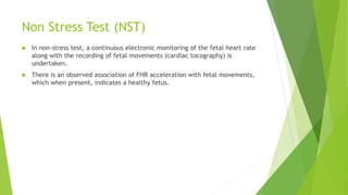
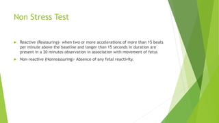
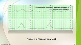
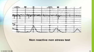
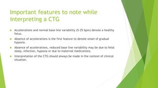
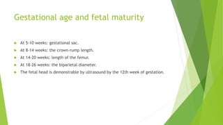
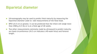
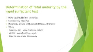
Asessment 0f Maternal and
Fetal Wellbeing
prepared by
Neethu P
1 st year Msc Nursing
Introduction
 Majority of fetal deaths occurs in the antepartum
period.
 There is progressive decline in the maternal
death all over the world. Currently more interest
is focused to evaluate the fetal health.
 The primary objective of antenatal assessment is
to avoid fetal death.
Aims of antenatal fetal monitoring
 To ensure satisfactory growth and wellbeing of
the fetus throughout pregnancy.
 To screen out the high risk factors that affect the
growth of the fetus.
 To detect those congenital abnormalities or
inborn metabolic disorders during early
pregnancy.
Indications for antepartum fetal monitoring
Maternal serum alpha fetoprotein
(MSAFP)
 AFP is a oncofatal protein.
 It is produced by yolk sac and fetal liver.
 Highest level of AFP in fetal serum and
amniotic fluid is reached around 13 weeks and
thereafter it decreases.
 Maternal serum level reaches a peak around
32 weeks.
MSAFP Contd..
MSAFP leelevated in number of conditions:
vel is Wrong gestational age
 Open neural tube defect
 Multiple pregnancy
 IUFD
 Anterior abdominal wall defects
 Renal anomalies
 Low levels are found in tisomies (Down‘s syndrome) and
gestatonal trophoblastic disease.
Triple test
 It is combined biochemical test which includes
MSAFP,HCG Q and UE3(unconjugated oestriol).
 It is used for detection of Down’s syndrome.
 It is an affected pregnancy level of MSAFP and ue3
tend to be low while that of HCG is high.
 It is performed at 15-18 weeks.
 ACETYLCHOLINE ESTERASE :(AChE) Amniotic
fluid AChE level is elevated in most cases
of open neural tube defects.
 It has got better diagnostic value than AFP.
INHIBIN A :is a dimeric glycoprotein.it is
produced by the corpus leuteum and the
placenta. Serum level of inhibin A is raised in
women carrying a fetus with Down‘s
syndrome.
AMNIOCENTESIS
 Aspiration of amniotic fluid from the
pregnant uterus for examination.
 Typically scheduled between the 14 th and
16 th weeks of pregnancy.
 An ultra sound is done to determine the
position of fetus and the location of a
pocket of amniotic fluid and the placenta.
AMNIOCENTESIS
 ALPHA FETO PROTEIN :
Increased levels of AFP:open body defect,
such as anencephaly,myelomeningocele , or
omphalocele.
Decreased level of AFP :Chromosomal defects
such as Down syndrome.
BILIRUBIN DETERMINATION:
If a blood incompactability is suspected.
AMNIOCENTESIS CONTD.
 CHROMOSOME ANALYSIS : Few fetal skin cells are
always present in amniotic fluid .these cells may be
cultured and stained for karyotyping for genetic
analysis.
 Colour:a strong yellow colour suggest a blood
incompactability. A green colour suggest meconium
staining ,A Phenomenon associated with fetal distress.
 FETAL FIBRONECTIN
 Fibronectin is a glycoprotein that plays a part in
helping the placenta attach to the uterine decidua.
 Detection of fibronectin in either the amniotic fluid
or in the mother’s vagina can serve as an
announcement that preterm labor may be beginning.
Amniocentesis contd..
 Inborn errors of metabolism:can be detected by
amniocentesis , for example :cystinosis and maple
syrup urine disease(amnio acid disorders).
 LECITHIN /SPHINGOMYELIN RATIO:
 L/s ratio are the protein components of the lung
enzyme surfactant that the alveoli begins to form
at the 22nd to 24 th weeks of prewgnancy.
 After amniocentesis, the L/S ratio may be
determined quckily by a shake test.
 An L/S ratio of 2:1 is traditionally accepted as
lung maturity.
CHORIONIC VILLUS SAMPLING
 CVS is performed for prenatal diagnosis of genetic
disorders.
 It is carried out trancervically between 10 to 12
weeks and trans abdominally from 10 weeks to
term.
 A few villi are cokllected from the chorion
frondosum under ultrasonic guidance.
 While it provides earlier diagnosis than amniotic
fluid studies, complications like fetal loss(1-
2%),oromandibular limb deformities or vaginal
bleeding are higher.
FETAL BLOOD SAMPLING(cordocentesis)
 This technique is used to take a sample of fetal
blood during pregnancy in order to screen for
chromosomal abnormalities, hemoglobinopathies
and other disorders affecting blood cells.
 It is performed under local anaesthestic usually
after 18 weeks gestation.
 Risks: The invasive procedure may lead to
abortion, preterm labour and intrauterine fetal
death. These may be due to bleeding, cord
haematoma formation, infection or preterm
rupture of membranes.
All the information as obtained in amniocentesis or
chorion vilius sampling, could be gathered.
Additional values are mentioned below:
 Hematological- For fetal anaemia, bleeding
disorders (autoimmine thrombocytopenia), rhesus
disease.
 Fetal infections – toxoplasmosis, viral infections
 Fetal blood gas and acid base status- in fetal
growth restrictions .
 Fetal therapy-blood transfusion .
FETAL BLOOD SAMPLING(cont..)
BIOPHYSICAL
 Ultrasonographic examination of the fetus in the
early (10-14 weeks) pregnancy can detect fetal
anomalies.
 Crown-rump length (CRL) smaller than the
gestational age is associated with the risk of
chromosomal anomalies (trisomy or triploidy).
 Increased nuchal translucency (soft tissue marker)
at 10-14 weeks is associated with many
chromosomal abnormalities (trisomy, monosomy,
triploidy).
Clinical
The clinical assessment of fetal growth can be
evaluated by the following parameters:
 Maternal weight gain
 Blood pressure
 Assessment of the size of the uterus and height of
the fundus
 Clinical assessment of excess liquor
Biophysical
 Biophysical profile is a screening test for
uteroplacental insufficiency.
 The following biophysical tests are used:
1. Fetal movement count
2. Cardiotocography
3. Non stress test (NST)
4. Fetal biophysical profile (BPP)
5. Doppler ultrasound
6. Vibroacoustic stimulation test
7. Contraction stress test (CST)
Fetal Movement Count
 A healthy fetus moves with a degree of consistency,
or at least 10 times a day.
 In contrast, a fetus not receiving enough nutrients
because of placental insufficiency has greatly
decreased movements.
 Based on this, asking a woman to observe and
record the number of movements the fetus is
making offers a gross assessment of fetal wellbeing.
Fetal Movement Count (cont..)
 Cardif count 10 formula
 Daily fetal movement count (DFMC)
 Mothers perceive 88% of the fetal movements detected by Doppler imaging.
 Loss of fetal movements is commonly followed by disappearance of FHR
within next 24 hours.
 In either of the above methods, if the results is ominous, the candidate is
subjected to NST.
Non Stress Test (NST)
 In non-stress test, a continuous electronic monitoring of the fetal heart rate
along with the recording of fetal movements (cardiac tocography) is
undertaken.
 There is an observed association of FHR acceleration with fetal movements,
which when present, indicates a healthy fetus.
Non Stress Test
 Reactive (Reassuring)- when two or more accelerations of more than 15 beats
per minute above the baseline and longer than 15 seconds in duration are
present in a 20 minutes observation in association with movement of fetus
 Non-reactive (Nonreassuring)- Absence of any fetal reactivity.
Important features to note while
interpreting a CTG
 Accelerations and normal base line variability (5-25 bpm) denote a healthy
fetus.
 Absence of accelerations is the first feature to denote onset of gradual
hypoxia.
 Absence of accelerations, reduced base line variability may be due to fetal
sleep, infection, hypoxia or due to maternal medications.
 Interpretation of the CTG should always be made in the context of clinical
situation.
Non stress test cont…
 Baseline FHR is the mean level of FHR between the peaks and the depressions
in beats per minute (bpm).
 Accelerations are increase in FHR by 15 bpm or more lasting for at least 15
seconds.
 Deceleration is decrease in FHR below the baseline by 15 bpm or more.
 Baseline variability is the oscillation of baseline FHR excluding the
accelerations and decelerations.
 A baseline variability of 5-25 bpm is a sign of fetal wellbeing
Contraction stress testing
 With contraction stress testing, the fetal heart rate is analyzed in conjunction
with contractions.
 A source of oxytocin for contraction stress testing currently is achieved by
nipple stimulation.
 Gentle stimulation of the nipples releases oxytocin in the same way as
happens with breastfeeding.
 With external uterine contraction and fetal heart rate monitors in place, the
baseline fetal heart rate is obtained.
Contraction stress test cont…
 Next, the woman rolls a nipple between her finger and thumb until uterine
contractions begin, which are recorded by a uterine monitor.
 Three contractions with a duration of 40 seconds or longer must be present in
a 10-minute window before the test can be interpreted.
 The test is negative (normal) if no fetal heart rate decelerations are present
with contractions.
 It is positive (abnormal) if 50% or more of contractions cause a late
deceleration
Oxytocin challenge test
 To elicit an abnormal FHR response
 Performed by iv infusion of dilute oxytocin until three contractions occur in
10 minutes
 Positive test: Late deceleration after each of the three contractions; FHR
baseline variability ; no FHR increase after FM
 Indicates decreased fetal reserve
 Negative test: No decelerations
 Fetus is safe within one week
 Suspicious: Repetitive variable decelerations
 Associated with abnormal FHR in labor, particularly in post-term gestation
Vibroacoustic stimulation
 A specially designed acoustic stimulator is applied to the mother’s abdomen
to produce a sharp sound of approximately 80 decibels at a frequency of 80
Hz, startling and waking the fetus.
 During a standard nonstress test, if a spontaneous acceleration has not
occurred within 5 minutes, apply a single 1- to 2-second sound stimulation to
the lower abdomen.
 This can be repeated again at the end of 10 minutes if no further spontaneous
movement occurs, so that two movements within the 20-minute window can
be evaluated.
Biophysical profile
 A biophysical profile combines five parameters
1. fetal reactivity
2. fetal breathing movements
3. fetal body movement
4. fetal tone
5. amniotic fluid volume
Biophysical profile
 With use of this system, each item has the potential for scoring a 2, so 10
would be the highest score possible.
 It is popularly called a fetal Apgar.
 Biophysical profiles may be done as often as daily during a high-risk
pregnancy.
 If the fetus score on a complete profile is 8–10, the fetus is considered to be
doing well.
 A score of 6 is considered suspicious; a score of 4 denotes a fetus probably in
jeopardy
Modified biophysical profile
 Use only two assessments (amniotic fluid index and a nonstress test).
 A healthy fetus should show a reactive nonstress test and an AFI range
between 5 and 25 cm.
 Modified BPP is considered abnormal (non reassuring) when the NST is non-
reactive and/or the AFI is <5.
Ultrasonography
 The production of high frequency sound waves which are reflected or echoed
when beamed into the body and an interface is encountered between
different types of tissues or structures with different densities.
 These echoes can be translated into visible images of the tissues or structures
encountered.
Uses of ultrasonography
 Diagnose pregnancy as early as 6 weeks gestation.
 Confirm the presence, size, and location of the placenta and amniotic fluid
 Establish that a fetus is growing and has no gross anomalies, such as
hydrocephalus, anencephaly, or spinal cord, heart, kidney, and bladder
defects
 Establish sex if a penis is revealed
 Establish the presentation and position of the fetus
 Predict maturity by measurement of the biparietal diameter of the head
Uses of ultrasonography cont…
 To discover complications of pregnancy.
 Fetal anomalies
 Fetal death
 After birth, an ultrasound may be used to detect a retained placenta or poor
uterine involution in the new mother.
Gestational age and fetal maturity
 At 5-10 weeks: gestational sac.
 At 8-14 weeks: the crown-rump length.
 At 14-20 weeks: length of the femur.
 At 18-26 weeks: the biparietal diameter.
 The fetal head is demonstrable by ultrasound by the 12th week of gestation.
Biparietal diameter
 Ultrasonography may be used to predict fetal maturity by measuring the
biparietal diameter (side-to- side measurement) of the fetal head.
 BPD is 8.5 cm or greater, it can be predicted that the infant will weigh more
than 2500 g (5.5 lb) or is at a fetal age of 40 weeks.
 Two other measurements commonly made by ultrasound to predict maturity
are head circumference (34.5 cm indicates a 40-week fetus) and femoral
length.
Doppler Umbilical Velocimetry
 Doppler ultrasonography measures the velocity at which red blood cells in the
uterine and fetal vessels travel.
 Assessment of the blood flow through uterine blood vessels is helpful to
determine the vascular resistance present in women with diabetes or
hypertension of pregnancy and whether resultant placental insufficiency is
occurring
Placental grading
 Based particularly on the amount of calcium deposits in the base of the
placenta.
 Placentas can be graded by ultrasound as
1. 0 (a placenta 12–24 weeks)
2. 1 (30–32 weeks)
3. 2 (36 weeks)
4. 3 (38 weeks).
Determination of fetal maturity by the
rapid surfactant test
1. Shake test or bubble test (clement’s)
2. Foam stabililty index( FSI)
3. Phosphatidyl Glycerol and Desaturated Phosphatidylcholine
4. Others
• Creatinine (Cr) – assess fetal renal maturity
• ΔOD450 – assess fetal liver maturity
• Lipocyte—assess fetal skin maturity
Matenal and fetal weebeing

Matenal and fetal weebeing

  • 1.
    Asessment 0f Maternaland Fetal Wellbeing prepared by Neethu P 1 st year Msc Nursing
  • 2.
    Introduction  Majority offetal deaths occurs in the antepartum period.  There is progressive decline in the maternal death all over the world. Currently more interest is focused to evaluate the fetal health.  The primary objective of antenatal assessment is to avoid fetal death.
  • 3.
    Aims of antenatalfetal monitoring  To ensure satisfactory growth and wellbeing of the fetus throughout pregnancy.  To screen out the high risk factors that affect the growth of the fetus.  To detect those congenital abnormalities or inborn metabolic disorders during early pregnancy.
  • 4.
  • 6.
    Maternal serum alphafetoprotein (MSAFP)  AFP is a oncofatal protein.  It is produced by yolk sac and fetal liver.  Highest level of AFP in fetal serum and amniotic fluid is reached around 13 weeks and thereafter it decreases.  Maternal serum level reaches a peak around 32 weeks.
  • 7.
    MSAFP Contd.. MSAFP leelevatedin number of conditions: vel is Wrong gestational age  Open neural tube defect  Multiple pregnancy  IUFD  Anterior abdominal wall defects  Renal anomalies  Low levels are found in tisomies (Down‘s syndrome) and gestatonal trophoblastic disease.
  • 8.
    Triple test  Itis combined biochemical test which includes MSAFP,HCG Q and UE3(unconjugated oestriol).  It is used for detection of Down’s syndrome.  It is an affected pregnancy level of MSAFP and ue3 tend to be low while that of HCG is high.  It is performed at 15-18 weeks.
  • 9.
     ACETYLCHOLINE ESTERASE:(AChE) Amniotic fluid AChE level is elevated in most cases of open neural tube defects.  It has got better diagnostic value than AFP. INHIBIN A :is a dimeric glycoprotein.it is produced by the corpus leuteum and the placenta. Serum level of inhibin A is raised in women carrying a fetus with Down‘s syndrome.
  • 11.
    AMNIOCENTESIS  Aspiration ofamniotic fluid from the pregnant uterus for examination.  Typically scheduled between the 14 th and 16 th weeks of pregnancy.  An ultra sound is done to determine the position of fetus and the location of a pocket of amniotic fluid and the placenta.
  • 13.
    AMNIOCENTESIS  ALPHA FETOPROTEIN : Increased levels of AFP:open body defect, such as anencephaly,myelomeningocele , or omphalocele. Decreased level of AFP :Chromosomal defects such as Down syndrome. BILIRUBIN DETERMINATION: If a blood incompactability is suspected.
  • 14.
    AMNIOCENTESIS CONTD.  CHROMOSOMEANALYSIS : Few fetal skin cells are always present in amniotic fluid .these cells may be cultured and stained for karyotyping for genetic analysis.  Colour:a strong yellow colour suggest a blood incompactability. A green colour suggest meconium staining ,A Phenomenon associated with fetal distress.  FETAL FIBRONECTIN  Fibronectin is a glycoprotein that plays a part in helping the placenta attach to the uterine decidua.  Detection of fibronectin in either the amniotic fluid or in the mother’s vagina can serve as an announcement that preterm labor may be beginning.
  • 15.
    Amniocentesis contd..  Inbornerrors of metabolism:can be detected by amniocentesis , for example :cystinosis and maple syrup urine disease(amnio acid disorders).  LECITHIN /SPHINGOMYELIN RATIO:  L/s ratio are the protein components of the lung enzyme surfactant that the alveoli begins to form at the 22nd to 24 th weeks of prewgnancy.  After amniocentesis, the L/S ratio may be determined quckily by a shake test.  An L/S ratio of 2:1 is traditionally accepted as lung maturity.
  • 16.
    CHORIONIC VILLUS SAMPLING CVS is performed for prenatal diagnosis of genetic disorders.  It is carried out trancervically between 10 to 12 weeks and trans abdominally from 10 weeks to term.  A few villi are cokllected from the chorion frondosum under ultrasonic guidance.  While it provides earlier diagnosis than amniotic fluid studies, complications like fetal loss(1- 2%),oromandibular limb deformities or vaginal bleeding are higher.
  • 18.
    FETAL BLOOD SAMPLING(cordocentesis) This technique is used to take a sample of fetal blood during pregnancy in order to screen for chromosomal abnormalities, hemoglobinopathies and other disorders affecting blood cells.  It is performed under local anaesthestic usually after 18 weeks gestation.  Risks: The invasive procedure may lead to abortion, preterm labour and intrauterine fetal death. These may be due to bleeding, cord haematoma formation, infection or preterm rupture of membranes.
  • 20.
    All the informationas obtained in amniocentesis or chorion vilius sampling, could be gathered. Additional values are mentioned below:  Hematological- For fetal anaemia, bleeding disorders (autoimmine thrombocytopenia), rhesus disease.  Fetal infections – toxoplasmosis, viral infections  Fetal blood gas and acid base status- in fetal growth restrictions .  Fetal therapy-blood transfusion . FETAL BLOOD SAMPLING(cont..)
  • 21.
    BIOPHYSICAL  Ultrasonographic examinationof the fetus in the early (10-14 weeks) pregnancy can detect fetal anomalies.  Crown-rump length (CRL) smaller than the gestational age is associated with the risk of chromosomal anomalies (trisomy or triploidy).  Increased nuchal translucency (soft tissue marker) at 10-14 weeks is associated with many chromosomal abnormalities (trisomy, monosomy, triploidy).
  • 23.
    Clinical The clinical assessmentof fetal growth can be evaluated by the following parameters:  Maternal weight gain  Blood pressure  Assessment of the size of the uterus and height of the fundus  Clinical assessment of excess liquor
  • 24.
    Biophysical  Biophysical profileis a screening test for uteroplacental insufficiency.  The following biophysical tests are used: 1. Fetal movement count 2. Cardiotocography 3. Non stress test (NST) 4. Fetal biophysical profile (BPP) 5. Doppler ultrasound 6. Vibroacoustic stimulation test 7. Contraction stress test (CST)
  • 25.
    Fetal Movement Count A healthy fetus moves with a degree of consistency, or at least 10 times a day.  In contrast, a fetus not receiving enough nutrients because of placental insufficiency has greatly decreased movements.  Based on this, asking a woman to observe and record the number of movements the fetus is making offers a gross assessment of fetal wellbeing.
  • 26.
    Fetal Movement Count(cont..)  Cardif count 10 formula  Daily fetal movement count (DFMC)  Mothers perceive 88% of the fetal movements detected by Doppler imaging.  Loss of fetal movements is commonly followed by disappearance of FHR within next 24 hours.  In either of the above methods, if the results is ominous, the candidate is subjected to NST.
  • 27.
    Non Stress Test(NST)  In non-stress test, a continuous electronic monitoring of the fetal heart rate along with the recording of fetal movements (cardiac tocography) is undertaken.  There is an observed association of FHR acceleration with fetal movements, which when present, indicates a healthy fetus.
  • 29.
    Non Stress Test Reactive (Reassuring)- when two or more accelerations of more than 15 beats per minute above the baseline and longer than 15 seconds in duration are present in a 20 minutes observation in association with movement of fetus  Non-reactive (Nonreassuring)- Absence of any fetal reactivity.
  • 32.
    Important features tonote while interpreting a CTG  Accelerations and normal base line variability (5-25 bpm) denote a healthy fetus.  Absence of accelerations is the first feature to denote onset of gradual hypoxia.  Absence of accelerations, reduced base line variability may be due to fetal sleep, infection, hypoxia or due to maternal medications.  Interpretation of the CTG should always be made in the context of clinical situation.
  • 33.
    Non stress testcont…  Baseline FHR is the mean level of FHR between the peaks and the depressions in beats per minute (bpm).  Accelerations are increase in FHR by 15 bpm or more lasting for at least 15 seconds.  Deceleration is decrease in FHR below the baseline by 15 bpm or more.  Baseline variability is the oscillation of baseline FHR excluding the accelerations and decelerations.  A baseline variability of 5-25 bpm is a sign of fetal wellbeing
  • 37.
    Contraction stress testing With contraction stress testing, the fetal heart rate is analyzed in conjunction with contractions.  A source of oxytocin for contraction stress testing currently is achieved by nipple stimulation.  Gentle stimulation of the nipples releases oxytocin in the same way as happens with breastfeeding.  With external uterine contraction and fetal heart rate monitors in place, the baseline fetal heart rate is obtained.
  • 38.
    Contraction stress testcont…  Next, the woman rolls a nipple between her finger and thumb until uterine contractions begin, which are recorded by a uterine monitor.  Three contractions with a duration of 40 seconds or longer must be present in a 10-minute window before the test can be interpreted.  The test is negative (normal) if no fetal heart rate decelerations are present with contractions.  It is positive (abnormal) if 50% or more of contractions cause a late deceleration
  • 39.
    Oxytocin challenge test To elicit an abnormal FHR response  Performed by iv infusion of dilute oxytocin until three contractions occur in 10 minutes  Positive test: Late deceleration after each of the three contractions; FHR baseline variability ; no FHR increase after FM  Indicates decreased fetal reserve  Negative test: No decelerations  Fetus is safe within one week  Suspicious: Repetitive variable decelerations  Associated with abnormal FHR in labor, particularly in post-term gestation
  • 40.
    Vibroacoustic stimulation  Aspecially designed acoustic stimulator is applied to the mother’s abdomen to produce a sharp sound of approximately 80 decibels at a frequency of 80 Hz, startling and waking the fetus.  During a standard nonstress test, if a spontaneous acceleration has not occurred within 5 minutes, apply a single 1- to 2-second sound stimulation to the lower abdomen.  This can be repeated again at the end of 10 minutes if no further spontaneous movement occurs, so that two movements within the 20-minute window can be evaluated.
  • 41.
    Biophysical profile  Abiophysical profile combines five parameters 1. fetal reactivity 2. fetal breathing movements 3. fetal body movement 4. fetal tone 5. amniotic fluid volume
  • 42.
    Biophysical profile  Withuse of this system, each item has the potential for scoring a 2, so 10 would be the highest score possible.  It is popularly called a fetal Apgar.  Biophysical profiles may be done as often as daily during a high-risk pregnancy.  If the fetus score on a complete profile is 8–10, the fetus is considered to be doing well.  A score of 6 is considered suspicious; a score of 4 denotes a fetus probably in jeopardy
  • 45.
    Modified biophysical profile Use only two assessments (amniotic fluid index and a nonstress test).  A healthy fetus should show a reactive nonstress test and an AFI range between 5 and 25 cm.  Modified BPP is considered abnormal (non reassuring) when the NST is non- reactive and/or the AFI is <5.
  • 46.
    Ultrasonography  The productionof high frequency sound waves which are reflected or echoed when beamed into the body and an interface is encountered between different types of tissues or structures with different densities.  These echoes can be translated into visible images of the tissues or structures encountered.
  • 48.
    Uses of ultrasonography Diagnose pregnancy as early as 6 weeks gestation.  Confirm the presence, size, and location of the placenta and amniotic fluid  Establish that a fetus is growing and has no gross anomalies, such as hydrocephalus, anencephaly, or spinal cord, heart, kidney, and bladder defects  Establish sex if a penis is revealed  Establish the presentation and position of the fetus  Predict maturity by measurement of the biparietal diameter of the head
  • 49.
    Uses of ultrasonographycont…  To discover complications of pregnancy.  Fetal anomalies  Fetal death  After birth, an ultrasound may be used to detect a retained placenta or poor uterine involution in the new mother.
  • 50.
    Gestational age andfetal maturity  At 5-10 weeks: gestational sac.  At 8-14 weeks: the crown-rump length.  At 14-20 weeks: length of the femur.  At 18-26 weeks: the biparietal diameter.  The fetal head is demonstrable by ultrasound by the 12th week of gestation.
  • 51.
    Biparietal diameter  Ultrasonographymay be used to predict fetal maturity by measuring the biparietal diameter (side-to- side measurement) of the fetal head.  BPD is 8.5 cm or greater, it can be predicted that the infant will weigh more than 2500 g (5.5 lb) or is at a fetal age of 40 weeks.  Two other measurements commonly made by ultrasound to predict maturity are head circumference (34.5 cm indicates a 40-week fetus) and femoral length.
  • 52.
    Doppler Umbilical Velocimetry Doppler ultrasonography measures the velocity at which red blood cells in the uterine and fetal vessels travel.  Assessment of the blood flow through uterine blood vessels is helpful to determine the vascular resistance present in women with diabetes or hypertension of pregnancy and whether resultant placental insufficiency is occurring
  • 53.
    Placental grading  Basedparticularly on the amount of calcium deposits in the base of the placenta.  Placentas can be graded by ultrasound as 1. 0 (a placenta 12–24 weeks) 2. 1 (30–32 weeks) 3. 2 (36 weeks) 4. 3 (38 weeks).
  • 54.
    Determination of fetalmaturity by the rapid surfactant test 1. Shake test or bubble test (clement’s) 2. Foam stabililty index( FSI) 3. Phosphatidyl Glycerol and Desaturated Phosphatidylcholine 4. Others • Creatinine (Cr) – assess fetal renal maturity • ΔOD450 – assess fetal liver maturity • Lipocyte—assess fetal skin maturity