Downloaded 190 times


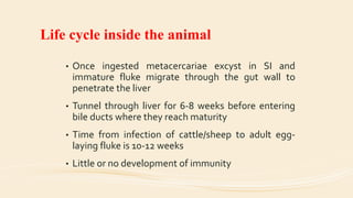
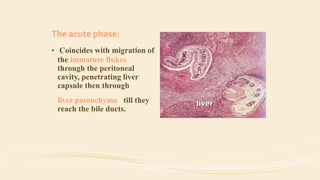
![Life cycle outside the animal
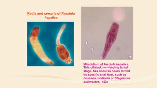
• Eggs hatch in spring (>10 °C) to release miracidia
which must penetrate a mud snail (Lymnaea [syn.
Galba] truncatula) within 3 hours
• Develop inside snail
• Cercariae emerge from snail
• Encyst on grass (metacercariae)
• Infection of a snail with one miracidium can produce
over 600 metacercariae](https://image.slidesharecdn.com/fasciolahepatica-140612025528-phpapp01/85/Fasiola-hepatica-round-worms-14-320.jpg)


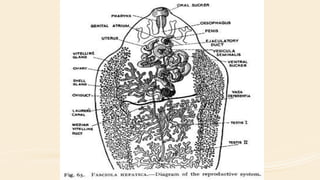


Fasciola hepatica, commonly known as the common liver fluke or sheep liver fluke, is a parasitic flatworm that infects the livers of various mammals. It has a complex life cycle involving an intermediate snail host and transmission through metacercariae encysted on aquatic plants. In humans, F. hepatica infection can cause acute, chronic, or obstructive phases of disease depending on the fluke's life stage and location. Diagnosis is typically made by identifying eggs in stool or bile samples, though serological tests can detect antibodies earlier. Treatment involves anthelmintic drugs while prevention focuses on limiting the parasite's transmission between hosts.


