Download as PPSX, PPTX

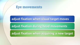
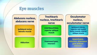
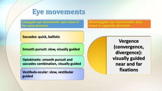
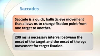
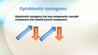
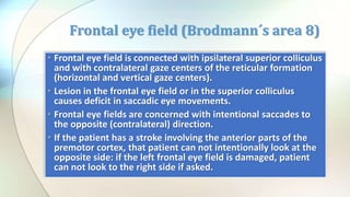
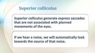

This document summarizes the anatomy and physiology of eye movements. It describes the six extraocular muscles that control eye movement and their innervation by three cranial nerves. It discusses the different types of eye movements including saccades, smooth pursuit, optokinetic nystagmus, vergence, and vestibulo-ocular movements. It also outlines the brain structures involved in motor control of eye movements, including the frontal eye fields, superior colliculus, brainstem gaze centers, and their connections.


