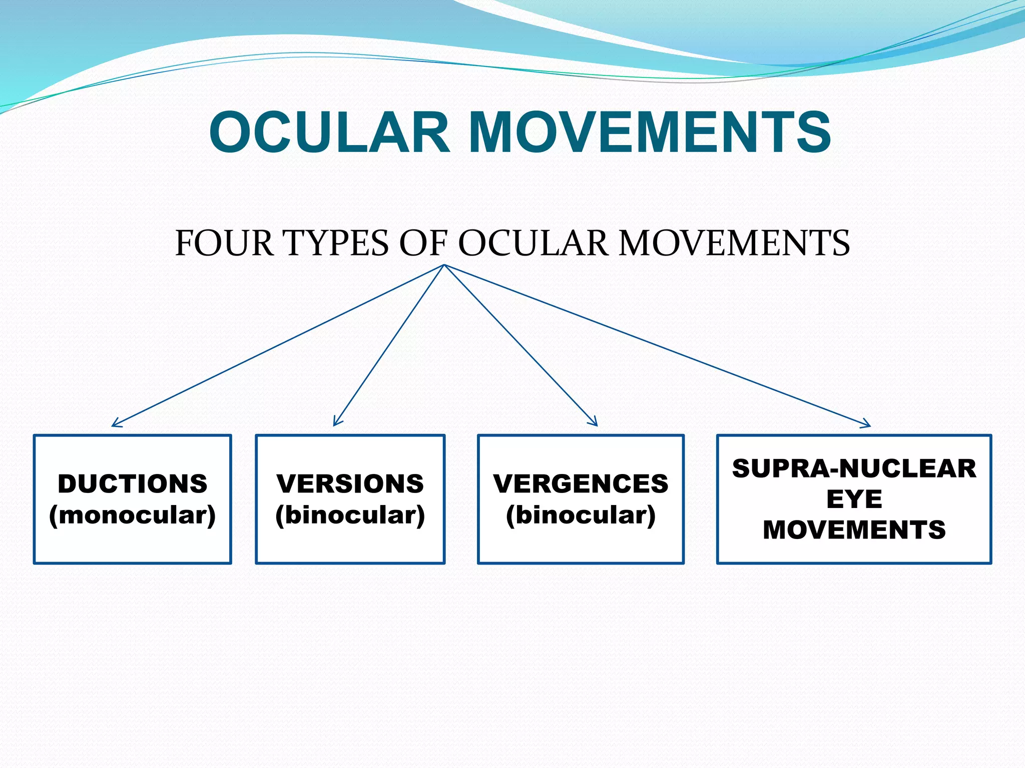
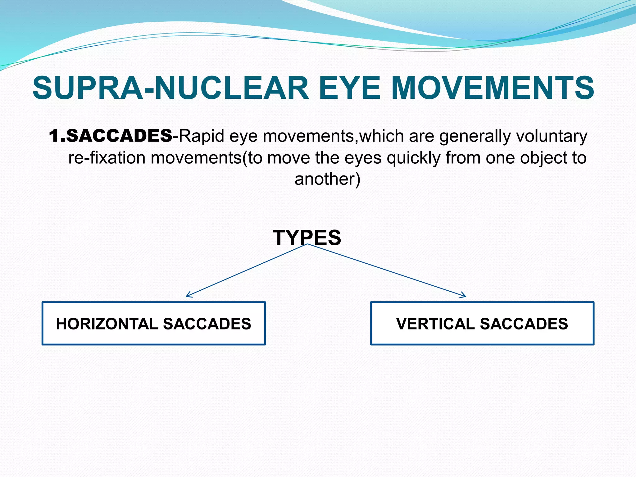
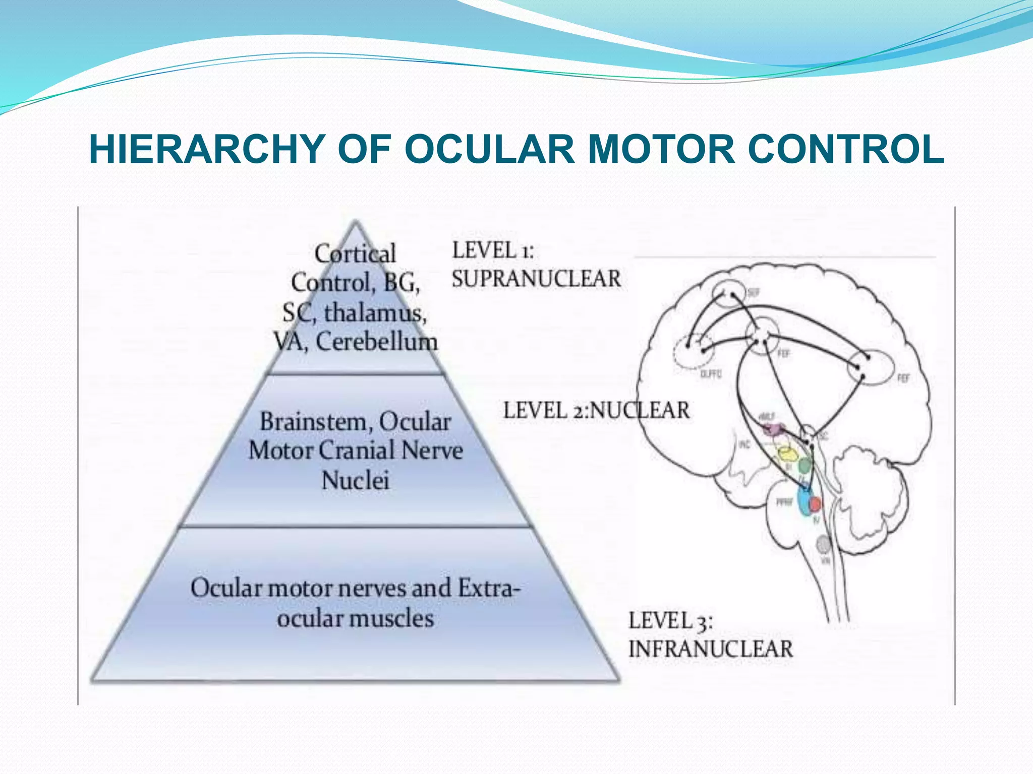
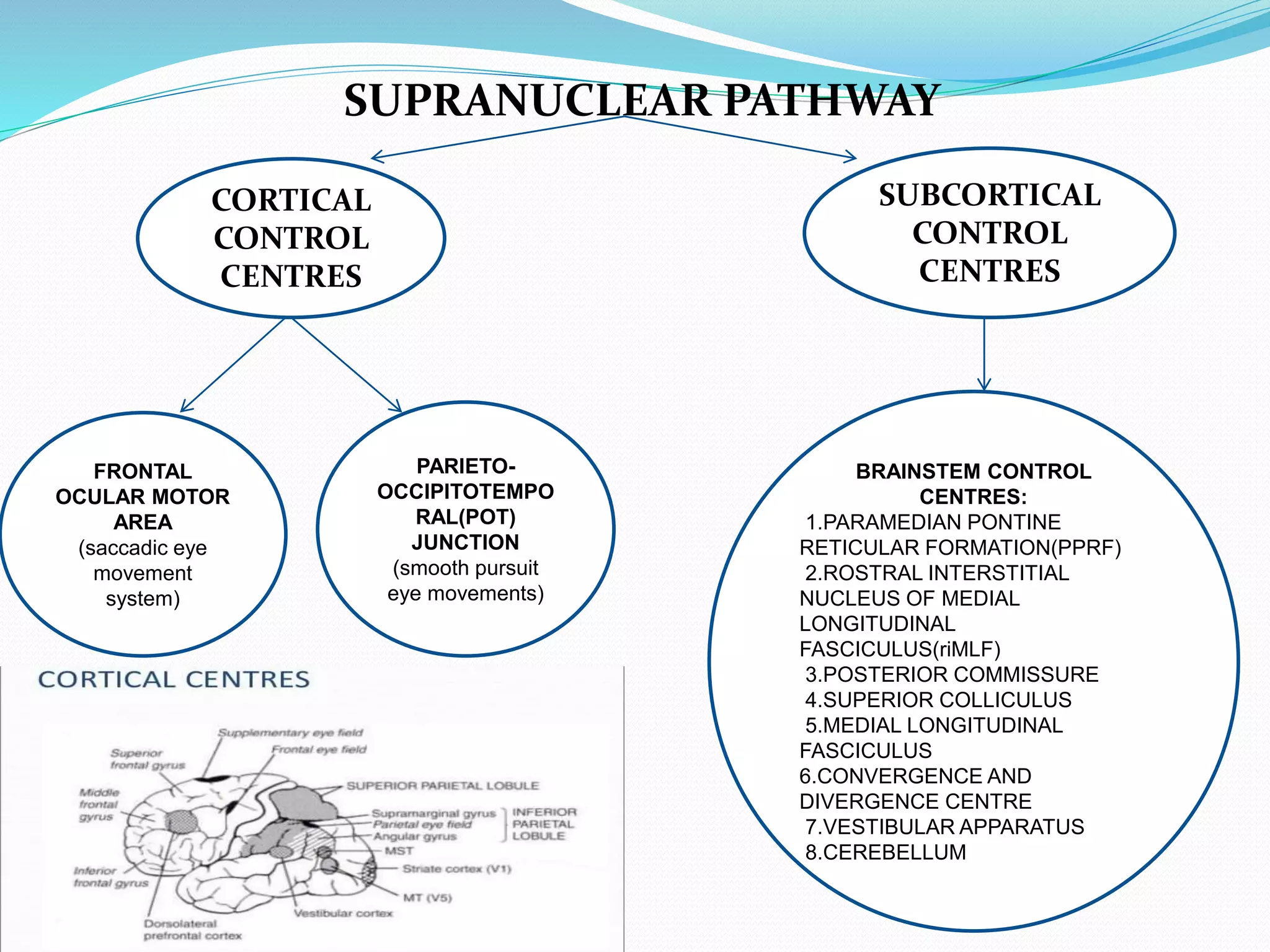
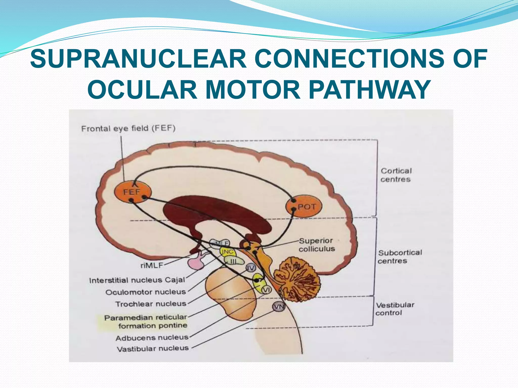
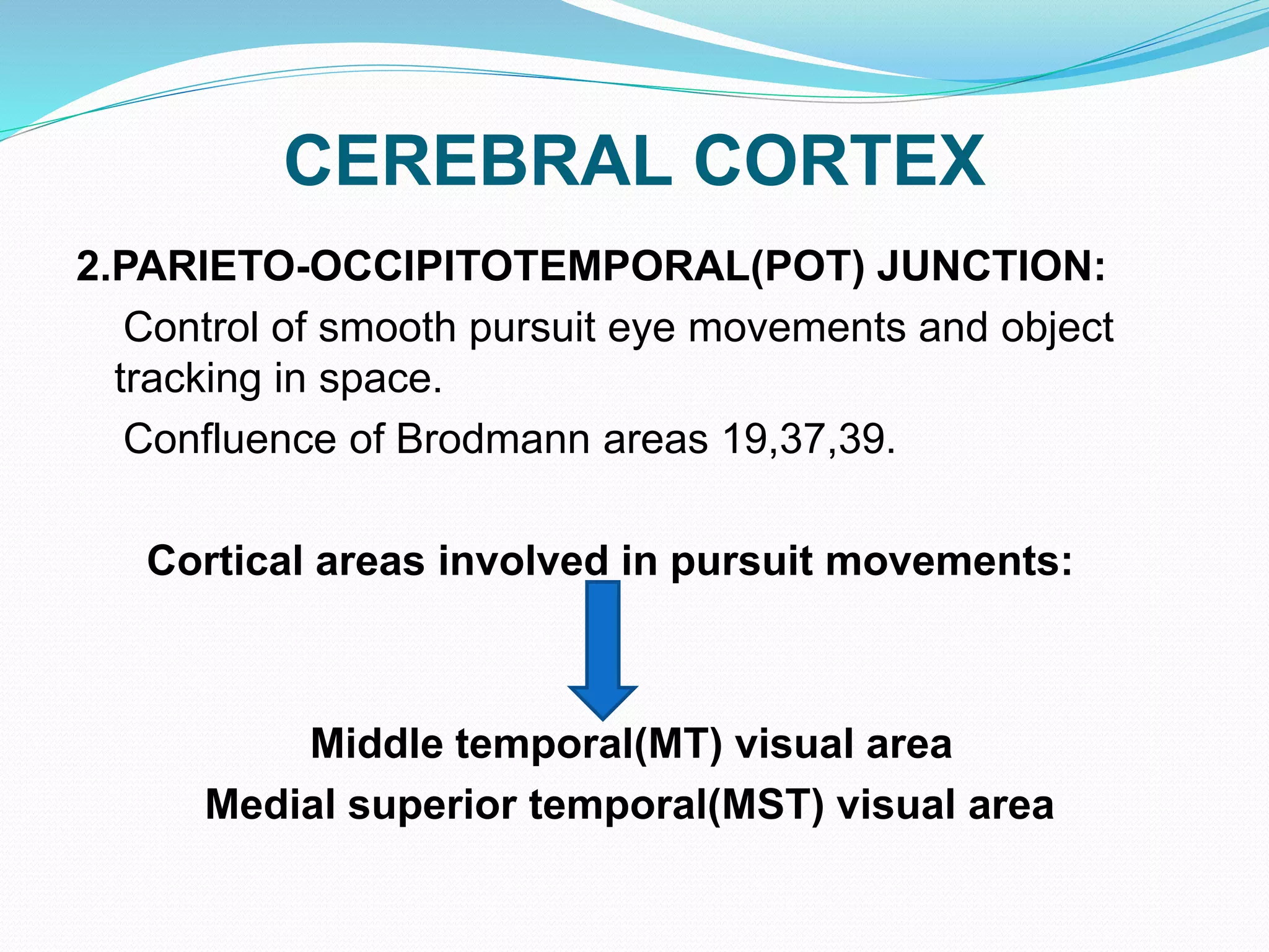
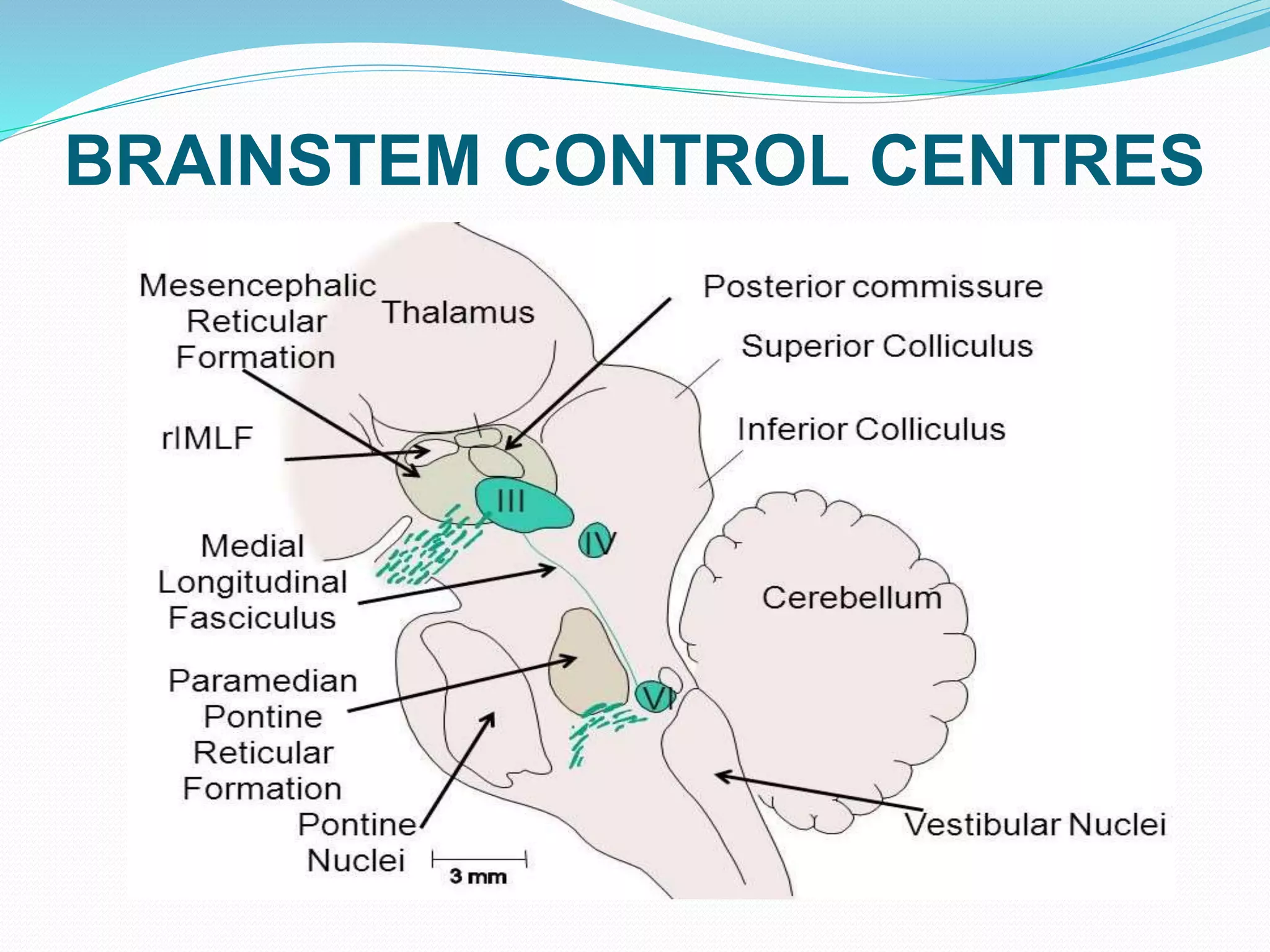
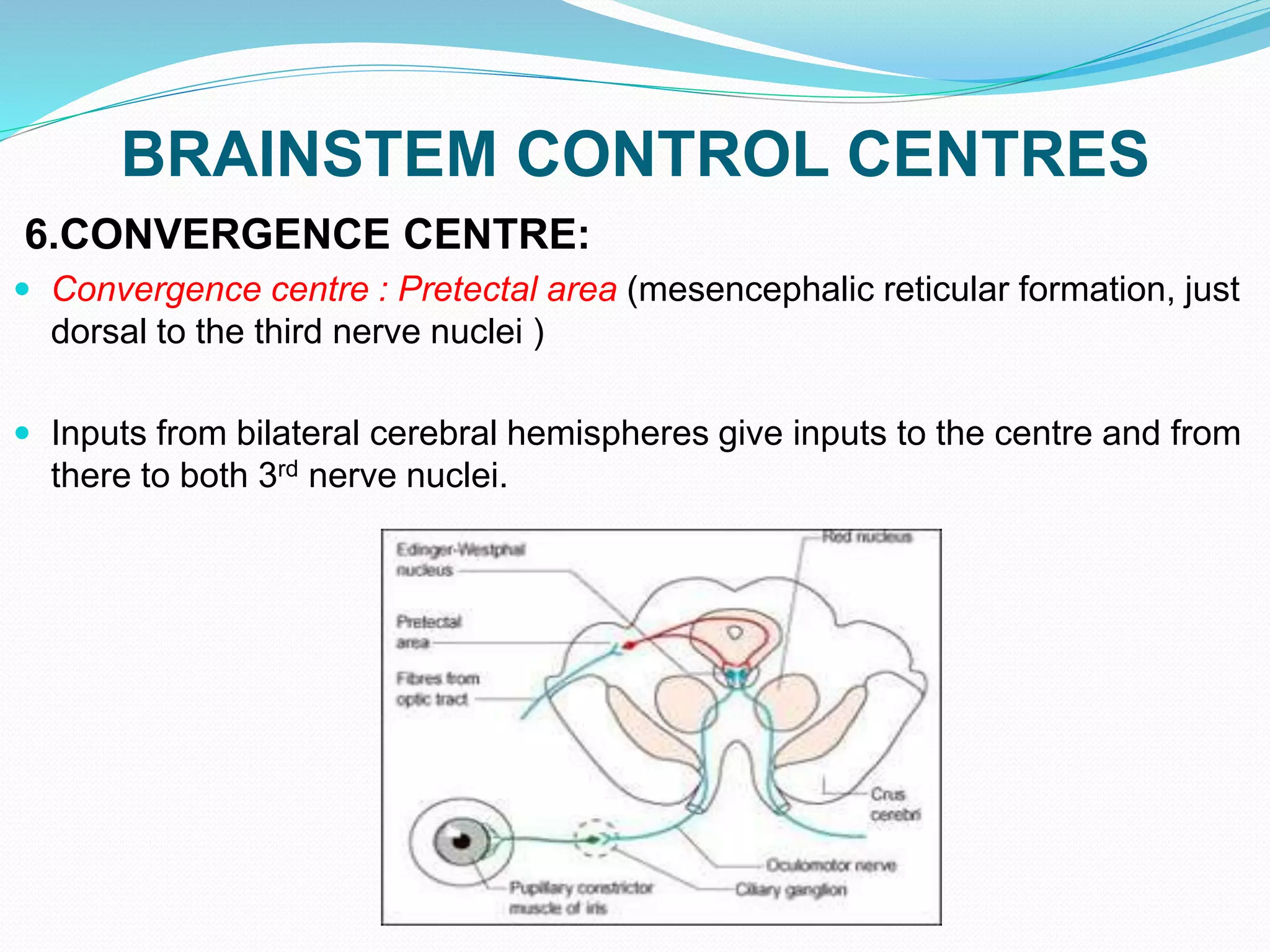
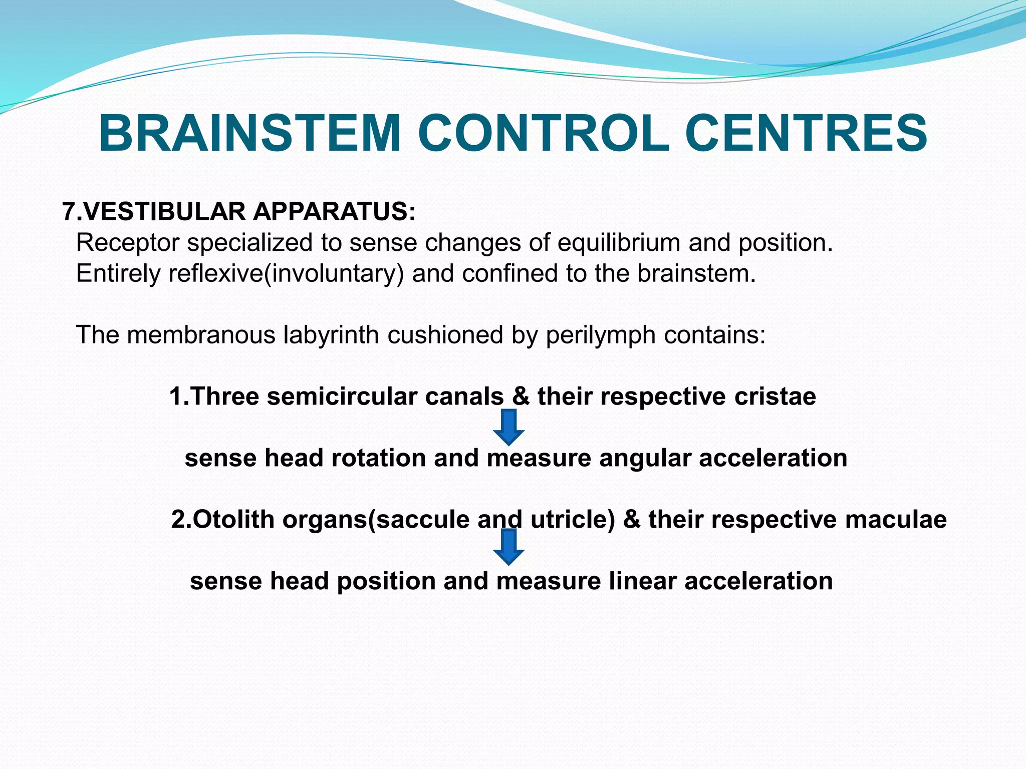
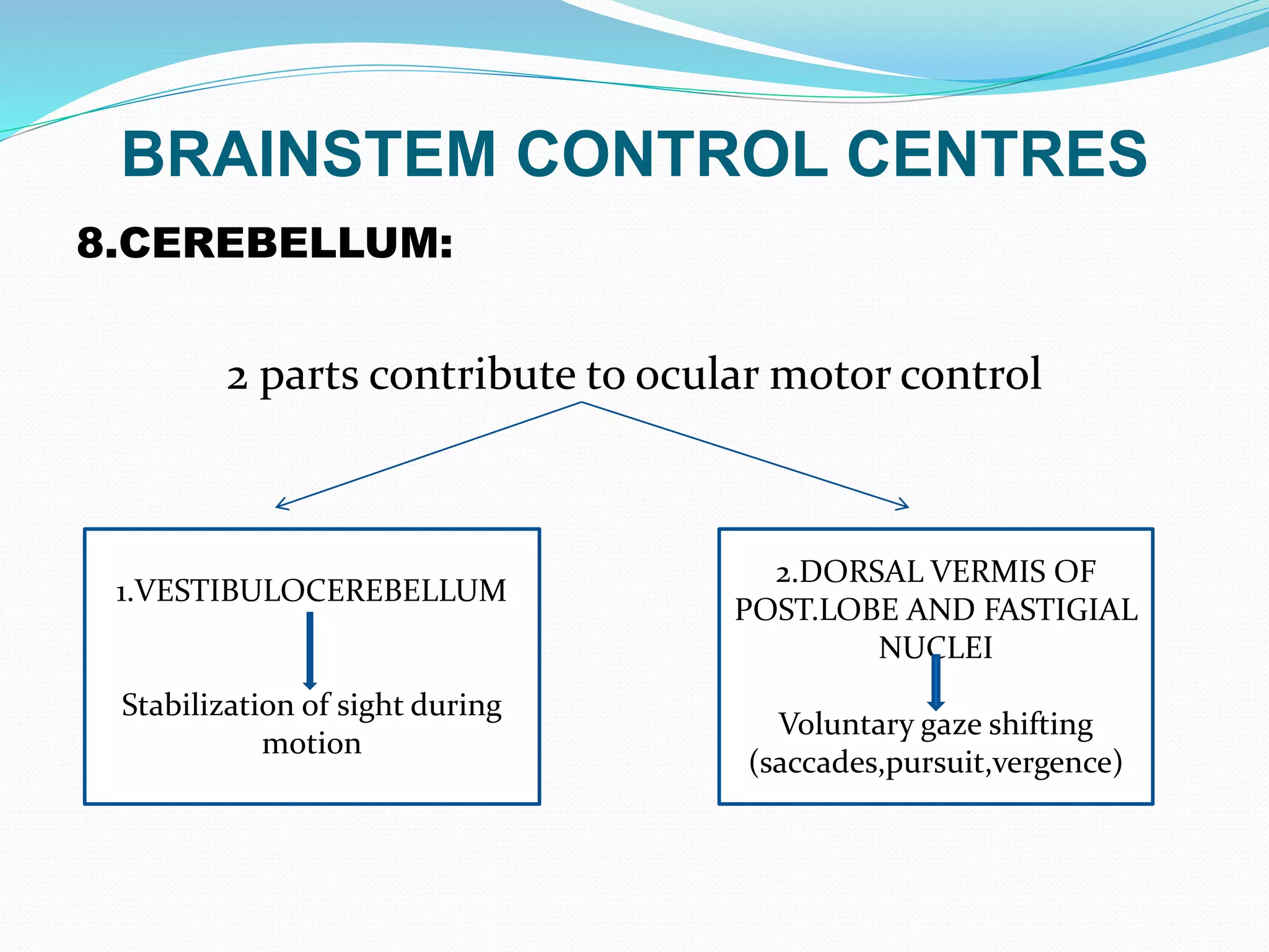
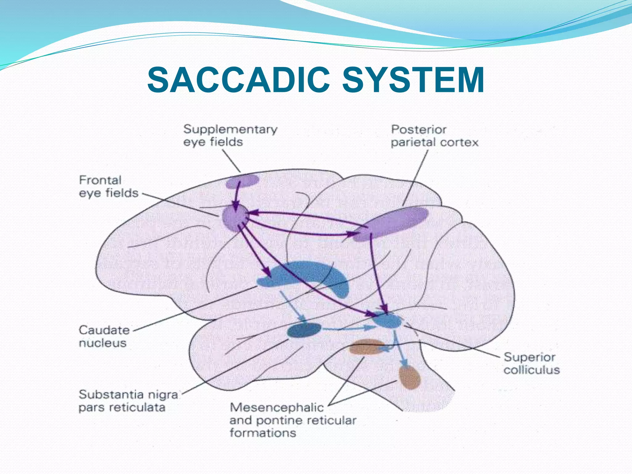
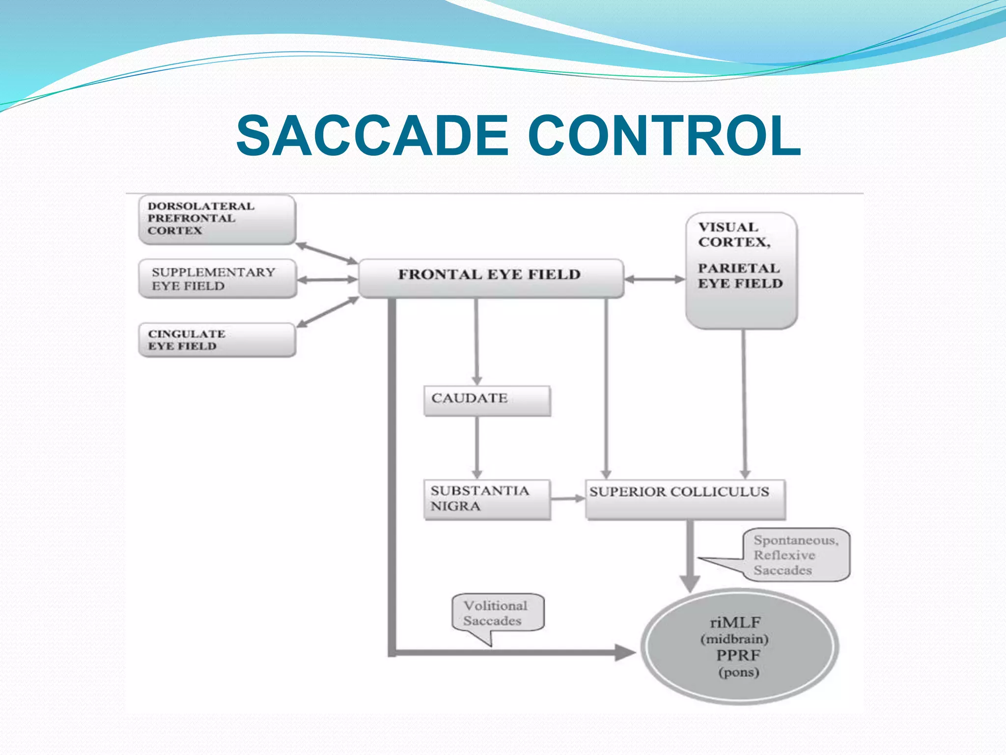
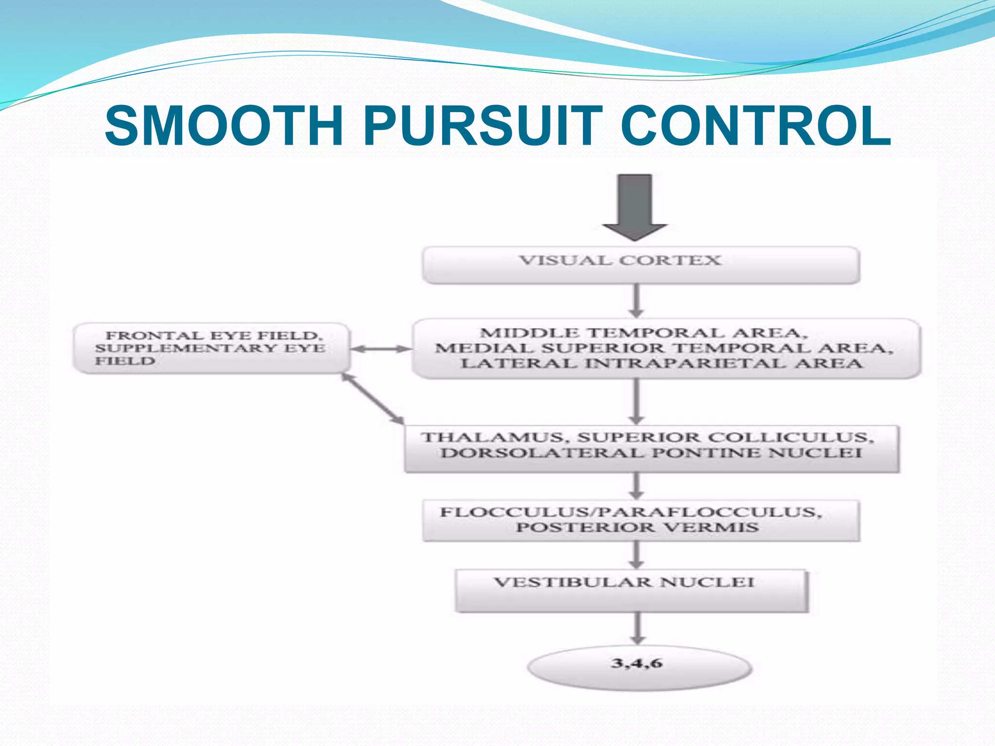
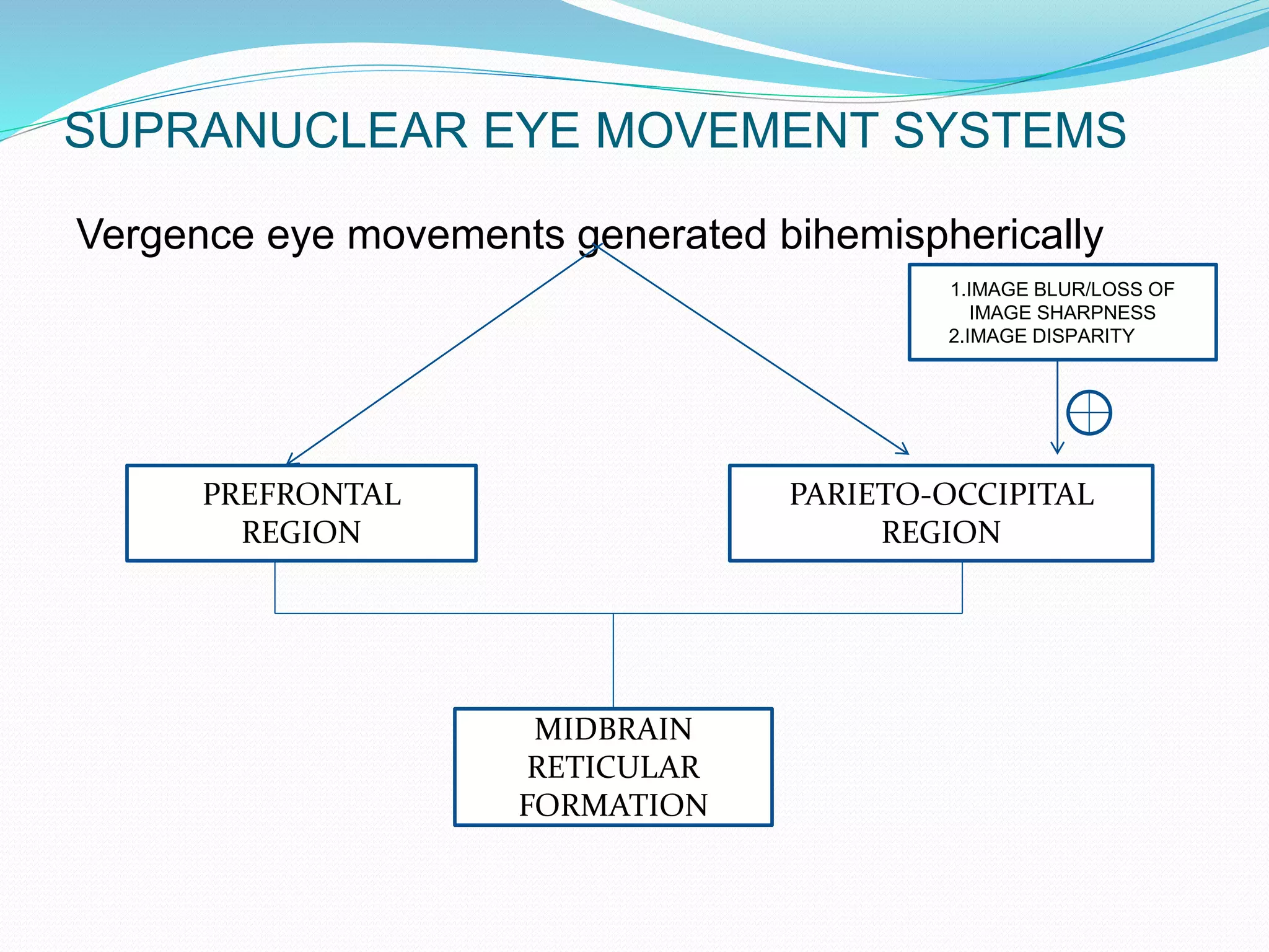
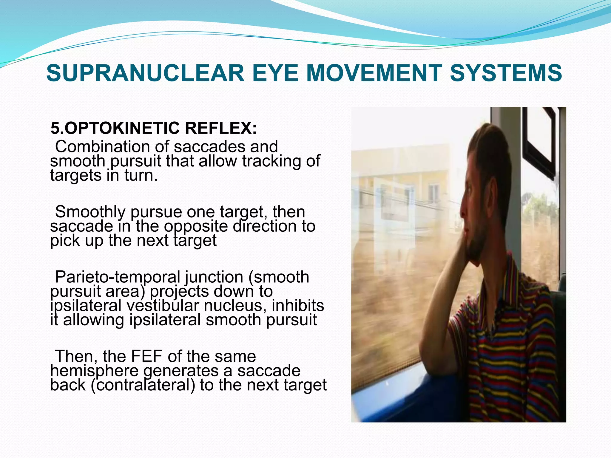
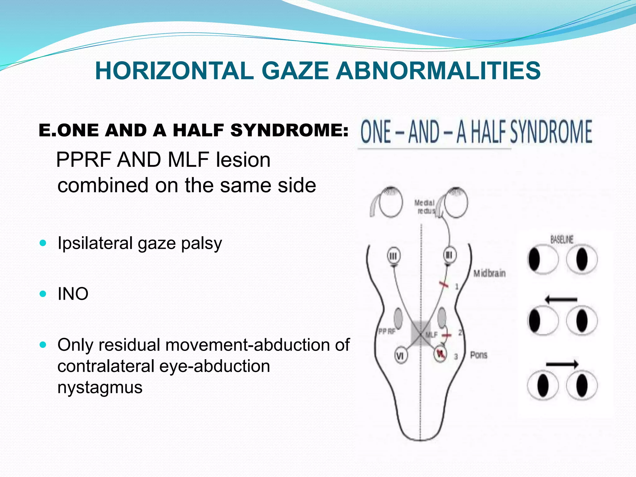
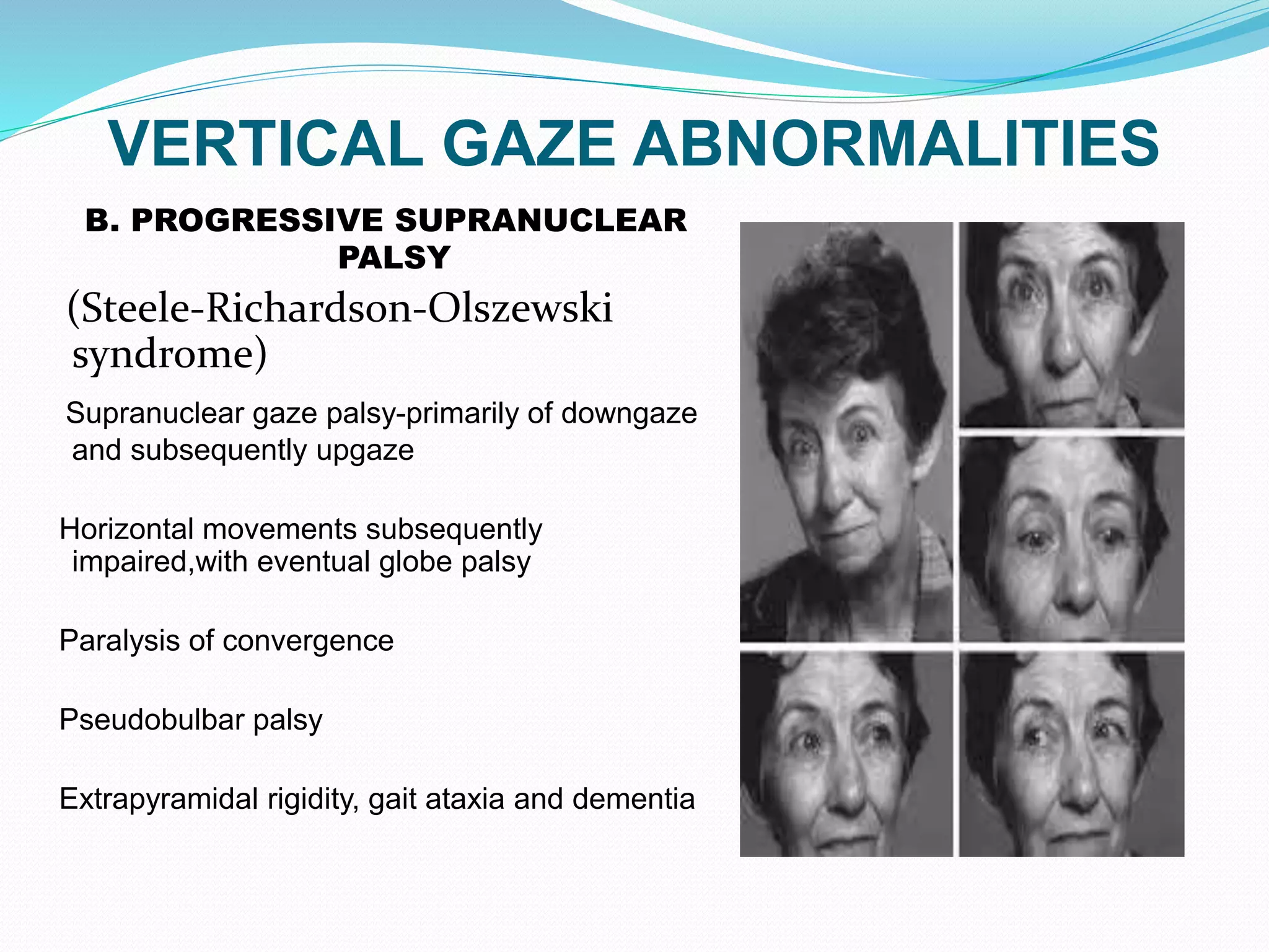
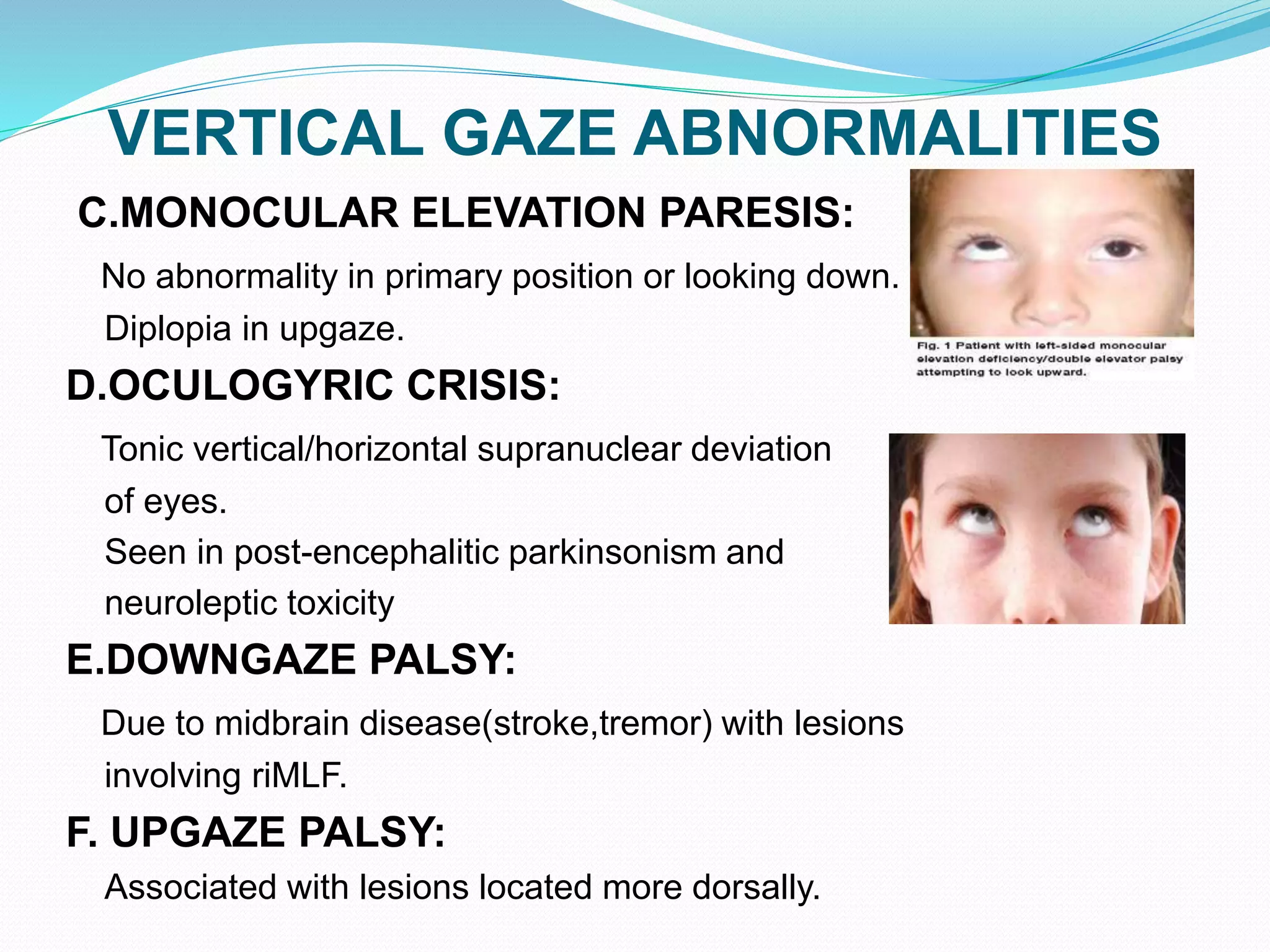
This document discusses ocular movements and their neural control pathways. It describes four types of ocular movements - versions, ductions, vergences, and supranuclear eye movements. Supranuclear eye movements include saccades, smooth pursuit, vestibulo-ocular, and optokinetic movements. The document outlines the cortical and brainstem control centers that generate each type of eye movement, including pathways like the medial longitudinal fasciculus. It also discusses various disorders that can occur with abnormalities in horizontal and vertical gaze, vergence, and other types of supranuclear eye movements.
The presentation includes physiological mechanism of different functional classes of eye movements such as horizontal & vertical eye movements, saccades, persuits, vestibuloocular reflex, Bell's phenomenon and it also includes different disorders that causes abnormal supranuclear eye movements e.g. skew deviation, Perinaud syndrome, INO.
Anatomy of Lateral Rectus, Anatomy of Abducens Nerve, Palsy of Abducens Nerve | by Optometrist Hasnain Pasha | Presented at Isra School of Optometry - Al-Ibrahim Eye Hospital
Ocular motility disorders: the Approach
Supra- vs infra- nuclear disorders and its related basic science
Other: synkinesis/aberrant regeneration, nystagmus
The presentation I have made and uploaded provides you with an in-depth insight into the patterns the strabismus may take following anomalies of extraocular muscles, deformities of the orbital structures,innnervational disturbances.
The author does not assume responsibility or legal liability for any errors in the text or for the misuse or misapplication of material in this work.
No copyright infringement, or plagiarism intended.
Amrit Pokharel
Slit lamp biomicroscopy and illumination techniquesLoknath Goswami
It is a presentation on slitlamp for beginner, shown the parts and different illumination techniques both for eye and contact lens and it have short history
It details about the sensory development, theories and the neural aspects of binocular vision development along with various tests involved to assess stereopsis.
Mechanism of balance & vestibular function test Dr Utkal MishraDr Utkal Mishra
This powerpoint elaborates the mechanism of balance & anatomy of vestibular apparutus. It also depicts the anatomy & physiology of haircells in detail. I also explained the vestibular function tests used for diagnosis of various vestibular disorders.
It is one of the most viewed document from Pgblaster India website: Disorders of ocular motility with an emphasis on squint. In this document I have tried to give some important concepts of the different types of squints in simple words.At a glance, it is a much harder and complex topic of ophthalmology but I had made it as simpler as I could. Hope it will help you..
ARTIFICIAL INTELLIGENCE IN HEALTHCARE.pdfAnujkumaranit
Artificial intelligence (AI) refers to the simulation of human intelligence processes by machines, especially computer systems. It encompasses tasks such as learning, reasoning, problem-solving, perception, and language understanding. AI technologies are revolutionizing various fields, from healthcare to finance, by enabling machines to perform tasks that typically require human intelligence.
The prostate is an exocrine gland of the male mammalian reproductive system
It is a walnut-sized gland that forms part of the male reproductive system and is located in front of the rectum and just below the urinary bladder
Function is to store and secrete a clear, slightly alkaline fluid that constitutes 10-30% of the volume of the seminal fluid that along with the spermatozoa, constitutes semen
A healthy human prostate measures (4cm-vertical, by 3cm-horizontal, 2cm ant-post ).
It surrounds the urethra just below the urinary bladder. It has anterior, median, posterior and two lateral lobes
It’s work is regulated by androgens which are responsible for male sex characteristics
Generalised disease of the prostate due to hormonal derangement which leads to non malignant enlargement of the gland (increase in the number of epithelial cells and stromal tissue)to cause compression of the urethra leading to symptoms (LUTS
Explore natural remedies for syphilis treatment in Singapore. Discover alternative therapies, herbal remedies, and lifestyle changes that may complement conventional treatments. Learn about holistic approaches to managing syphilis symptoms and supporting overall health.
Report Back from SGO 2024: What’s the Latest in Cervical Cancer?bkling
Are you curious about what’s new in cervical cancer research or unsure what the findings mean? Join Dr. Emily Ko, a gynecologic oncologist at Penn Medicine, to learn about the latest updates from the Society of Gynecologic Oncology (SGO) 2024 Annual Meeting on Women’s Cancer. Dr. Ko will discuss what the research presented at the conference means for you and answer your questions about the new developments.
Ethanol (CH3CH2OH), or beverage alcohol, is a two-carbon alcohol
that is rapidly distributed in the body and brain. Ethanol alters many
neurochemical systems and has rewarding and addictive properties. It
is the oldest recreational drug and likely contributes to more morbidity,
mortality, and public health costs than all illicit drugs combined. The
5th edition of the Diagnostic and Statistical Manual of Mental Disorders
(DSM-5) integrates alcohol abuse and alcohol dependence into a single
disorder called alcohol use disorder (AUD), with mild, moderate,
and severe subclassifications (American Psychiatric Association, 2013).
In the DSM-5, all types of substance abuse and dependence have been
combined into a single substance use disorder (SUD) on a continuum
from mild to severe. A diagnosis of AUD requires that at least two of
the 11 DSM-5 behaviors be present within a 12-month period (mild
AUD: 2–3 criteria; moderate AUD: 4–5 criteria; severe AUD: 6–11 criteria).
The four main behavioral effects of AUD are impaired control over
drinking, negative social consequences, risky use, and altered physiological
effects (tolerance, withdrawal). This chapter presents an overview
of the prevalence and harmful consequences of AUD in the U.S.,
the systemic nature of the disease, neurocircuitry and stages of AUD,
comorbidities, fetal alcohol spectrum disorders, genetic risk factors, and
pharmacotherapies for AUD.
These lecture slides, by Dr Sidra Arshad, offer a quick overview of physiological basis of a normal electrocardiogram.
Learning objectives:
1. Define an electrocardiogram (ECG) and electrocardiography
2. Describe how dipoles generated by the heart produce the waveforms of the ECG
3. Describe the components of a normal electrocardiogram of a typical bipolar leads (limb II)
4. Differentiate between intervals and segments
5. Enlist some common indications for obtaining an ECG
Study Resources:
1. Chapter 11, Guyton and Hall Textbook of Medical Physiology, 14th edition
2. Chapter 9, Human Physiology - From Cells to Systems, Lauralee Sherwood, 9th edition
3. Chapter 29, Ganong’s Review of Medical Physiology, 26th edition
4. Electrocardiogram, StatPearls - https://www.ncbi.nlm.nih.gov/books/NBK549803/
5. ECG in Medical Practice by ABM Abdullah, 4th edition
6. ECG Basics, http://www.nataliescasebook.com/tag/e-c-g-basics
Title: Sense of Taste
Presenter: Dr. Faiza, Assistant Professor of Physiology
Qualifications:
MBBS (Best Graduate, AIMC Lahore)
FCPS Physiology
ICMT, CHPE, DHPE (STMU)
MPH (GC University, Faisalabad)
MBA (Virtual University of Pakistan)
Learning Objectives:
Describe the structure and function of taste buds.
Describe the relationship between the taste threshold and taste index of common substances.
Explain the chemical basis and signal transduction of taste perception for each type of primary taste sensation.
Recognize different abnormalities of taste perception and their causes.
Key Topics:
Significance of Taste Sensation:
Differentiation between pleasant and harmful food
Influence on behavior
Selection of food based on metabolic needs
Receptors of Taste:
Taste buds on the tongue
Influence of sense of smell, texture of food, and pain stimulation (e.g., by pepper)
Primary and Secondary Taste Sensations:
Primary taste sensations: Sweet, Sour, Salty, Bitter, Umami
Chemical basis and signal transduction mechanisms for each taste
Taste Threshold and Index:
Taste threshold values for Sweet (sucrose), Salty (NaCl), Sour (HCl), and Bitter (Quinine)
Taste index relationship: Inversely proportional to taste threshold
Taste Blindness:
Inability to taste certain substances, particularly thiourea compounds
Example: Phenylthiocarbamide
Structure and Function of Taste Buds:
Composition: Epithelial cells, Sustentacular/Supporting cells, Taste cells, Basal cells
Features: Taste pores, Taste hairs/microvilli, and Taste nerve fibers
Location of Taste Buds:
Found in papillae of the tongue (Fungiform, Circumvallate, Foliate)
Also present on the palate, tonsillar pillars, epiglottis, and proximal esophagus
Mechanism of Taste Stimulation:
Interaction of taste substances with receptors on microvilli
Signal transduction pathways for Umami, Sweet, Bitter, Sour, and Salty tastes
Taste Sensitivity and Adaptation:
Decrease in sensitivity with age
Rapid adaptation of taste sensation
Role of Saliva in Taste:
Dissolution of tastants to reach receptors
Washing away the stimulus
Taste Preferences and Aversions:
Mechanisms behind taste preference and aversion
Influence of receptors and neural pathways
Impact of Sensory Nerve Damage:
Degeneration of taste buds if the sensory nerve fiber is cut
Abnormalities of Taste Detection:
Conditions: Ageusia, Hypogeusia, Dysgeusia (parageusia)
Causes: Nerve damage, neurological disorders, infections, poor oral hygiene, adverse drug effects, deficiencies, aging, tobacco use, altered neurotransmitter levels
Neurotransmitters and Taste Threshold:
Effects of serotonin (5-HT) and norepinephrine (NE) on taste sensitivity
Supertasters:
25% of the population with heightened sensitivity to taste, especially bitterness
Increased number of fungiform papillae
New Directions in Targeted Therapeutic Approaches for Older Adults With Mantl...i3 Health
i3 Health is pleased to make the speaker slides from this activity available for use as a non-accredited self-study or teaching resource.
This slide deck presented by Dr. Kami Maddocks, Professor-Clinical in the Division of Hematology and
Associate Division Director for Ambulatory Operations
The Ohio State University Comprehensive Cancer Center, will provide insight into new directions in targeted therapeutic approaches for older adults with mantle cell lymphoma.
STATEMENT OF NEED
Mantle cell lymphoma (MCL) is a rare, aggressive B-cell non-Hodgkin lymphoma (NHL) accounting for 5% to 7% of all lymphomas. Its prognosis ranges from indolent disease that does not require treatment for years to very aggressive disease, which is associated with poor survival (Silkenstedt et al, 2021). Typically, MCL is diagnosed at advanced stage and in older patients who cannot tolerate intensive therapy (NCCN, 2022). Although recent advances have slightly increased remission rates, recurrence and relapse remain very common, leading to a median overall survival between 3 and 6 years (LLS, 2021). Though there are several effective options, progress is still needed towards establishing an accepted frontline approach for MCL (Castellino et al, 2022). Treatment selection and management of MCL are complicated by the heterogeneity of prognosis, advanced age and comorbidities of patients, and lack of an established standard approach for treatment, making it vital that clinicians be familiar with the latest research and advances in this area. In this activity chaired by Michael Wang, MD, Professor in the Department of Lymphoma & Myeloma at MD Anderson Cancer Center, expert faculty will discuss prognostic factors informing treatment, the promising results of recent trials in new therapeutic approaches, and the implications of treatment resistance in therapeutic selection for MCL.
Target Audience
Hematology/oncology fellows, attending faculty, and other health care professionals involved in the treatment of patients with mantle cell lymphoma (MCL).
Learning Objectives
1.) Identify clinical and biological prognostic factors that can guide treatment decision making for older adults with MCL
2.) Evaluate emerging data on targeted therapeutic approaches for treatment-naive and relapsed/refractory MCL and their applicability to older adults
3.) Assess mechanisms of resistance to targeted therapies for MCL and their implications for treatment selection
Title: Sense of Smell
Presenter: Dr. Faiza, Assistant Professor of Physiology
Qualifications:
MBBS (Best Graduate, AIMC Lahore)
FCPS Physiology
ICMT, CHPE, DHPE (STMU)
MPH (GC University, Faisalabad)
MBA (Virtual University of Pakistan)
Learning Objectives:
Describe the primary categories of smells and the concept of odor blindness.
Explain the structure and location of the olfactory membrane and mucosa, including the types and roles of cells involved in olfaction.
Describe the pathway and mechanisms of olfactory signal transmission from the olfactory receptors to the brain.
Illustrate the biochemical cascade triggered by odorant binding to olfactory receptors, including the role of G-proteins and second messengers in generating an action potential.
Identify different types of olfactory disorders such as anosmia, hyposmia, hyperosmia, and dysosmia, including their potential causes.
Key Topics:
Olfactory Genes:
3% of the human genome accounts for olfactory genes.
400 genes for odorant receptors.
Olfactory Membrane:
Located in the superior part of the nasal cavity.
Medially: Folds downward along the superior septum.
Laterally: Folds over the superior turbinate and upper surface of the middle turbinate.
Total surface area: 5-10 square centimeters.
Olfactory Mucosa:
Olfactory Cells: Bipolar nerve cells derived from the CNS (100 million), with 4-25 olfactory cilia per cell.
Sustentacular Cells: Produce mucus and maintain ionic and molecular environment.
Basal Cells: Replace worn-out olfactory cells with an average lifespan of 1-2 months.
Bowman’s Gland: Secretes mucus.
Stimulation of Olfactory Cells:
Odorant dissolves in mucus and attaches to receptors on olfactory cilia.
Involves a cascade effect through G-proteins and second messengers, leading to depolarization and action potential generation in the olfactory nerve.
Quality of a Good Odorant:
Small (3-20 Carbon atoms), volatile, water-soluble, and lipid-soluble.
Facilitated by odorant-binding proteins in mucus.
Membrane Potential and Action Potential:
Resting membrane potential: -55mV.
Action potential frequency in the olfactory nerve increases with odorant strength.
Adaptation Towards the Sense of Smell:
Rapid adaptation within the first second, with further slow adaptation.
Psychological adaptation greater than receptor adaptation, involving feedback inhibition from the central nervous system.
Primary Sensations of Smell:
Camphoraceous, Musky, Floral, Pepperminty, Ethereal, Pungent, Putrid.
Odor Detection Threshold:
Examples: Hydrogen sulfide (0.0005 ppm), Methyl-mercaptan (0.002 ppm).
Some toxic substances are odorless at lethal concentrations.
Characteristics of Smell:
Odor blindness for single substances due to lack of appropriate receptor protein.
Behavioral and emotional influences of smell.
Transmission of Olfactory Signals:
From olfactory cells to glomeruli in the olfactory bulb, involving lateral inhibition.
Primitive, less old, and new olfactory systems with different path
TEST BANK for Operations Management, 14th Edition by William J. Stevenson, Ve...kevinkariuki227
TEST BANK for Operations Management, 14th Edition by William J. Stevenson, Verified Chapters 1 - 19, Complete Newest Version.pdf
Ozempic: Preoperative Management of Patients on GLP-1 Receptor Agonists Saeid Safari
Preoperative Management of Patients on GLP-1 Receptor Agonists like Ozempic and Semiglutide
ASA GUIDELINE
NYSORA Guideline
2 Case Reports of Gastric Ultrasound
- Video recording of this lecture in English language: https://youtu.be/lK81BzxMqdo
- Video recording of this lecture in Arabic language: https://youtu.be/Ve4P0COk9OI
- Link to download the book free: https://nephrotube.blogspot.com/p/nephrotube-nephrology-books.html
- Link to NephroTube website: www.NephroTube.com
- Link to NephroTube social media accounts: https://nephrotube.blogspot.com/p/join-nephrotube-on-social-media.html
2. OCULAR MOVEMENTS
FOUR TYPES OF OCULAR MOVEMENTS
DUCTIONS
(monocular)
VERSIONS
(binocular)
VERGENCES
(binocular)
SUPRA-NUCLEAR
EYE
MOVEMENTS
3. SUPRANUCLEAR EYE MOVEMENTS
Basic supranuclear control systems of ocular
movements,which must function simultaneously
I.SACCADES
II.PURSUITS
III.VESTIBULO-OCULAR MOVEMENTS
IV.OPTOKINETIC MOVEMENTS
4. SUPRA-NUCLEAR EYE MOVEMENTS
1.SACCADES-Rapid eye movements,which are generally voluntary
re-fixation movements(to move the eyes quickly from one object to
another)
TYPES
HORIZONTAL SACCADES VERTICAL SACCADES
5. SUPRA-NUCLEAR EYE MOVEMENTS
2.PURSUITS-Smooth following movements which
maintain visual axes on any slow moving object.
3.VESTIBULO-OCULAR MOVEMENTS-Co-ordinated
eye movements with respect to gravity and head
positions.
4.OPTOKINETIC MOVEMENTS-Co-ordinated eye
movements when the environment moves.
6. EYE MOVEMENTS-FUNCTIONAL CLASSIFICATION
GAZE SHIFT GAZE MAINTENENCE
1.SACCADES- To bring images of
objects of interest onto the fovea
1.VESTIBULAR- To hold images
on the retina during brief
head rotation
2.SMOOTH PURSUIT- To keep
the image of a moving target on
the fovea
2.OPTOKINETIC- To hold images
on the retina during sustained
head rotation
3.VERGENCE- To move the eyes
in opposite direction
3.FIXATION- To hold eyes
conjugately in a particular position
11. CEREBRAL CORTEX
1.FRONTAL OCULAR MOTOR AREA:
Involved in the saccadic eye movement system.
Four main cortical areas involved in generation
of saccades:
i)Frontal eye field(FEF)-Brodman’s area 8
ii)Supplementary eye field(SEF)
iii)Dorsolateral prefrontal cortex (DLPFC)
iv)Posterior eye field(PEF)
12. CEREBRAL CORTEX
2.PARIETO-OCCIPITOTEMPORAL(POT) JUNCTION:
Control of smooth pursuit eye movements and object
tracking in space.
Confluence of Brodmann areas 19,37,39.
Cortical areas involved in pursuit movements:
Middle temporal(MT) visual area
Medial superior temporal(MST) visual area
15. BRAINSTEM CONTROL CENTRES
6.CONVERGENCE CENTRE:
Convergence centre : Pretectal area (mesencephalic reticular formation, just
dorsal to the third nerve nuclei )
Inputs from bilateral cerebral hemispheres give inputs to the centre and from
there to both 3rd nerve nuclei.
16. BRAINSTEM CONTROL CENTRES
7.VESTIBULAR APPARATUS:
Receptor specialized to sense changes of equilibrium and position.
Entirely reflexive(involuntary) and confined to the brainstem.
The membranous labyrinth cushioned by perilymph contains:
1.Three semicircular canals & their respective cristae
sense head rotation and measure angular acceleration
2.Otolith organs(saccule and utricle) & their respective maculae
sense head position and measure linear acceleration
18. DYNAMIC EYE RESPONSE
This system repositions the eyes during acceleration and
deceleration of the head.
The endolymph within the semicircular canals is
displaced when the head is moved. This results in a
change in pressure on the ciliated cells of the crista
ampullaris, resulting in a stimulus to the brain.
Once the head movement reaches a stable unchanging
velocity, the pressure gradient disappears, and the
peripheral vestibular signal disappears 30 to 45 seconds
later.
Thus, the semicircular canals make no contribution to
the maintenance of static ocular position.
19. BRAINSTEM CONTROL CENTRES
8.CEREBELLUM:
2 parts contribute to ocular motor control
1.VESTIBULOCEREBELLUM
Stabilization of sight during
motion
2.DORSAL VERMIS OF
POST.LOBE AND FASTIGIAL
NUCLEI
Voluntary gaze shifting
(saccades,pursuit,vergence)
20. SUPRA-NUCLEAR EYE MOVEMENT SYSTEMS
1.SACCADIC SYSTEM:
Rapid conjugate eye movements performed to bring
image of an object quickly on the fovea.
CHARACTERISTIC FEATURES OF SACCADES:
1.Place the image on the fovea
2.Alertness required.
3.Saccadic omission
4.Ballistic movements- Once initiated,they cannot be
stopped or modified during the course of movement
5.Long delay of 200msec from stimulus to execution.
6.Velocity-100degrees/sec to 700degrees/sec
25. SUPRANUCLEAR EYE MOVEMENT SYSTEMS
2.SMOOTH PURSUIT SYSTEM
Tracking movements of the eye as they follow moving objects.
Ipsilateral cortical control
CHARACTERISTIC FEATURES OF PURSUITS:
1.Images moving away from the fovea - strongest stimuli for pursuit
movements
2. Latency- 125msec
3.Can keep up with targets moving up to 30 to 40 degrees/sec.
Beyond that point, saccades have to be made to catch up.
4.Limited ability to follow targets moving back and forth;beyond 2Hz, it
breaks down.
5.Only one image can be tracked normally.
6.Can track even an after image placed on the retina of some real
object in space or bright light
7.Effectiveness - dependent on the degree of alertness
28. SUPRANUCLEAR EYE MOVEMENT SYSTEMS
3.VERGENCE MOVEMENT SYSTEMS
Drive the eyes in opposite directions to maintain the image of an
object on the fovea of both eyes as the object moves towards or
away from the observer.
Disparity between the location of images on the retina of each eye
FUSIONAL VERGENCE
Loss of focus of images on the retina(retinal blur)
ACCOMODATIVE VERGENCE
CHARACTERISTICS:
1.Very slow(20 degrees/sec)
2.Latency -160 msec
29. SUPRANUCLEAR EYE MOVEMENT SYSTEMS
Vergence eye movements generated bihemispherically
PREFRONTAL
REGION
PARIETO-OCCIPITAL
REGION
1.IMAGE BLUR/LOSS OF
IMAGE SHARPNESS
2.IMAGE DISPARITY
MIDBRAIN
RETICULAR
FORMATION
30. SUPRANUCLEAR EYE MOVEMENT SYSTEMS
4.VESTIBULAR OCULAR REFLEX:
Brief, high frequency rotation of the head
The maintainence of eye position without conscious input following a
change of head or body position.
Centre: Vestibular nuclei
Efferent: fibres carried via MLF to cranial nerve nuclei.
32. SUPRANUCLEAR EYE MOVEMENT SYSTEMS
5.OPTOKINETIC REFLEX:
Combination of saccades and
smooth pursuit that allow tracking of
targets in turn.
Smoothly pursue one target, then
saccade in the opposite direction to
pick up the next target
Parieto-temporal junction (smooth
pursuit area) projects down to
ipsilateral vestibular nucleus, inhibits
it allowing ipsilateral smooth pursuit
Then, the FEF of the same
hemisphere generates a saccade
back (contralateral) to the next target
33. 1.HORIZONTAL GAZE ABNORMALITIES
A.SUPRANUCLEAR
1.Acute cerbrovascular accident(CVA)affecting frontal or parietal lobes.
2.Congenital ocular motor apraxia
3.Balint syndrome:acquired ocular motor apraxia
4.Spasticity of conjugate gaze(Cogan’s sign)
B.PONTINE CONJUGATE GAZE PALSY
Unilateral VI nerve nucleus or PPRF lesion
C.PARALYTIC PONTINE EXOTROPIA
A transient phenomenon seen in one and a half syndrome during the
first few days after onset.
34. HORIZONTAL GAZE ABNORMALITIES
D.INTERNUCLEAR OPHTHALMOPLEGIA
Lesion in MLF.
UNILATERAL INO:
Straight eyes in the primary position
Defective adduction of the eye on the side of the lesion
and nystagmus of the contralateral eye on abduction.
Gaze to the side of the lesion is normal.
Convergence is intact.
BILATERAL INO:
Limitation of left adduction and ataxic nystagmus of the
right eye on right gaze.
Limitation of right adduction and ataxic nystagmus of left
eye on left gaze.
Convergence may be intact or impaired.
WEBINO(wall eyed bilateral INO)-Rostral midbrain lesion
with convergence deficit with bilateral exotropia and
abducting nystagmus.
35. HORIZONTAL GAZE ABNORMALITIES
E.ONE AND A HALF SYNDROME:
PPRF AND MLF lesion
combined on the same side
Ipsilateral gaze palsy
INO
Only residual movement-abduction of
contralateral eye-abduction
nystagmus
36. 2.VERTICAL GAZE ABNORMALITIES
A.DORSAL MIDBRAIN SYNDROME-PARINAUD
SYNDROME
(Pretectal syndrome, Sylvian aqueduct syndrome)
Straight eyes in primary position
Supranuclear upgaze palsy
Defective convergence
Large pupils with light-near dissociation
Lid retraction(collier sign)
Convergence-retraction nystagmus
37. VERTICAL GAZE ABNORMALITIES
B. PROGRESSIVE SUPRANUCLEAR
PALSY
(Steele-Richardson-Olszewski
syndrome)
Supranuclear gaze palsy-primarily of downgaze
and subsequently upgaze
Horizontal movements subsequently
impaired,with eventual globe palsy
Paralysis of convergence
Pseudobulbar palsy
Extrapyramidal rigidity, gait ataxia and dementia
38. VERTICAL GAZE ABNORMALITIES
C.MONOCULAR ELEVATION PARESIS:
No abnormality in primary position or looking down.
Diplopia in upgaze.
D.OCULOGYRIC CRISIS:
Tonic vertical/horizontal supranuclear deviation
of eyes.
Seen in post-encephalitic parkinsonism and
neuroleptic toxicity
E.DOWNGAZE PALSY:
Due to midbrain disease(stroke,tremor) with lesions
involving riMLF.
F. UPGAZE PALSY:
Associated with lesions located more dorsally.
39. 3.DISORDERS OF VERGENCE
A.Spasm of near reflex-Triad of convergence,miosis,
accomodation
B.Convergence paresis/paralysis-Diplopia at near or
easy fatiguability while reading
orthophoria at near
C.Divergence paresis/paralysis comitant esotropia at distance
full extraocular movements
40. 4.SKEW DEVIATION:
Supranuclear motility disorder
Eyes deviated vertically and exhibit
cyclotorsional disturbance.
Most frequent cause-brainstem or
cerebellar stroke.
5.OCULAR TILT REACTION:
Lesion affecting peripheral(utricular) or
central otolithic pathways(vestibular nuclei,
MLF,INC)