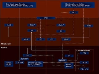
Eye Movements
Anatomy, Physiology and Clinical
Implications
Introduction
Eye movements
Outline for the session
To understand and describe
The extraocular muscles and neural circuitry involved in
moving the eyes.
The different types of eye movements: their purpose, neural
structure, and how they differ.
Eye movements
Think of this function not as vision, but as an effector system
required to move the eyes, therefore a legitimate area of
motor control research.
This relatively simple motor system can be compared to
other muscular systems, and the stimulus can be defined
precisely.
Eye movements involve rotation of the eyes in the socket.
Why ???
Why do the eyes move?
We need our eyes to increase the visual range that can be covered.
Why do the eyes move?
Eye movements bring the image onto the fovea.
Receptors for vision located on back of eyeball, on the
retina.
Visual
axis
Why do the eyes move?
Concentration of receptors providing high
resolution (clear image) = fovea.
More cortical area devoted to foveal
region, so need to have image focused
here.
How…
The III, IV & VI
MLF (medial longitudinal
fasciculus)
• Internuclear connection
• Nonvestibular pathways
(among CN nuclei)
– VI-contralateral III
– III-VII, VII-V, V-XII, XII-VII
• Vestibular pathways:
– Eye
– Ear
– Neck
– Limb extensors
p389
Conjugate Eye Movements
• Yoking mechanism
• Via MLF
E.g. CN VI  contralat. CN III
Extraocular Muscles: three complimentary pairs
Muscle properties
• More complex than somatomotor muscle
fibers
–5 distinct fiber types (vs 2 - fast & slow)
•Unclear why
–More proprioceptors (?)
•but proprioception is (too) slow
–Much higher innervation ratio (nerve endings/fiber)
• Built for speed, not for comfort
–8 ms twitch time (2-3 times faster than fast
somatomotor fibers)
Muscle innervation
(oculomotor nerves)
• At rest, firing rate of an
individual nerve is linear with
eye position
• Different nerves have different
slopes and offsets
–Sum to a non-linear increasing function
that matches passive muscle properties
MidlineFar left Far
right
Eye position
Innervation
of right l. r.
(Firing rate
in sp/s)
When the eyes move, you need still
more force - activity is proportional to
position and velocity (stay tuned)
Innervation…
Types….
Five types of eye movements
Each eye movement:
1) serves a unique function and
2) has properties particularly suited to that function
Five types:
Gaze shifting
1) Saccades
2) Vergence
3) Smooth pursuit
Gaze holding
4) Vestibular ocular reflex
5) Optokinetic reflex (OKR)
Saccades…
Saccades
Rapid rotation of the eyes that bring images onto the fovea.
Saccades are made spontaneously in response to a sudden appearing
object, or to scan a scene or to read.
Thus, saccades can be either voluntary or reflexive.
Saccades
Saccades allow us to scan the visual field on parts of the scene that convey the
most significant information.
We make about 3
saccades a second, and
> 150,000 saccadic eye
movements a day.
Saccades
The trigger for a saccade is position error, the difference
where your looking and where you want to look.
So when the target isn’t centred on the fovea, a saccade
brings the eyes onto the target.
200 ms
Target position
Eye position
Time
Right
Left
Saccades
Saccade amplitude ranges from miniature eye movements (0.1o
) to
movements ~45o
amplitude from the straight ahead position.
Saccade amplitude (deg)
Peakvelocity(deg/s)
Saccade are fast (peak velocity
500o
/sec), but peak velocity
varies with saccade amplitude.
Neural control of saccades
The discharge frequency of extraocular motor neurons is
directly proportional to the position and velocity of the eye.
Saccade
onset
5 ms
Horizontal eye position
Abducens motor
neuron
Action potential
Neural control of saccades
The saccade signal of motor neurons has the form of a pulse-
step.
Eye position
Spikes
Eye velocity
Pulse
Step
Spikes/sec
Height of the step determines the amplitude of the saccade
Height of the pulse determines the speed of the saccade.
Neural control of saccades
The saccade signal of motor neurons has the form of a pulse-
step.
Eye position
Spikes
Eye velocity
Pulse
Step
Spikes/sec
The pulse is the phasic signal that commands the eyes to move.
The step is the tonic signal that commands the eyes to hold in an eccentric position.
Neural control of saccades
The saccade signal of motor neurons has the form of
a pulse-step.
Eye position
Spikes
Eye velocity
Pulse
Step
Spikes/sec
The duration of the pulse
determines the duration of the
saccade.
Saccadic Eye Movements
(‘saccades’)
Subtypes often referred to:
1. Volitional (‘purposive’)
-predictive, anticipatory
-memory-guided
-antisaccades
2. Reflexive
3. Express saccades
4. Spontaneous
5. Quick phase of nystagmus
Velocity, Duration and the ‘Main Sequence’
Visually Guided Saccades
Deviations from main sequence:
-saccades in complete darkness
-saccades to auditory stimuli
-saccades to remembered targets
-saccades made in the opposite
direction (antisaccades)
[abducens, trochlear, om nucleus]
[cerebellum, brainstem]
[pprf, mrf]
[dorsal raphe]
Neural control of saccades
1) The horizontal gaze centre is in
the paramedian pontine reticular
formation (PPRF) next to the
abducens nucleus.
The direction of saccades is dictated by premotor neurons in two gaze
centres in the reticular formation.
Neural control of saccades
2) The vertical gaze center is in the rostral interstitial nucleus of the medial
longitudinal fasciculus (rostral iMLF) in the mesencephalic reticular formation
near the oculo-motor nucleus.
Major Pathways for Saccadic Eye Movements
Superficial Layers
Intermediate and
Deep Layers
Retina
Major Connections of the Superior Colliculus
Striate cortex (V1) Extrastriate cortex
(e.g. V4, MT)
Parietal cortex (e.g. LIP)
Frontal Eye Field
Inferior Pulvinar
Brainstem Saccade
generator
Dorsal lateral geniculate
nucleus (dLGN)
Medio-dorsal thalamus
SC
Visual and Motor Related Properties of Cells in the
Superior Colliculus
Superficial Layers:
Intermediate:
Deep Layers
SC
Visual Receptive Fields,
Some enhanced Visual Responses, but
no Presaccadic (motor) bursts; ‘visual’
cells
Visual Receptive Fields and Presaccadic
Bursts before saccades to ‘movement field’;
‘visuomotor cells’, ‘visually-triggered motor
cells’
No visual RFs, just movement fields,
Presaccadic burst gets earlier as you go
deeper
Sparks and Mays, 1980
Tuning of SC burst neuron to direction and amplitude of saccades
Enhancement of Superior Colliculus Visual Responses and
the Need to Dissociate Behavioral Components
Passive
fixation
Saccade to
RF target
Saccade to
Control target
‘Movement field’ of Superior Colliculus neuron
Map of Stimulation Evoked Saccades
amplitude
elevation
Rostral
Caudal
Major Pathways for Saccadic Eye Movements
Lateral Intraparietal Area (LIP): visual, saccade-related
and mnemonic responses
Incidence of ‘light-sensitive’, ‘saccade-coincident’
and ‘memory’ activity in LIP
Pursuit …
Smooth pursuit
Saccades involve fixating on a point then jumping to the next object
of interest.
Smooth pursuit involved keeping a visible moving target on the
fovea.
Although voluntary, smooth pursuit requires a stimulus to track;
they cannot be executed in the absence of some environmental
stimulus.
The trigger for a smooth pursuit movement is a velocity difference
between the eyes and the target.
Smooth pursuit
The pursuit system needs to compute the speed of the moving
stimulus to produce the proper eye velocity.
Fast moving stimuli (30o
/s) cannot be tracked with precision, and
they usually elicit a saccade.
Smooth pursuit
If a target starts to move
1) a pursuit movement is generated after
a short delay or latency (~100 ms)
2) a saccade is often used to catch up to
the target
3) finally if the pursuit is perfect, your
eye tracks the moving object
Target
movement
Eye
movement
100 ms
Catch-up
saccade
Time
Amplitude 1
2
3
Smooth pursuit
How well do pursuit movements match the movement of the object
being tracked?
Slow targets are matched perfectly; less than 0.33 mm retinal
slip/sec.
Target moving at higher speeds – large retinal slips.
Retinal slip is the distance between the image of the target on the
retina and the fovea.
Smooth pursuit vs. Saccade
Smooth pursuit isn’t ballistic, like saccades, and instead moves
smoothly.
Agonists and antagonists are activated
simultaneously – in saccades, only
muscle agonists are used.
So smooth pursuit movements are produced by creating small
differences in the tensions of the opposing ocular muscles.
Neural control of smooth pursuit
The sequence of structures that are used to generate pursuit eye
movement:
Striate Cortex
↓
MT & MST
↓
Pontine nuclei
↓
Cerebellum
↓
Brainstem
Neural control of smooth pursuit
The brainstem
structures that are
used to generate
pursuit eye
movement:
Abducens
nucleus
Oculo-
motor
nucleus
Medial
longitudinal
fasciculus
Vestibulo-
cerebellum
Trochlear
nucleus
Pontine
nucleus
Vestibular nucleus
and PPRF
The FEF
Microstimulation of the Frontal Eye Field
Continuum of Visual and Motor Responses in the FEF
Stimulation-Evoked Smooth Pursuit Movements
Gaze-holding eye movements
Gaze holding eye movements include the vestibular ocular reflex and
the optokinetic reflex.
Their purpose is to keep the image of the whole scene still on the entire
retina when the head moves (or the scene moves).
Vergence…
Vergence eye movements
Vergence eye movements aligns the fovea of each eye with targets
located at different distances from the observer.
Vergence eye movements
They are just disconjugate movements, i.e., eyes move in opposite
directions, producing a convergence or divergence of each eye’s visual
field to focus an object that is near or far.
Vergence eye movements
Convergence is one of the three reflexive responses elicited by a near
target.
The other two include
accommodation of the lens,
which brings the object into
focus, and pupil constriction,
which increases the depth of
field and sharpen the retinal
image.
Accommodation
Vergence eye movements
Either blur or retina disparity will generate vergence.
Latency for vergence
movements is ~160
ms.
Maximum velocity is
20o
/sec.
VOR …
Vestibulo-
ocular Reflex
• Contralateral CN VI n.
• From CN VI n
–  ipsi. CN III n
Vestibular ocular reflex
Vestibular ocular reflex (VOR)
stabilizes the eyes relative to the
external world, compensating for
head movements, by rotating the
eyes in opposite direction.
Vestibular ocular reflex
This permits the visual axis, or gaze, to remain on the newly
foveated stimulus (but, visual stimulus is not required!)
This reflex prevents visual images from slipping on the surface of
the retina (retinal slip) as head position varies.
The latency of the VOR is 14 ms.
It can accurately follow head velocities up to 300o
/s.
Can be produce without a stimulus (not visual).
Vestibular ocular reflex
The VOR also acts during the coordinated eye-head movements
(gaze shifts), compensating for the portion of the head movement
that lags the more rapid displacements of the eye.
Vestibular ocular reflex sensors
Head movements are sensed by the labyrinth of the inner ear which
acts as an accelerometer.
Acceleration and deceleration are the triggering stimuli (not
velocity, so unaffected by a constant rate).
Vestibular ocular reflex
Three semicircular canals at right angles to each other.
They each contain fluid (endolymph)
and a transducer (cupula).
Vestibular ocular reflex
The fluid transmits the direction and force of acceleration or
deceleration of the head via the cupula to the oculomotor system to
drive the eyes.
Vestibular ocular reflex pathway
The horizontal VOR is a short tri-synaptic
path (3-neuron arc) at
1) vestibular nucleus
2) abducens nucleus
3) lateral rectus muscle
Abducens
nucleusVestibular
nucleus
Oculomotor
nucleus
Vestibular ocular reflex pathway
The medial rectus
muscle is activated by
BOTH the abducens
nucleus and
oculomotor nucleus.
M R
L R
Abducens
nucleus
Oculo-
motor
nucleus
Medial
longitudinal
fasciculus
Head
turning
Optokinetic reflex
Sometimes also called Optokinetic nystagmus.
VOR doesn’t work well for slow, prolonged movements, so vision
through the optokinetic reflex (OKR) assists the VOR.
OKR is activated when the image of the world slips on a large
portion of the retina and produces a sense of self motion.
Optokinetic reflex
Sometimes consider to be a combination of smooth pursuit
(following the visual field) and a saccade to return the eyes
back to center – see a rhythmic back and forth movement
of the eyes.
Plasticity and Development
The VOR gain (eye amplitude/head amplitude) can change, for ex.
with glasses.
VOR adaptation are controlled by the cerebellum.
Prenatal development of eye movements:
15 weeks 20
Eyelid
movements
Slow eye movements
BIRTH
3525 30
Rapid eye
movements
Different development times suggest different neural systems.
Summary …
Abnormalities…
EOG
Measuring Eye Movements/Position
Scleral search coil Infrared Eye Tracking
Temporal resolution: analog
Spatial resolution: <0.1 deg.
Temporal resolution: video frame rate, <500 Hz
Spatial resolution: <0.25 deg.
scleral
coil
magnetic field
(2 axes)
ENG & VNG
Data Collection
Nystagmus
• Nystagmus is an involuntary, to-and-fro,
repetitive, rhythmic and generally conjugate
eye movement.
• Nystagmus may be pendular or jerky
•
• Pendular nystagmus is usually congenital
• Congenital nystagmus is often horizontal, does
not induce oscillopsia, increases in amplitude
during fixation and decreases during eyelid
closure.
• Jerk nystagmus is more common and of great
variety.
• Downbeat nystagmus is especially suggestive
of a cervicomedullary junction abnormality;
may also be observed in cerebellar
degeneration or lithium intoxication
• Convergence-retraction and retractorius
nystagmus (fast eyeball retractions into the
orbit) strongly suggests a tectal lesion
• some forms of nystagmus have little localizing
value, such as upbeat nystagmus, periodic
alternating nystagmus (the direction of
nystagmus is alternately inverted) and
circumduction nystagmus (rotator movement
around the eyeball axis, sweeping a circle or
an ellipse).
• Monocular nystagmus is most often seen in
internuclear ophthalmoplegia
Non Nystagmic Disorders
• Ocular flutter - consists of bursts (6-12 Hz) of
horizontal saccadic oscillations (2_5°
amplitude), without intersaccadic interval
• Opsoclonus - saccades are the same as in
ocular flutter, except that they are
omnidirectional and frequently associated with
axial myoclonus.
•
•
• Flutter and opsoclonus may be congenital or,
in childhood, reveal a neuroblastoma
• In adults, they may appear after several
infectious diseases (salmonella, coxsackie),
during brain stem encephalitis or malignant
pathology (paraneoplastic syndrome).
• They may be induced by drugs (lithium,
haloperidol) or by fluid balance and electrolyte
• Mention must also be made of microsaccadic
flutter, a rare micro saccadic oscillation (0.1-
0.5°) causing blurred vision, but without any
associated neurological disease.
• It could be due to malfunction of the brain
stem omnipause neurones
•
• Square wave jerks (SW]) consist of
consecutive to-and-fro, horizontal saccades of
small amplitude (O.S-3"), with a 200-ms inter
saccadic interval.
• They usually increase during smooth pursuit
and fixation. SW} are found in cerebellar
pathology, degenerative diseases, particularly
in PSP, and, rarely, in hemispheric diseases.
•
• Ocular bobbing - consists of an initial rapid
downward eye movement, followed after a
few milliseconds by a slow return to the initial
position, with a frequency of 10-1 S per
minute.
• It suggests a cerebellar or pontine lesion.
• Inverse ocular bobbing (or ocular dipping) consists
of an initial low downward movement, followed by
a rapid return to the baseline
•
• Reverse ocular bobbing consists of a rapid upward
eye movement, followed by a slow return. These
other forms of ocular bobbing have been described
in widespread diseases (metabolic encephalopathy,
bilateral hemispheric lesions).
•
• Ping-Pong gaze consists of alternating (2-
1SJmin) large-amplitude (60-80°) horizontal
slow eye movements, and is observed in
comatose: patients suffering from bilateral
mesodiencephalic lesions
• iSuperior oblique myokymia is a monocular
vertico-rotatory fast eye movement, appearing
spontaneously in midlife or rarely revealing a
tumour, and may be reduced by
carbamazepine
Peripheral Gaze Nystagmus:
• strongest on gaze in
direction of beating
• never vertical
• declines quickly (within
days to a couple of
weeks)
• Alexander's Law:
1st degree Nystagmus:
present only on lat.
gaze
2nd deg: both on
center and lat. side of
beat
3rd deg: on center, and
both lateral gazes.
• Video Periph Gaze
Central Nervous System
Lesions:
• Often bilateral beating
• Can have vertical beating
• declines slowly if at all
Some Central Gaze
Nystagmi:
• Bilateral Horiz. Gaze (Brun's) Nystagmus:
• Rebound Nystagmus:
• Periodic Alternating Nystagmus:
• Vertical Nystagmus:
• Congenital Nystagmus:
What is Going on here?:Voluntary Nystagmus
Bilateral Horiz. Gaze (Brun's)
Nystagmus:
• in large CPA tumors.
• Gaze ipsi to lesion generates large slow nyst,
with exp. decay in slow phase.
• Gaze contra to lesion generates small fast nyst,
in opposite direction of ipsi resp.
• Video Bruns
Rebound Nystagmus:
• Cerebellar disease
• movement-generated,
decays rapidly (10-20s)
• Beats in direction of
movement
• Video Rebound
Periodic Alternating
Nystagmus:
• Medullary disease. Periodic Alternating Video
• cyclic, 90 s one direction,
• 10 s nothing or vertical,
• then 90s in other direction, 10 s down time,
• and back again.
• present w/ eyes open or closed.
• strongest in middle of phases>>visual impairment.
Vertical Nystagmus:
• Brainstem/Cerebellar or Inf. olivary disease
• Can be generated by alcohol, drugs, too.
• Upbeat Video
• Downbeat Video
Congenital Nystagmus:
• From fixed brain defect either genetic or
developmental in origin.
• Pendular and/or jerk-type
• Disorder of slow eye movement sub-
system.
• Null points or periods.
• Convergence inhibition
• Congenital Video
Nystagmus: 2 types
1) Jerk nystagmus
2) Pendular nystagmus
500 ms
Pendular nystagmus
congenital
yes no
Evaluate for
Visual loss
Binocular
Visual loss
yes no yes no
Congenital
Sensory
nystagmus
Congenital
Motor
nystagmus
Binocular
Visual loss
MRI
Structural
lesion
No structural
lesion
Other etiologies
Treat and evaluate
etiology of
visual loss
Monocular or Asymmetric Oscillations
Age?
child adult
Spasmus
nutans
yes no
MRI
normal abnormal
Spasmus nutans R |o Cerebral lesion
Monocular visual
loss
yes no
W-up
ophthalmology
Monocular pendular
Monocular downbeat
INO
Sup. oblique myokymia
MRI
To Summarize…
• 5 distinct eye movements
• Saccades
• Smooth pursuit
• VOR
• OKN
• Vergence
• Interconnections hard wired with extreme
precision, predominantly controlled by the pre-
motor neural integrators in the pons and mid-
brain, ably assisted by vestibulo cerebellar
inputs
• The precision, the need and the type is
precisely analysed from the sensory input at
PPC and FEP and associated areas and
translated into meaningful triggers to the
subserving neural integrator at brainstem.
• Distinct pathology at different points in the
neuro axis can produce distinct and sometime
varied ocular movement abnormalities
• Some of them are highly localizable and some
are not.
Further Reading…
Eye movements
Eye movements

Eye movements

  • 1.
    Eye Movements Anatomy, Physiologyand Clinical Implications
  • 2.
  • 4.
    Eye movements Outline forthe session To understand and describe The extraocular muscles and neural circuitry involved in moving the eyes. The different types of eye movements: their purpose, neural structure, and how they differ.
  • 5.
    Eye movements Think ofthis function not as vision, but as an effector system required to move the eyes, therefore a legitimate area of motor control research. This relatively simple motor system can be compared to other muscular systems, and the stimulus can be defined precisely. Eye movements involve rotation of the eyes in the socket.
  • 6.
  • 7.
    Why do theeyes move? We need our eyes to increase the visual range that can be covered.
  • 8.
    Why do theeyes move? Eye movements bring the image onto the fovea. Receptors for vision located on back of eyeball, on the retina. Visual axis
  • 9.
    Why do theeyes move? Concentration of receptors providing high resolution (clear image) = fovea. More cortical area devoted to foveal region, so need to have image focused here.
  • 10.
  • 12.
  • 13.
    MLF (medial longitudinal fasciculus) •Internuclear connection • Nonvestibular pathways (among CN nuclei) – VI-contralateral III – III-VII, VII-V, V-XII, XII-VII • Vestibular pathways: – Eye – Ear – Neck – Limb extensors p389
  • 14.
    Conjugate Eye Movements •Yoking mechanism • Via MLF E.g. CN VI  contralat. CN III
  • 15.
    Extraocular Muscles: threecomplimentary pairs
  • 16.
    Muscle properties • Morecomplex than somatomotor muscle fibers –5 distinct fiber types (vs 2 - fast & slow) •Unclear why –More proprioceptors (?) •but proprioception is (too) slow –Much higher innervation ratio (nerve endings/fiber) • Built for speed, not for comfort –8 ms twitch time (2-3 times faster than fast somatomotor fibers)
  • 18.
    Muscle innervation (oculomotor nerves) •At rest, firing rate of an individual nerve is linear with eye position • Different nerves have different slopes and offsets –Sum to a non-linear increasing function that matches passive muscle properties MidlineFar left Far right Eye position Innervation of right l. r. (Firing rate in sp/s) When the eyes move, you need still more force - activity is proportional to position and velocity (stay tuned)
  • 19.
  • 20.
  • 22.
    Five types ofeye movements Each eye movement: 1) serves a unique function and 2) has properties particularly suited to that function Five types: Gaze shifting 1) Saccades 2) Vergence 3) Smooth pursuit Gaze holding 4) Vestibular ocular reflex 5) Optokinetic reflex (OKR)
  • 23.
  • 24.
    Saccades Rapid rotation ofthe eyes that bring images onto the fovea. Saccades are made spontaneously in response to a sudden appearing object, or to scan a scene or to read. Thus, saccades can be either voluntary or reflexive.
  • 25.
    Saccades Saccades allow usto scan the visual field on parts of the scene that convey the most significant information. We make about 3 saccades a second, and > 150,000 saccadic eye movements a day.
  • 26.
    Saccades The trigger fora saccade is position error, the difference where your looking and where you want to look. So when the target isn’t centred on the fovea, a saccade brings the eyes onto the target. 200 ms Target position Eye position Time Right Left
  • 27.
    Saccades Saccade amplitude rangesfrom miniature eye movements (0.1o ) to movements ~45o amplitude from the straight ahead position. Saccade amplitude (deg) Peakvelocity(deg/s) Saccade are fast (peak velocity 500o /sec), but peak velocity varies with saccade amplitude.
  • 28.
    Neural control ofsaccades The discharge frequency of extraocular motor neurons is directly proportional to the position and velocity of the eye. Saccade onset 5 ms Horizontal eye position Abducens motor neuron Action potential
  • 29.
    Neural control ofsaccades The saccade signal of motor neurons has the form of a pulse- step. Eye position Spikes Eye velocity Pulse Step Spikes/sec Height of the step determines the amplitude of the saccade Height of the pulse determines the speed of the saccade.
  • 30.
    Neural control ofsaccades The saccade signal of motor neurons has the form of a pulse- step. Eye position Spikes Eye velocity Pulse Step Spikes/sec The pulse is the phasic signal that commands the eyes to move. The step is the tonic signal that commands the eyes to hold in an eccentric position.
  • 31.
    Neural control ofsaccades The saccade signal of motor neurons has the form of a pulse-step. Eye position Spikes Eye velocity Pulse Step Spikes/sec The duration of the pulse determines the duration of the saccade.
  • 32.
    Saccadic Eye Movements (‘saccades’) Subtypesoften referred to: 1. Volitional (‘purposive’) -predictive, anticipatory -memory-guided -antisaccades 2. Reflexive 3. Express saccades 4. Spontaneous 5. Quick phase of nystagmus
  • 33.
    Velocity, Duration andthe ‘Main Sequence’ Visually Guided Saccades Deviations from main sequence: -saccades in complete darkness -saccades to auditory stimuli -saccades to remembered targets -saccades made in the opposite direction (antisaccades)
  • 35.
    [abducens, trochlear, omnucleus] [cerebellum, brainstem] [pprf, mrf] [dorsal raphe]
  • 36.
    Neural control ofsaccades 1) The horizontal gaze centre is in the paramedian pontine reticular formation (PPRF) next to the abducens nucleus. The direction of saccades is dictated by premotor neurons in two gaze centres in the reticular formation.
  • 37.
    Neural control ofsaccades 2) The vertical gaze center is in the rostral interstitial nucleus of the medial longitudinal fasciculus (rostral iMLF) in the mesencephalic reticular formation near the oculo-motor nucleus.
  • 38.
    Major Pathways forSaccadic Eye Movements
  • 40.
    Superficial Layers Intermediate and DeepLayers Retina Major Connections of the Superior Colliculus Striate cortex (V1) Extrastriate cortex (e.g. V4, MT) Parietal cortex (e.g. LIP) Frontal Eye Field Inferior Pulvinar Brainstem Saccade generator Dorsal lateral geniculate nucleus (dLGN) Medio-dorsal thalamus SC
  • 41.
    Visual and MotorRelated Properties of Cells in the Superior Colliculus Superficial Layers: Intermediate: Deep Layers SC Visual Receptive Fields, Some enhanced Visual Responses, but no Presaccadic (motor) bursts; ‘visual’ cells Visual Receptive Fields and Presaccadic Bursts before saccades to ‘movement field’; ‘visuomotor cells’, ‘visually-triggered motor cells’ No visual RFs, just movement fields, Presaccadic burst gets earlier as you go deeper
  • 42.
    Sparks and Mays,1980 Tuning of SC burst neuron to direction and amplitude of saccades
  • 43.
    Enhancement of SuperiorColliculus Visual Responses and the Need to Dissociate Behavioral Components Passive fixation Saccade to RF target Saccade to Control target
  • 45.
    ‘Movement field’ ofSuperior Colliculus neuron
  • 46.
    Map of StimulationEvoked Saccades amplitude elevation Rostral Caudal
  • 47.
    Major Pathways forSaccadic Eye Movements
  • 48.
    Lateral Intraparietal Area(LIP): visual, saccade-related and mnemonic responses
  • 49.
    Incidence of ‘light-sensitive’,‘saccade-coincident’ and ‘memory’ activity in LIP
  • 50.
  • 51.
    Smooth pursuit Saccades involvefixating on a point then jumping to the next object of interest. Smooth pursuit involved keeping a visible moving target on the fovea. Although voluntary, smooth pursuit requires a stimulus to track; they cannot be executed in the absence of some environmental stimulus. The trigger for a smooth pursuit movement is a velocity difference between the eyes and the target.
  • 52.
    Smooth pursuit The pursuitsystem needs to compute the speed of the moving stimulus to produce the proper eye velocity. Fast moving stimuli (30o /s) cannot be tracked with precision, and they usually elicit a saccade.
  • 53.
    Smooth pursuit If atarget starts to move 1) a pursuit movement is generated after a short delay or latency (~100 ms) 2) a saccade is often used to catch up to the target 3) finally if the pursuit is perfect, your eye tracks the moving object Target movement Eye movement 100 ms Catch-up saccade Time Amplitude 1 2 3
  • 54.
    Smooth pursuit How welldo pursuit movements match the movement of the object being tracked? Slow targets are matched perfectly; less than 0.33 mm retinal slip/sec. Target moving at higher speeds – large retinal slips. Retinal slip is the distance between the image of the target on the retina and the fovea.
  • 55.
    Smooth pursuit vs.Saccade Smooth pursuit isn’t ballistic, like saccades, and instead moves smoothly. Agonists and antagonists are activated simultaneously – in saccades, only muscle agonists are used. So smooth pursuit movements are produced by creating small differences in the tensions of the opposing ocular muscles.
  • 56.
    Neural control ofsmooth pursuit The sequence of structures that are used to generate pursuit eye movement: Striate Cortex ↓ MT & MST ↓ Pontine nuclei ↓ Cerebellum ↓ Brainstem
  • 57.
    Neural control ofsmooth pursuit The brainstem structures that are used to generate pursuit eye movement: Abducens nucleus Oculo- motor nucleus Medial longitudinal fasciculus Vestibulo- cerebellum Trochlear nucleus Pontine nucleus Vestibular nucleus and PPRF
  • 58.
  • 59.
    Microstimulation of theFrontal Eye Field
  • 60.
    Continuum of Visualand Motor Responses in the FEF
  • 61.
  • 62.
    Gaze-holding eye movements Gazeholding eye movements include the vestibular ocular reflex and the optokinetic reflex. Their purpose is to keep the image of the whole scene still on the entire retina when the head moves (or the scene moves).
  • 63.
  • 64.
    Vergence eye movements Vergenceeye movements aligns the fovea of each eye with targets located at different distances from the observer.
  • 65.
    Vergence eye movements Theyare just disconjugate movements, i.e., eyes move in opposite directions, producing a convergence or divergence of each eye’s visual field to focus an object that is near or far.
  • 66.
    Vergence eye movements Convergenceis one of the three reflexive responses elicited by a near target. The other two include accommodation of the lens, which brings the object into focus, and pupil constriction, which increases the depth of field and sharpen the retinal image. Accommodation
  • 67.
    Vergence eye movements Eitherblur or retina disparity will generate vergence. Latency for vergence movements is ~160 ms. Maximum velocity is 20o /sec.
  • 68.
  • 69.
    Vestibulo- ocular Reflex • ContralateralCN VI n. • From CN VI n –  ipsi. CN III n
  • 70.
    Vestibular ocular reflex Vestibularocular reflex (VOR) stabilizes the eyes relative to the external world, compensating for head movements, by rotating the eyes in opposite direction.
  • 71.
    Vestibular ocular reflex Thispermits the visual axis, or gaze, to remain on the newly foveated stimulus (but, visual stimulus is not required!) This reflex prevents visual images from slipping on the surface of the retina (retinal slip) as head position varies. The latency of the VOR is 14 ms. It can accurately follow head velocities up to 300o /s. Can be produce without a stimulus (not visual).
  • 72.
    Vestibular ocular reflex TheVOR also acts during the coordinated eye-head movements (gaze shifts), compensating for the portion of the head movement that lags the more rapid displacements of the eye.
  • 73.
    Vestibular ocular reflexsensors Head movements are sensed by the labyrinth of the inner ear which acts as an accelerometer. Acceleration and deceleration are the triggering stimuli (not velocity, so unaffected by a constant rate).
  • 74.
    Vestibular ocular reflex Threesemicircular canals at right angles to each other. They each contain fluid (endolymph) and a transducer (cupula).
  • 75.
    Vestibular ocular reflex Thefluid transmits the direction and force of acceleration or deceleration of the head via the cupula to the oculomotor system to drive the eyes.
  • 76.
    Vestibular ocular reflexpathway The horizontal VOR is a short tri-synaptic path (3-neuron arc) at 1) vestibular nucleus 2) abducens nucleus 3) lateral rectus muscle Abducens nucleusVestibular nucleus Oculomotor nucleus
  • 77.
    Vestibular ocular reflexpathway The medial rectus muscle is activated by BOTH the abducens nucleus and oculomotor nucleus. M R L R Abducens nucleus Oculo- motor nucleus Medial longitudinal fasciculus Head turning
  • 78.
    Optokinetic reflex Sometimes alsocalled Optokinetic nystagmus. VOR doesn’t work well for slow, prolonged movements, so vision through the optokinetic reflex (OKR) assists the VOR. OKR is activated when the image of the world slips on a large portion of the retina and produces a sense of self motion.
  • 79.
    Optokinetic reflex Sometimes considerto be a combination of smooth pursuit (following the visual field) and a saccade to return the eyes back to center – see a rhythmic back and forth movement of the eyes.
  • 80.
    Plasticity and Development TheVOR gain (eye amplitude/head amplitude) can change, for ex. with glasses. VOR adaptation are controlled by the cerebellum. Prenatal development of eye movements: 15 weeks 20 Eyelid movements Slow eye movements BIRTH 3525 30 Rapid eye movements Different development times suggest different neural systems.
  • 81.
  • 82.
  • 83.
  • 84.
    Measuring Eye Movements/Position Scleralsearch coil Infrared Eye Tracking Temporal resolution: analog Spatial resolution: <0.1 deg. Temporal resolution: video frame rate, <500 Hz Spatial resolution: <0.25 deg. scleral coil magnetic field (2 axes)
  • 85.
  • 86.
  • 88.
    Nystagmus • Nystagmus isan involuntary, to-and-fro, repetitive, rhythmic and generally conjugate eye movement. • Nystagmus may be pendular or jerky • • Pendular nystagmus is usually congenital • Congenital nystagmus is often horizontal, does not induce oscillopsia, increases in amplitude during fixation and decreases during eyelid closure.
  • 89.
    • Jerk nystagmusis more common and of great variety. • Downbeat nystagmus is especially suggestive of a cervicomedullary junction abnormality; may also be observed in cerebellar degeneration or lithium intoxication • Convergence-retraction and retractorius nystagmus (fast eyeball retractions into the orbit) strongly suggests a tectal lesion
  • 90.
    • some formsof nystagmus have little localizing value, such as upbeat nystagmus, periodic alternating nystagmus (the direction of nystagmus is alternately inverted) and circumduction nystagmus (rotator movement around the eyeball axis, sweeping a circle or an ellipse). • Monocular nystagmus is most often seen in internuclear ophthalmoplegia
  • 91.
    Non Nystagmic Disorders •Ocular flutter - consists of bursts (6-12 Hz) of horizontal saccadic oscillations (2_5° amplitude), without intersaccadic interval • Opsoclonus - saccades are the same as in ocular flutter, except that they are omnidirectional and frequently associated with axial myoclonus. • •
  • 92.
    • Flutter andopsoclonus may be congenital or, in childhood, reveal a neuroblastoma • In adults, they may appear after several infectious diseases (salmonella, coxsackie), during brain stem encephalitis or malignant pathology (paraneoplastic syndrome). • They may be induced by drugs (lithium, haloperidol) or by fluid balance and electrolyte
  • 93.
    • Mention mustalso be made of microsaccadic flutter, a rare micro saccadic oscillation (0.1- 0.5°) causing blurred vision, but without any associated neurological disease. • It could be due to malfunction of the brain stem omnipause neurones •
  • 94.
    • Square wavejerks (SW]) consist of consecutive to-and-fro, horizontal saccades of small amplitude (O.S-3"), with a 200-ms inter saccadic interval. • They usually increase during smooth pursuit and fixation. SW} are found in cerebellar pathology, degenerative diseases, particularly in PSP, and, rarely, in hemispheric diseases. •
  • 95.
    • Ocular bobbing- consists of an initial rapid downward eye movement, followed after a few milliseconds by a slow return to the initial position, with a frequency of 10-1 S per minute. • It suggests a cerebellar or pontine lesion.
  • 96.
    • Inverse ocularbobbing (or ocular dipping) consists of an initial low downward movement, followed by a rapid return to the baseline • • Reverse ocular bobbing consists of a rapid upward eye movement, followed by a slow return. These other forms of ocular bobbing have been described in widespread diseases (metabolic encephalopathy, bilateral hemispheric lesions). •
  • 97.
    • Ping-Pong gazeconsists of alternating (2- 1SJmin) large-amplitude (60-80°) horizontal slow eye movements, and is observed in comatose: patients suffering from bilateral mesodiencephalic lesions • iSuperior oblique myokymia is a monocular vertico-rotatory fast eye movement, appearing spontaneously in midlife or rarely revealing a tumour, and may be reduced by carbamazepine
  • 98.
    Peripheral Gaze Nystagmus: •strongest on gaze in direction of beating • never vertical • declines quickly (within days to a couple of weeks) • Alexander's Law: 1st degree Nystagmus: present only on lat. gaze 2nd deg: both on center and lat. side of beat 3rd deg: on center, and both lateral gazes. • Video Periph Gaze
  • 99.
    Central Nervous System Lesions: •Often bilateral beating • Can have vertical beating • declines slowly if at all
  • 100.
    Some Central Gaze Nystagmi: •Bilateral Horiz. Gaze (Brun's) Nystagmus: • Rebound Nystagmus: • Periodic Alternating Nystagmus: • Vertical Nystagmus: • Congenital Nystagmus: What is Going on here?:Voluntary Nystagmus
  • 101.
    Bilateral Horiz. Gaze(Brun's) Nystagmus: • in large CPA tumors. • Gaze ipsi to lesion generates large slow nyst, with exp. decay in slow phase. • Gaze contra to lesion generates small fast nyst, in opposite direction of ipsi resp. • Video Bruns
  • 102.
    Rebound Nystagmus: • Cerebellardisease • movement-generated, decays rapidly (10-20s) • Beats in direction of movement • Video Rebound
  • 103.
    Periodic Alternating Nystagmus: • Medullarydisease. Periodic Alternating Video • cyclic, 90 s one direction, • 10 s nothing or vertical, • then 90s in other direction, 10 s down time, • and back again. • present w/ eyes open or closed. • strongest in middle of phases>>visual impairment.
  • 104.
    Vertical Nystagmus: • Brainstem/Cerebellaror Inf. olivary disease • Can be generated by alcohol, drugs, too. • Upbeat Video • Downbeat Video
  • 105.
    Congenital Nystagmus: • Fromfixed brain defect either genetic or developmental in origin. • Pendular and/or jerk-type • Disorder of slow eye movement sub- system. • Null points or periods. • Convergence inhibition • Congenital Video
  • 106.
    Nystagmus: 2 types 1)Jerk nystagmus 2) Pendular nystagmus 500 ms
  • 107.
    Pendular nystagmus congenital yes no Evaluatefor Visual loss Binocular Visual loss yes no yes no Congenital Sensory nystagmus Congenital Motor nystagmus Binocular Visual loss MRI Structural lesion No structural lesion Other etiologies Treat and evaluate etiology of visual loss
  • 108.
    Monocular or AsymmetricOscillations Age? child adult Spasmus nutans yes no MRI normal abnormal Spasmus nutans R |o Cerebral lesion Monocular visual loss yes no W-up ophthalmology Monocular pendular Monocular downbeat INO Sup. oblique myokymia MRI
  • 116.
    To Summarize… • 5distinct eye movements • Saccades • Smooth pursuit • VOR • OKN • Vergence
  • 117.
    • Interconnections hardwired with extreme precision, predominantly controlled by the pre- motor neural integrators in the pons and mid- brain, ably assisted by vestibulo cerebellar inputs • The precision, the need and the type is precisely analysed from the sensory input at PPC and FEP and associated areas and translated into meaningful triggers to the subserving neural integrator at brainstem.
  • 118.
    • Distinct pathologyat different points in the neuro axis can produce distinct and sometime varied ocular movement abnormalities • Some of them are highly localizable and some are not.
  • 120.

Editor's Notes

  • #17 Proprios: no joint capsule for joint receptors, and you really want precision control, but too slow to be useful. innervation ratio: for better control?