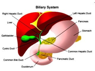
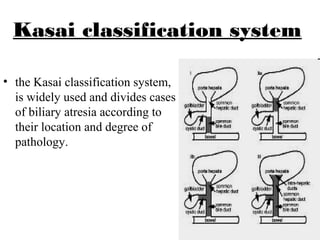
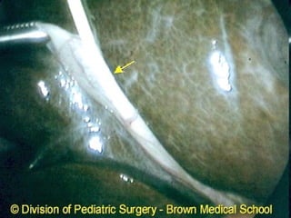
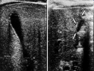
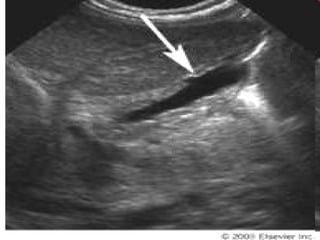
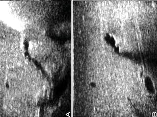
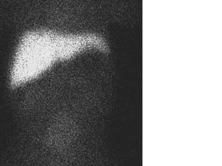
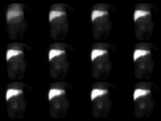
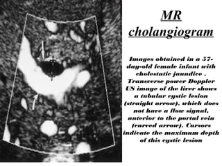
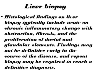
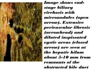
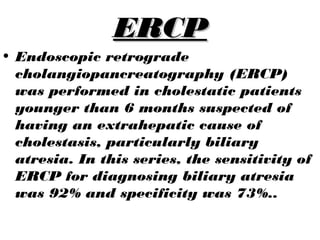
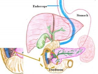
STARTING WITH THE NAME OF GOD WHO IS MOST
BENEFICIENT AND MERCIFUL
BILIARY ATRESIABILIARY ATRESIA
Presented by:
Mukesh Kumar
Flow of presentation
• Introduction
• Normal anatomy of hepatobiliary
system
• Causes of biliary atresia
• Clinical presentation
• Importance of early diagnosis of
biliary atresia.
• Diagnostic approach
Introduction
• Biliary atresia is the failure of a fetus
to develop an adequate pathway for
bile to drain from the liver to the
intestine.
• Biliary atresia is a rare disease of
unknown etiology which leads to
progressive fibrosis and destruction of
both the intra- and extra-hepatic
biliary duct system with resultant
cholestasis, cirrhosis and portal
hypertension.
causes
• More common in females than males.
• Incidence highest in Asian
populations
• It is possible that a viral infection is
responsible for this disease, but
evidence is not yet convincing. The
cause remains unknown.
• 10-20% of affected neonates have
associated congenital defects,
including situs inversus, polysplenia,
malrotation, intestinal atresia, and
Clinical presentation
• Prolonged jaundice (ie. beyond 14 days age)Prolonged jaundice (ie. beyond 14 days age)
• Plus:
– Pale stools
– dark urine
– Hepatomegaly
– Failure to thrive
• Other presentations:
• Coagulopathy
• Abnormal antenatal scan ~5%
• Splenomegaly / portal hypertension (late signs)
Why we are worried?
•It is the most
common lethal
liver disease in
children.
• Untreated, this condition leads to
cirrhosis and DEATH within the
first years of life.
Kasai classification system
• the Kasai classification system,
is widely used and divides cases
of biliary atresia according to
their location and degree of
pathology.
Diagnostic
Approach
To
Biliary Atresia
• No single biochemical marker
can be used to distinguish
biliary atresia from other
causes of neonatal cholestasis.
• Neonatal jaundice may be due
to a variety of causes,
including hemolysis, sepsis,
and metabolic disease. When
jaundice persists longer than
14 days, biliary atresia and
idiopathic neonatal hepatitis
• Hematological tests
• Ultra sound
• Hepatobiliary scintigraphy
• MR cholangiogram
• Liver biopsy
• ERCP
(Intraoperative
cholangiography)
CBC in advanced disease; low platelets, low
WCC
Serum bilirubin T/D conjugated bilirubin > 2.0 mg/
deciliter or >20% of total bilirubin.
New born screening
(TORCH) ( galactosemia,
thyroid dysfunction, cystic
fibrosis and a variety of
metabolic diseases
usually normal in biliary atresia
PT, INR Usually normal but in late stages
abnormal
Serum AST, ALP, ALT,
gamma GT
Dispropotionately high
gamma-GT
Abdominal ultra sound
abdominal ultrasound liver usually
normal texture, possibly enlarged,
unlikely ductal dilatation; absent or
multiple spleen; ascites.
MR
cholangiogram
Images obtained in a 57-
day-old female infant with
cholestatic jaundice .
Transverse power Doppler
US image of the liver shows
a tubular cystic lesion
(straight arrow), which does
not have a flow signal,
anterior to the portal vein
(curved arrow). Cursors
indicate the maximum depth
of this cystic lesion
Liver biopsy
• Histological findings on liver
biopsy typically include acute on
chronic inflammatory change with
obstruction, fibrosis, and the
proliferation of ductal and
glandular elements. Findings may
not be definitive early in the
course of the disease, and repeat
biopsy may be required to reach a
definitive diagnosis.
Image shows end-
stage biliary
cirrhosis with
micronodules (open
arrow). Extensive
perivascular fibrosis
(arrowhead) and
dilated inspissated
cystic areas (closed
arrow) are seen at
the hepatic hilum
about 5-10 mm from
remnants of the
obstructed bile duct
ERCPERCP
• Endoscopic retrograde
cholangiopancreatography (ERCP)
was performed in cholestatic patients
younger than 6 months suspected of
having an extrahepatic cause of
cholestasis, particularly biliary
atresia. In this series, the sensitivity of
ERCP for diagnosing biliary atresia
was 92% and specificity was 73%..
Biliary atresia, type B: cholangiographic features.
Intraoperative cholangiography is performed by catheterization
of the gallbladder (asterisk) and injection of contrast. This
infant has an extremely hypoplastic but patent common bile
duct (arrow). There is no filling of the common hepatic duct or
intrahepatic ducts. This is consistent with Type B biliary atresia.
Importance of ERCP
• ERCP is not an alternative to
noninvasive imaging but can avoid
unnecessary surgical procedures
in approximately 25% of cases.
They therefore recommended that
ERCP be performed before
explorative laparotomy in all
patients suspected of having
biliary atresia.
Thank you
Any
question
??

biliary atresia

  • 1.
    STARTING WITH THENAME OF GOD WHO IS MOST BENEFICIENT AND MERCIFUL
  • 2.
  • 3.
    Flow of presentation •Introduction • Normal anatomy of hepatobiliary system • Causes of biliary atresia • Clinical presentation • Importance of early diagnosis of biliary atresia. • Diagnostic approach
  • 4.
    Introduction • Biliary atresiais the failure of a fetus to develop an adequate pathway for bile to drain from the liver to the intestine. • Biliary atresia is a rare disease of unknown etiology which leads to progressive fibrosis and destruction of both the intra- and extra-hepatic biliary duct system with resultant cholestasis, cirrhosis and portal hypertension.
  • 6.
    causes • More commonin females than males. • Incidence highest in Asian populations • It is possible that a viral infection is responsible for this disease, but evidence is not yet convincing. The cause remains unknown. • 10-20% of affected neonates have associated congenital defects, including situs inversus, polysplenia, malrotation, intestinal atresia, and
  • 7.
    Clinical presentation • Prolongedjaundice (ie. beyond 14 days age)Prolonged jaundice (ie. beyond 14 days age) • Plus: – Pale stools – dark urine – Hepatomegaly – Failure to thrive • Other presentations: • Coagulopathy • Abnormal antenatal scan ~5% • Splenomegaly / portal hypertension (late signs)
  • 8.
    Why we areworried?
  • 9.
    •It is themost common lethal liver disease in children. • Untreated, this condition leads to cirrhosis and DEATH within the first years of life.
  • 10.
    Kasai classification system •the Kasai classification system, is widely used and divides cases of biliary atresia according to their location and degree of pathology.
  • 12.
  • 13.
    • No singlebiochemical marker can be used to distinguish biliary atresia from other causes of neonatal cholestasis. • Neonatal jaundice may be due to a variety of causes, including hemolysis, sepsis, and metabolic disease. When jaundice persists longer than 14 days, biliary atresia and idiopathic neonatal hepatitis
  • 14.
    • Hematological tests •Ultra sound • Hepatobiliary scintigraphy • MR cholangiogram • Liver biopsy • ERCP (Intraoperative cholangiography)
  • 15.
    CBC in advanceddisease; low platelets, low WCC Serum bilirubin T/D conjugated bilirubin > 2.0 mg/ deciliter or >20% of total bilirubin. New born screening (TORCH) ( galactosemia, thyroid dysfunction, cystic fibrosis and a variety of metabolic diseases usually normal in biliary atresia PT, INR Usually normal but in late stages abnormal Serum AST, ALP, ALT, gamma GT Dispropotionately high gamma-GT Abdominal ultra sound abdominal ultrasound liver usually normal texture, possibly enlarged, unlikely ductal dilatation; absent or multiple spleen; ascites.
  • 21.
    MR cholangiogram Images obtained ina 57- day-old female infant with cholestatic jaundice . Transverse power Doppler US image of the liver shows a tubular cystic lesion (straight arrow), which does not have a flow signal, anterior to the portal vein (curved arrow). Cursors indicate the maximum depth of this cystic lesion
  • 22.
    Liver biopsy • Histologicalfindings on liver biopsy typically include acute on chronic inflammatory change with obstruction, fibrosis, and the proliferation of ductal and glandular elements. Findings may not be definitive early in the course of the disease, and repeat biopsy may be required to reach a definitive diagnosis.
  • 23.
    Image shows end- stagebiliary cirrhosis with micronodules (open arrow). Extensive perivascular fibrosis (arrowhead) and dilated inspissated cystic areas (closed arrow) are seen at the hepatic hilum about 5-10 mm from remnants of the obstructed bile duct
  • 24.
    ERCPERCP • Endoscopic retrograde cholangiopancreatography(ERCP) was performed in cholestatic patients younger than 6 months suspected of having an extrahepatic cause of cholestasis, particularly biliary atresia. In this series, the sensitivity of ERCP for diagnosing biliary atresia was 92% and specificity was 73%..
  • 27.
    Biliary atresia, typeB: cholangiographic features. Intraoperative cholangiography is performed by catheterization of the gallbladder (asterisk) and injection of contrast. This infant has an extremely hypoplastic but patent common bile duct (arrow). There is no filling of the common hepatic duct or intrahepatic ducts. This is consistent with Type B biliary atresia.
  • 28.
    Importance of ERCP •ERCP is not an alternative to noninvasive imaging but can avoid unnecessary surgical procedures in approximately 25% of cases. They therefore recommended that ERCP be performed before explorative laparotomy in all patients suspected of having biliary atresia.
  • 29.
  • 30.