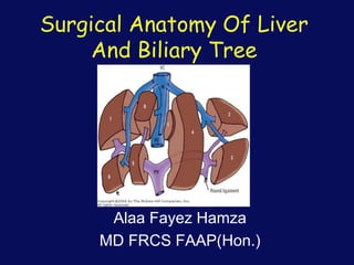
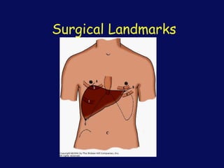
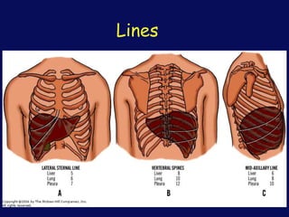
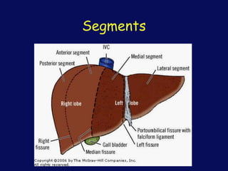
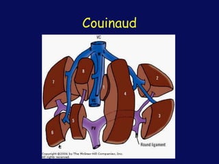
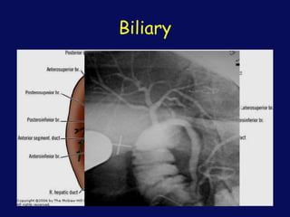
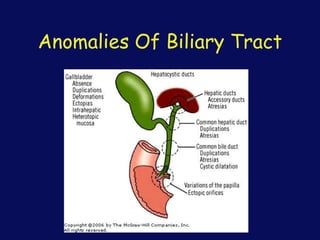
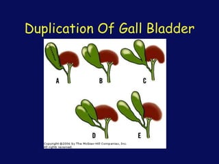
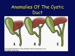
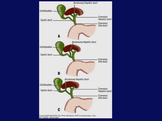
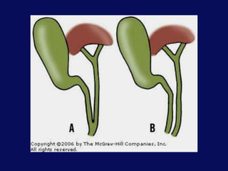
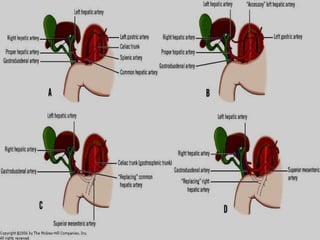
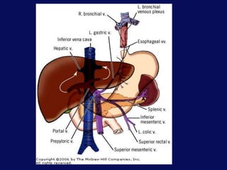
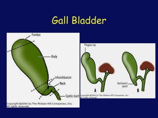
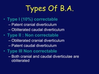
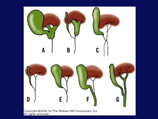
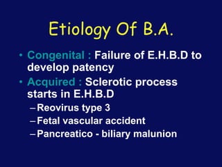
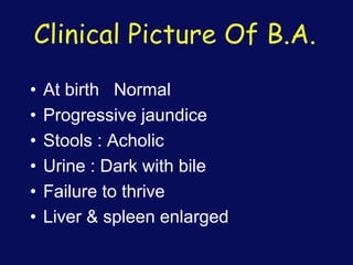
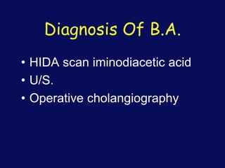
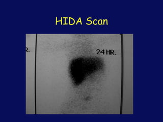
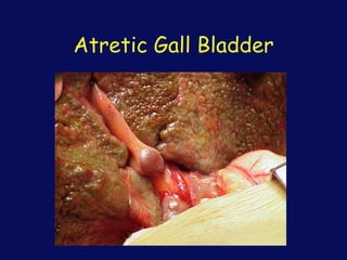
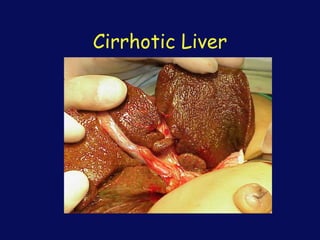
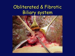
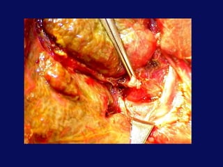
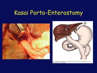
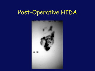
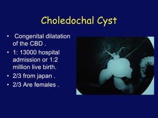
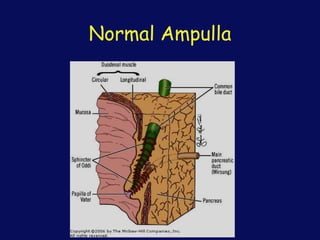
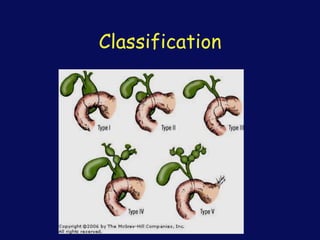
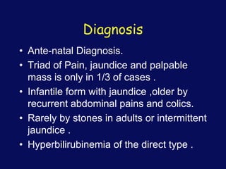
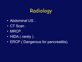
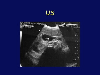
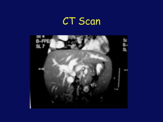
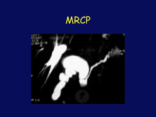
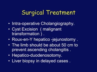
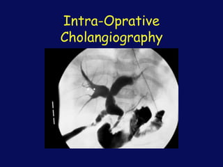
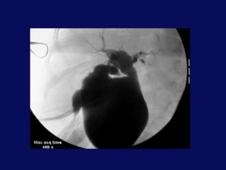
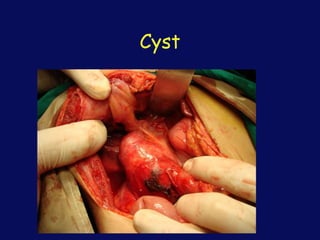
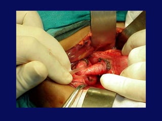
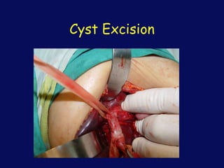
Surgical Anatomy Of Liver
And Biliary Tree
Alaa Fayez Hamza
MD FRCS FAAP(Hon.)
Liver Embryology
• The transverse septum (septum
transversum) arises at an embryonic
junctional site. The junctional region
externally is where the ectoderm of the
amnion meets the endoderm of the yolk sac.
The junctional region internally is where the
foregut meets the midgut. The mesenchymal
structure of the transverse septum
provides a support within which both blood
vessels and the liver begin to form. This
structure grows rapidly.
• The transverse septum then differentiates
to form the hepatic diverticulum and the
hepatic primordium, these two structures
together will go on to form different
components of the mature liver and gall
bladder.
• The rapidly developing liver forms a visible
surface bulge on the embryo directly under
the heart bulge.
Embryology
• At 4 wks of gestation, the biliary duct
system originate from hepatic
diverticulum of the foregut &
differentiate into
–Cranial component : Prox E.H.B.D &
most of the I.H.B.D
–Caudal component : G.B, cystic duct
C.B.D
Parenchyma
• By Day 24, the hepatic diverticulum is
growing into the transverse septum that,
at this stage, contains the vitelline
and umbilical veins.
• As the hepatic bud appears, the
hepatic and cardiac mesenchyme
become segregated.
• The second and third inductions occur
when the hepatic mesenchyme
stimulates the cells of the endodermal
cords to differentiate into hepatocytes,
and simultaneously the endodermal
hepatocytes stimulate the mesenchyme
to form the endothelial cells of the liver
sinusoids.
• Growth of the liver makes it bulge out of
the transverse septum so that the liver
becomes a truly abdominal organ lying
in the ventral mesentery. The bare area
of the liver and diaphragm remains as
an indication of the origin of the liver
from the transverse septum. The
asymmetry of the organ increases.
• The intrahepatic bile ducts were long
assumed to develop by extension of the
extrahepatic ducts. It is now believed that the
ducts differentiate from hepatic cells and join
the extrahepatic duct system secondarily. The
ducts appear first at the hilum and spread
peripherally. Bile may appear as early as the
third month and is often in the intestine by the
fifth month. By the ninth week, the liver
embraces as much as 10% of body volume.
Its relative size decreases to 5% by term.
Surgical Landmarks
Lines
Segments
Couinaud
Ant.& Post. Segments
Artery
Portal Vein
Hepatic veins
Biliary
Anomalies Of Biliary Tract
Duplication Of Gall Bladder
Anomalies Of The Cystic
Duct
Gall Bladder
Biliary Atresia
“B.A”
• Incidence
–1 : 15,000 live birth
• Sex
–F > M
Types Of B.A.
• Type I (10%) correctable
– Patent cranial diverticulum
– Obliterated caudal diverticulum
• Type II : Non correctable
– Obliterated cranial diverticulum
– Patent caudal diverticulum
• Type III Non correctable
– Both cranial and caudal diverticulae are
obliterated
Etiology Of B.A.
• Congenital : Failure of E.H.B.D to
develop patency
• Acquired : Sclerotic process
starts in E.H.B.D
–Reovirus type 3
–Fetal vascular accident
–Pancreatico - biliary malunion
Clinical Picture Of B.A.
• At birth Normal
• Progressive jaundice
• Stools : Acholic
• Urine : Dark with bile
• Failure to thrive
• Liver & spleen enlarged
Diagnosis Of B.A.
• HIDA scan iminodiacetic acid
• U/S.
• Operative cholangiography
HIDA Scan
Treatment Of B.A.
• Correctable:
–Roux en Y intest. anast. to prox B.D.
• Non correctable:
–Hepatic Porto - enterostomy (Kasai’s
operation)
• Liver transplantation
Prognostic factors
• Surgery below 45 days .
• Neonatal forms .
• Liver condition before surgery .
• Post-operative cholangitis .
• 25% will have normal life .
Atretic Gall Bladder
Cirrhotic Liver
Obliterated & Fibrotic
Biliary system
Kasai Porto-Enterostomy
Follow Up
• I.V. Antibiotics for 3-6 months .
• Liver assessment .
• Colour of stools.
• HIDA at 6 months .
• Treatment of cholangitis ,
Post-Operative HIDA
Post Operative
Complications Of B.A.
• Cholangitis
• Portal hypertension
• Liver failure
Choledochal Cyst
• Congenital dilatation
of the CBD .
• 1: 13000 hospital
admission or 1:2
million live birth.
• 2/3 from japan .
• 2/3 Are females .
Normal Ampulla
Etiology Of Choledochal Cyst
• Congenital abnormal insertion of the
pancreatic duct into the common bile
duct resulting in reflux of trypsin and
other pancreatic enzymes into the
common bile duct (CBD).
• Stenosis of the distal duct at the level of
duodenum or failure of canalization of
the distal part Of the CBD .
Classification
Diagnosis
• Ante-natal Diagnosis.
• Triad of Pain, jaundice and palpable
mass is only in 1/3 of cases .
• Infantile form with jaundice ,older by
recurrent abdominal pains and colics.
• Rarely by stones in adults or intermittent
jaundice .
• Hyperbilirubinemia of the direct type .
Radiology
• Abdominal US .
• CT Scan .
• MRCP .
• HIDA ( rarely ) .
• ERCP ( Dangerous for pancreatitis).
US
CT Scan
MRCP
Surgical Treatment
• Intra-operative Cholangiography.
• Cyst Excision ( malignant
transformation ) .
• Roux-en-Y hepatico -jejunostomy .
• The limb should be about 50 cm to
prevent ascending cholangitis .
• Hepatico-duodenosotomy.
• Liver biopsy in delayed cases .
Intra-Oprative
Cholangiography
Cyst
Cyst Excision
Hepatico-Duodenostomy
Post-Operative
• Intestinal Aseptic for long time .
• US & HIDA follow up .
• Liver assessment for delayed cases .

Liver& biliary

  • 1.
    Surgical Anatomy OfLiver And Biliary Tree Alaa Fayez Hamza MD FRCS FAAP(Hon.)
  • 2.
    Liver Embryology • Thetransverse septum (septum transversum) arises at an embryonic junctional site. The junctional region externally is where the ectoderm of the amnion meets the endoderm of the yolk sac. The junctional region internally is where the foregut meets the midgut. The mesenchymal structure of the transverse septum provides a support within which both blood vessels and the liver begin to form. This structure grows rapidly.
  • 3.
    • The transverseseptum then differentiates to form the hepatic diverticulum and the hepatic primordium, these two structures together will go on to form different components of the mature liver and gall bladder. • The rapidly developing liver forms a visible surface bulge on the embryo directly under the heart bulge.
  • 4.
    Embryology • At 4wks of gestation, the biliary duct system originate from hepatic diverticulum of the foregut & differentiate into –Cranial component : Prox E.H.B.D & most of the I.H.B.D –Caudal component : G.B, cystic duct C.B.D
  • 5.
    Parenchyma • By Day24, the hepatic diverticulum is growing into the transverse septum that, at this stage, contains the vitelline and umbilical veins. • As the hepatic bud appears, the hepatic and cardiac mesenchyme become segregated.
  • 6.
    • The secondand third inductions occur when the hepatic mesenchyme stimulates the cells of the endodermal cords to differentiate into hepatocytes, and simultaneously the endodermal hepatocytes stimulate the mesenchyme to form the endothelial cells of the liver sinusoids.
  • 7.
    • Growth ofthe liver makes it bulge out of the transverse septum so that the liver becomes a truly abdominal organ lying in the ventral mesentery. The bare area of the liver and diaphragm remains as an indication of the origin of the liver from the transverse septum. The asymmetry of the organ increases.
  • 8.
    • The intrahepaticbile ducts were long assumed to develop by extension of the extrahepatic ducts. It is now believed that the ducts differentiate from hepatic cells and join the extrahepatic duct system secondarily. The ducts appear first at the hilum and spread peripherally. Bile may appear as early as the third month and is often in the intestine by the fifth month. By the ninth week, the liver embraces as much as 10% of body volume. Its relative size decreases to 5% by term.
  • 9.
  • 10.
  • 11.
  • 12.
  • 13.
  • 14.
  • 15.
  • 16.
  • 17.
  • 18.
  • 19.
  • 20.
    Anomalies Of TheCystic Duct
  • 25.
  • 26.
  • 27.
    • Incidence –1 :15,000 live birth • Sex –F > M
  • 28.
    Types Of B.A. •Type I (10%) correctable – Patent cranial diverticulum – Obliterated caudal diverticulum • Type II : Non correctable – Obliterated cranial diverticulum – Patent caudal diverticulum • Type III Non correctable – Both cranial and caudal diverticulae are obliterated
  • 30.
    Etiology Of B.A. •Congenital : Failure of E.H.B.D to develop patency • Acquired : Sclerotic process starts in E.H.B.D –Reovirus type 3 –Fetal vascular accident –Pancreatico - biliary malunion
  • 31.
    Clinical Picture OfB.A. • At birth Normal • Progressive jaundice • Stools : Acholic • Urine : Dark with bile • Failure to thrive • Liver & spleen enlarged
  • 32.
    Diagnosis Of B.A. •HIDA scan iminodiacetic acid • U/S. • Operative cholangiography
  • 33.
  • 34.
    Treatment Of B.A. •Correctable: –Roux en Y intest. anast. to prox B.D. • Non correctable: –Hepatic Porto - enterostomy (Kasai’s operation) • Liver transplantation
  • 35.
    Prognostic factors • Surgerybelow 45 days . • Neonatal forms . • Liver condition before surgery . • Post-operative cholangitis . • 25% will have normal life .
  • 36.
  • 37.
  • 38.
  • 40.
  • 41.
    Follow Up • I.V.Antibiotics for 3-6 months . • Liver assessment . • Colour of stools. • HIDA at 6 months . • Treatment of cholangitis ,
  • 42.
  • 43.
    Post Operative Complications OfB.A. • Cholangitis • Portal hypertension • Liver failure
  • 44.
    Choledochal Cyst • Congenitaldilatation of the CBD . • 1: 13000 hospital admission or 1:2 million live birth. • 2/3 from japan . • 2/3 Are females .
  • 45.
  • 46.
    Etiology Of CholedochalCyst • Congenital abnormal insertion of the pancreatic duct into the common bile duct resulting in reflux of trypsin and other pancreatic enzymes into the common bile duct (CBD). • Stenosis of the distal duct at the level of duodenum or failure of canalization of the distal part Of the CBD .
  • 47.
  • 48.
    Diagnosis • Ante-natal Diagnosis. •Triad of Pain, jaundice and palpable mass is only in 1/3 of cases . • Infantile form with jaundice ,older by recurrent abdominal pains and colics. • Rarely by stones in adults or intermittent jaundice . • Hyperbilirubinemia of the direct type .
  • 49.
    Radiology • Abdominal US. • CT Scan . • MRCP . • HIDA ( rarely ) . • ERCP ( Dangerous for pancreatitis).
  • 50.
  • 51.
  • 52.
  • 53.
    Surgical Treatment • Intra-operativeCholangiography. • Cyst Excision ( malignant transformation ) . • Roux-en-Y hepatico -jejunostomy . • The limb should be about 50 cm to prevent ascending cholangitis . • Hepatico-duodenosotomy. • Liver biopsy in delayed cases .
  • 54.
  • 56.
  • 58.
  • 60.
  • 61.
    Post-Operative • Intestinal Asepticfor long time . • US & HIDA follow up . • Liver assessment for delayed cases .