CONTENTS
1. Exercise physiology
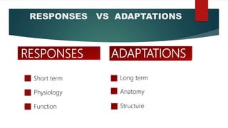
2.Responses Vs adaptations
3. Cardiovascular changes- short term and long term
4. Respiratory changes- short term and long term
5. Therapeutic benefits of exercise
6. References
3.
EXERCISE PHYSIOLOGY
EXERCISE: defined as intentional increased muscular activities, planned structured
and basically repetitive contraction and relaxation of group of muscles.
EXERCISE PHYSIOLOGY: Study of physio-chemical processes in the body that allow
conversion of chemical energy into mechanical work and the changes in the organ
systems in response to the effects of the work.
Continued skeletal muscle activity utilises energy that depends on the rate of
nutrients and oxygen supply to the exercising muscles.
4.
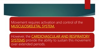
Movement requires activationand control of the
MUSCULOSKELETAL SYSTEM.
However, the CARDIOVASCULAR AND RESPIRATORY
SYSTEMS provide the ability to sustain this movement
over extended periods.
5.
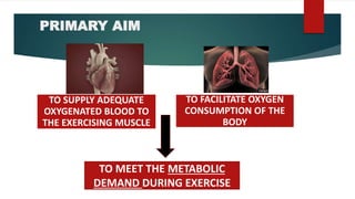
PRIMARY AIM
TO SUPPLYADEQUATE
OXYGENATED BLOOD TO
THE EXERCISING MUSCLE
TO FACILITATE OXYGEN
CONSUMPTION OF THE
BODY
TO MEET THE METABOLIC
DEMAND DURING EXERCISE
6.
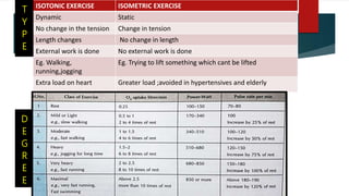
ISOTONIC EXERCISE ISOMETRICEXERCISE
Dynamic Static
No change in the tension Change in tension
Length changes No change in length
External work is done No external work is done
Eg. Walking,
running,jogging
Eg. Trying to lift something which cant be lifted
Extra load on heart Greater load ;avoided in hypertensives and elderly
T
Y
P
E
D
E
G
R
E
E
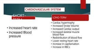
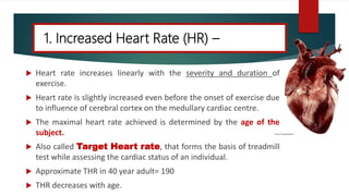
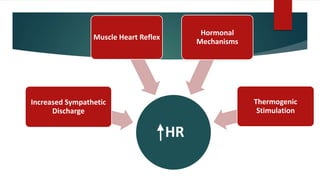
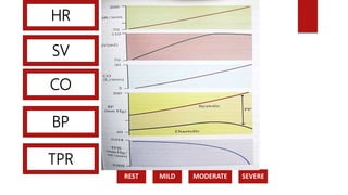
1. Increased HeartRate (HR) –
Heart rate increases linearly with the severity and duration of
exercise.
Heart rate is slightly increased even before the onset of exercise due
to influence of cerebral cortex on the medullary cardiac centre.
The maximal heart rate achieved is determined by the age of the
subject.
Also called Target Heart rate, that forms the basis of treadmill
test while assessing the cardiac status of an individual.
Approximate THR in 40 year adult= 190
THR decreases with age.
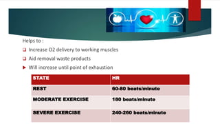
Helps to :
Increase O2 delivery to working muscles
Aid removal waste products
Will increase until point of exhaustion
STATE HR
REST 60-80 beats/minute
MODERATE EXERCISE 180 beats/minute
SEVERE EXERCISE 240-260 beats/minute
17.
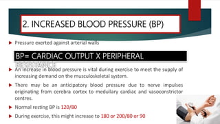
Pressure exertedagainst arterial walls
An increase in blood pressure is vital during exercise to meet the supply of
increasing demand on the musculoskeletal system.
There may be an anticipatory blood pressure due to nerve impulses
originating from cerebra cortex to medullary cardiac and vasoconstrictor
centres.
Normal resting BP is 120/80
During exercise, this might increase to 180 or 200/80 or 90
2. INCREASED BLOOD PRESSURE (BP)
BP= CARDIAC OUTPUT X PERIPHERAL
RESISTANCE
18.
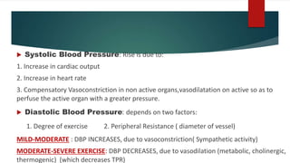
Systolic BloodPressure: Rise is due to:
1. Increase in cardiac output
2. Increase in heart rate
3. Compensatory Vasoconstriction in non active organs,vasodilatation on active so as to
perfuse the active organ with a greater pressure.
Diastolic Blood Pressure: depends on two factors:
1. Degree of exercise 2. Peripheral Resistance ( diameter of vessel)
MILD-MODERATE : DBP INCREASES, due to vasoconstriction( Sympathetic activity)
MODERATE-SEVERE EXERCISE: DBP DECREASES, due to vasodilation (metabolic, cholinergic,
thermogenic) (which decreases TPR)
19.
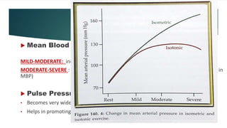
Mean BloodPressure:
MILD-MODERATE: increases
MODERATE-SEVERE : decreases as DBP decreases ( except isometric- overall increase in
MBP)
Pulse Pressure:
• Becomes very wide.
• Helps in promoting perfusion of skeletal muscles.
20.
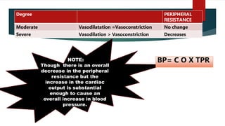
NOTE:
Though there isan overall
decrease in the peripheral
resistance but the
increase in the cardiac
output is substantial
enough to cause an
overall increase in blood
pressure.
Degree PERIPHERAL
RESISTANCE
Moderate Vasodilatation =Vasoconstriction No change
Severe Vasodilation > Vasoconstriction Decreases
BP= C O X TPR
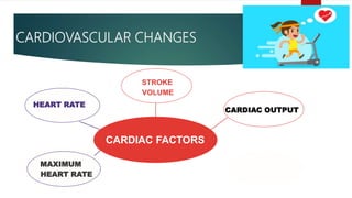
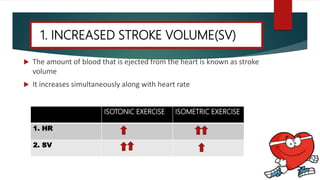
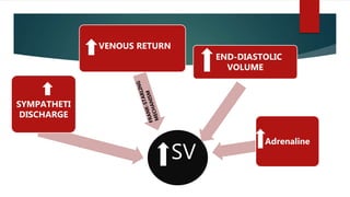
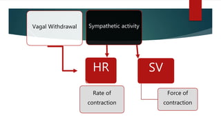
The amountof blood that is ejected from the heart is known as stroke
volume
It increases simultaneously along with heart rate
1. INCREASED STROKE VOLUME(SV)
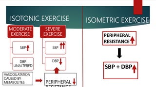
ISOTONIC EXERCISE ISOMETRIC EXERCISE
1. HR
2. SV
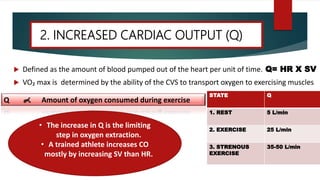
Defined asthe amount of blood pumped out of the heart per unit of time. Q= HR X SV
VO₂ max is determined by the ability of the CVS to transport oxygen to exercising muscles
2. INCREASED CARDIAC OUTPUT (Q)
Q Amount of oxygen consumed during exercise
STATE Q
1. REST 5 L/min
2. EXERCISE 25 L/min
3. STRENOUS
EXERCISE
35-50 L/min
• The increase in Q is the limiting
step in oxygen extraction.
• A trained athlete increases CO
mostly by increasing SV than HR.
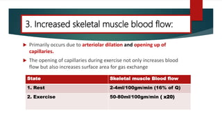
Primarily occursdue to arteriolar dilation and opening up of
capillaries.
The opening of capillaries during exercise not only increases blood
flow but also increases surface area for gas exchange
3. Increased skeletal muscle blood flow:
State Skeletal muscle Blood flow
1. Rest 2-4ml/100gm/min (16% of Q)
2. Exercise 50-80ml/100gm/min ( x20)
28.
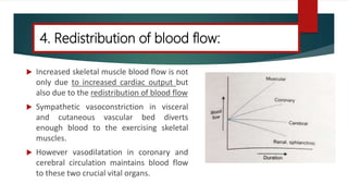
Increased skeletalmuscle blood flow is not
only due to increased cardiac output but
also due to the redistribution of blood flow
Sympathetic vasoconstriction in visceral
and cutaneous vascular bed diverts
enough blood to the exercising skeletal
muscles.
However vasodilatation in coronary and
cerebral circulation maintains blood flow
to these two crucial vital organs.
4. Redistribution of blood flow:
29.
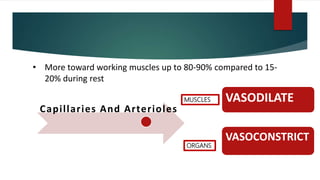
Capillaries And Arterioles
•More toward working muscles up to 80-90% compared to 15-
20% during rest
VASODILATE
VASOCONSTRICT
MUSCLES
ORGANS
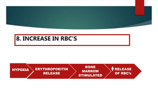
30.
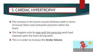
The increaseIn the hearts muscle thickness both in terms
of muscle fibres and contractile elements within the
heart.
This happens only to cope with the excessive work load
imposed upon the heart during work.
This is in order to increase the Stroke Volume
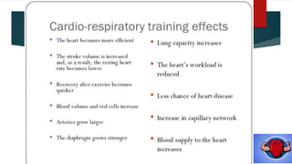
5. CARDIAC HYPERTROPHY
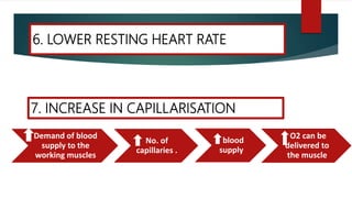
6. LOWER RESTINGHEART RATE
Demand of blood
supply to the
working muscles
No. of
capillaries .
blood
supply
O2 can be
delivered to
the muscle
7. INCREASE IN CAPILLARISATION
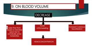
FLUID
LOSS/sweatingwater flow from
vesselsto interstitial
space due to
osmotically active
metabolites
HEMOCONCENTRATION
Increased Cutaneous
Vasodilatation
9. ON BLOOD VOLUME
DECREASE
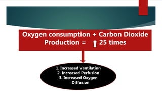
Oxygen consumption +Carbon Dioxide
Production = 25 times
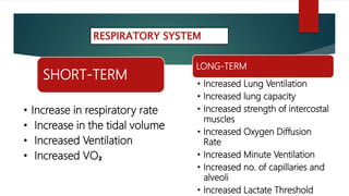
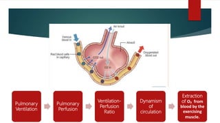
1. Increased Ventilation
2. Increased Perfusion
3. Increased Oxygen
Diffusion
39.
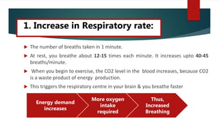
The numberof breaths taken in 1 minute.
At rest, you breathe about 12-15 times each minute. It increases upto 40-45
breaths/minute.
When you begin to exercise, the CO2 level in the blood increases, because CO2
is a waste product of energy production.
This triggers the respiratory centre in your brain & you breathe faster
Energy demand
increases
More oxygen
intake
required
Thus,
Increased
Breathing
1. Increase in Respiratory rate:
40.
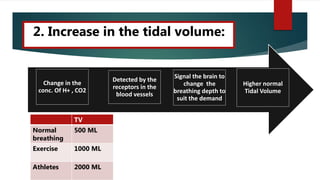
A. Increase inthe tidal volume:
Higher normal
Tidal Volume
Signal the brain to
change the
breathing depth to
suit the demand
Detected by the
receptors in the
blood vessels
Change in the
conc. Of H+ , CO2
2. Increase in the tidal volume:
TV
Normal
breathing
500 ML
Exercise 1000 ML
Athletes 2000 ML
41.
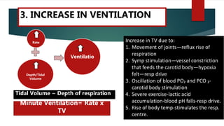
Rate
Depth/Tidal
Volume
Ventilatio
Increase in TVdue to:
1. Movement of joints—reflux rise of
respiration
2. Symp stimulation—vessel constriction
that feeds the carotid body—hypoxia
felt—resp drive
3. Oscillation of blood PO₂ and PCO ₂-
carotid body stimulation
4. Severe exercise-lactic acid
accumulation-blood pH falls-resp drive.
5. Rise of body temp-stimulates the resp.
centre.
Tidal Volume ~ Depth of respiration
Minute Ventilation= Rate x
TV
3. INCREASE IN VENTILATION
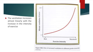
42.
The ventilationincreases
almost linearly with the
increase in the intensity
of exercise
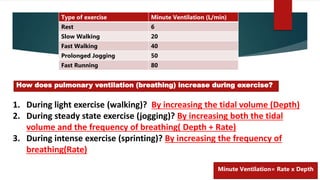
43.
1. During lightexercise (walking)? By increasing the tidal volume (Depth)
2. During steady state exercise (jogging)? By increasing both the tidal
volume and the frequency of breathing( Depth + Rate)
3. During intense exercise (sprinting)? By increasing the frequency of
breathing(Rate)
How does pulmonary ventilation (breathing) increase during exercise?
Type of exercise Minute Ventilation (L/min)
Rest 6
Slow Walking 20
Fast Walking 40
Prolonged Jogging 50
Fast Running 80
Minute Ventilation= Rate x Depth
44.
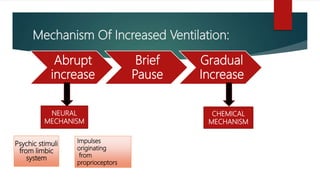
Mechanism Of IncreasedVentilation:
Abrupt
increase
Brief
Pause
Gradual
Increase
NEURAL
MECHANISM
CHEMICAL
MECHANISM
Psychic stimuli
from limbic
system
Impulses
originating
from
proprioceptors
45.
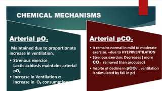
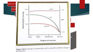
CHEMICAL MECHANISMS
Arterial pO₂
Maintaineddue to proportionate
increase in ventilation.
• Strenous exercise
Lactic acidosis maintains arterial
pO₂
• Increase in Ventilation α
Increase in O₂ consumption
Arterial pCO₂
• it remains normal in mild to moderate
exercise. –due to HYEPRVENTILATION
• Strenous exercise: Decreases ( more
CO₂ removed than produced)
• Inspite of decline in pCO₂ , ventilation
is stimulated by fall in pH
46.
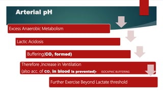
Arterial pH
Excess AnaerobicMetabolism
Lactic Acidosis
Buffering(CO₂ formed)
Therefore ,Increase in Ventilation
(also acc. of CO₂ in blood is prevented)- ISOCAPNIC BUFFERING
Further Exercise Beyond Lactate threshold
47.
Blood pH reduces
MetabolicAcidosis
Supralinear Stimulation of Ventilation
Ventilation stimulated (activation of peripheral chemoreceptors in
carotid and aortic bodies)
pCO₂ falls significantly
Decline in arterial pCO₂ induced by excessive ventilation
becomes the physiological mechanism for respiratory
compensation of metabolic acidosis.
48.
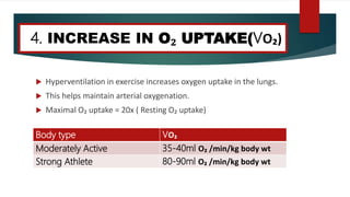
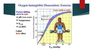
Hyperventilation inexercise increases oxygen uptake in the lungs.
This helps maintain arterial oxygenation.
Maximal O₂ uptake = 20x ( Resting O₂ uptake)
Body type VO₂
Moderately Active 35-40ml O₂ /min/kg body wt
Strong Athlete 80-90ml O₂ /min/kg body wt
4. INCREASE IN O₂ UPTAKE(VO₂)
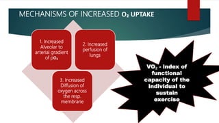
MECHANISMS OF INCREASEDO₂ UPTAKE
1. Increased
Alveolar to
arterial gradient
of pO₂
2. Increased
perfusion of
lungs
3. Increased
Diffusion of
oxygen across
the resp.
membrane
VO₂ - index of
functional
capacity of the
individual to
sustain
exercise
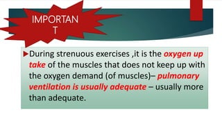
51.
During strenuous exercises,it is the oxygen up
take of the muscles that does not keep up with
the oxygen demand (of muscles)– pulmonary
ventilation is usually adequate – usually more
than adequate.
IMPORTAN
T
52.
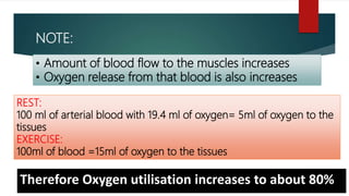
NOTE:
• Amount ofblood flow to the muscles increases
• Oxygen release from that blood is also increases
REST:
100 ml of arterial blood with 19.4 ml of oxygen= 5ml of oxygen to the
tissues
EXERCISE:
100ml of blood =15ml of oxygen to the tissues
Therefore Oxygen utilisation increases to about 80%
54.
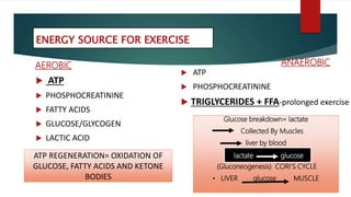
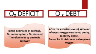
O₂ DEFICIT O₂ DEBT
In the beginning of exercise,
O ₂ consumption < O ₂ demand
Therefore met by anerobic
pathway
After the exercise(severe), Amount
of excess oxygen consumed during
recovery phase.
Cause: Lactic Acid removal requires
oxygen
55.
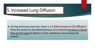
During strenuousexercise, there is a 3-fold increase in O2 diffusion
from the alveoli to the blood because of a massive increase in blood
flow to the lungs & dilation of the capillaries surrounding the
alveoli.
5. Increased Lung Diffusion
57.
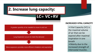
2. Increase lungcapacity:
More expansion provides more efficient inhalation and expiration
Lung Expansion occurs to meet the demand
A greater quantity of air needed to move in and out
LC= VC+RV
Vital Capacity (VC) is
the maximal volume
of air that can be
expired after maximal
inspiration in one
breath
Mainly due to the
increased strength of
intercostal muscles .
INCREASED VITAL CAPACITY
58.
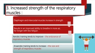
Diaphragm and intercostalmuscles increase in strength
Results in an improved ability to breathe in more air,
for longer with less fatigue .
Aerobic training tends to improve-- the endurance of
respiratory muscles
Anaerobic training tends to increase --the size and
strength of respiratory muscles
3. Increased strength of the respiratory
muscles :
59.
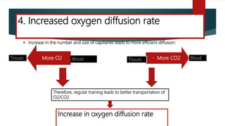
More O2 MoreCO2BloodTissues Tissues Blood
Therefore, regular training leads to better transportation of
O2/CO2
Increase in oxygen diffusion rate
Increase in the number and size of capillaries leads to more efficient diffusion:
4. Increased oxygen diffusion rate
60.
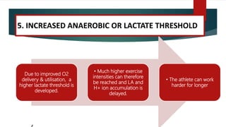
Due to improvedO2
delivery & utilisation, a
higher lactate threshold is
developed.
• Much higher exercise
intensities can therefore
be reached and LA and
H+ ion accumulation is
delayed.
• The athlete can work
harder for longer
5. INCREASED ANAEROBIC OR LACTATE THRESHOLD
REFERENCES
1. Textbook ofMedical Physiology- Prof G. K. Pal
2. Textbook of Medical Physiology- Sembulingam
3. Textbook of Medical Physiology-A. P Krishna
4. Textbook of Medical Physiology- Guyton
5. Healthy & Active Lifestyles- Book
6. Physiologic responses and long-term adaptations to
exercise-Article
7. Effects of exercise on the cardiovascular system-Article