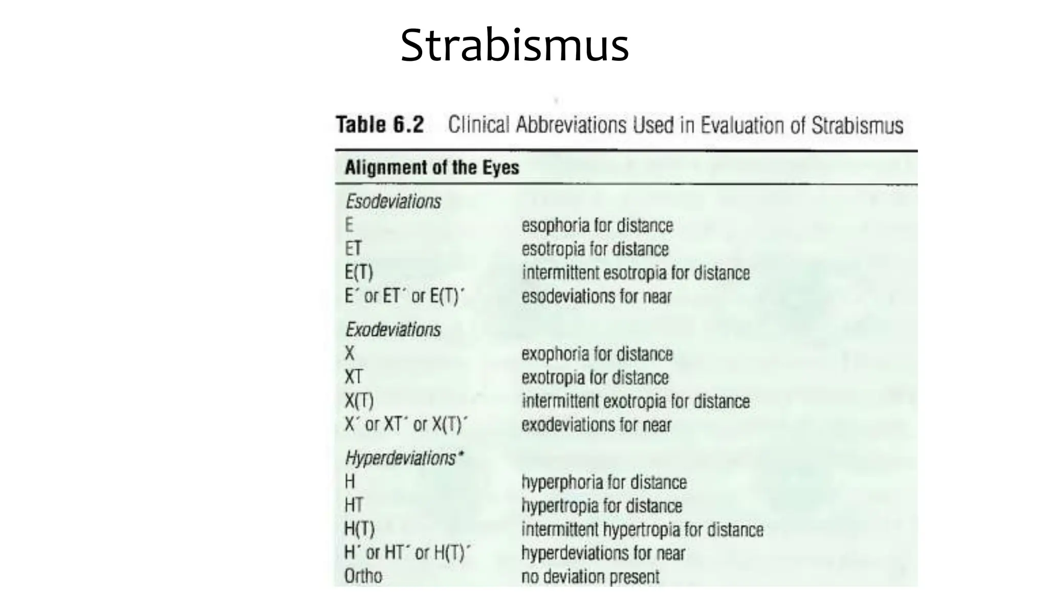
Strabismus
Eso-deviations
• Most common: >50% of the ocular deviations in the pediatric population
• Esodeviations occur with equal frequency in males and females
• Causes/Risk factors :-
– genetic, accommodative, innervational and anatomic…
– anisometropia, hyperopia,neurodevelopmental impairment, prematurity, low birth
weightmaternal smoking during pregnancy, and family history of strabismus
• The prevalence of esotropia increases with
– age (higher prevalence at 48–72 months compared with 6–11 months),
– moderate anisometropia, and
– moderate hyperopia.
• Insome families, a mendelian inheritance pattern has been observed.
• Amblyopia developsin approximately 50% of children who have esotropia.
Pseudoesotropia
• refers to the appearance of ET when the visual axes are in fact aligned.
• The appearance may be caused by
– a flat and broad nasal bridge,
– prominent epicanthal folds, a
– narrow interpupillary distance, or
– a negative angle kappa
• especially noticeable when the child gazes to either side
Congenital ( Infantile ) Esotropia Syndrome
• Infantile esotropia is defined as an esotropia that is present by 6 months of age
• If an ET is present after age 2 months, is constant, & measures 30 PD or more, it is
unlikely to resolve will probably require surgical intervention
• ocular instability of infancy
– Variable, transient, intermittent strabismus is commonly noted in the first 2–3 months of life.
– it is common to see both intermittent esotropia and exotropia in the same infant
– condition should resolve by 3 months of age but stimes persists, especially in premature infants.
• Initially small and intermittent =>increases in frequency & magnitude
=>constant > or equal to 35 PD.
Characteristics:-
- Equal visual acuity
- Large angle deviation usually >35 PD
- Cross - fixation is frequent
• Patients with infantile ET often have a family Hxof ET or other strabismus, but
well-defined genetic patterns are unusual.
• Infantile ET occurs more frequently in children born prematurely and in up to 30%
of children with
– neurologic &developmental problems, including cerebral palsy and hydrocephalus.
• Infantile ET has been associated with an increased risk of development of mental
illness by early adulthood
• The eyes may have equal vision, in which case alternate fixation or cross-
fixation will be present.
• Cross-fixation, the use of the adducted eye for fixation of objects in the
contralateral visual field, is associated with large-angle esotropias
• Amblyopia is commonly associated with infantile esotropia, and when it is
present, a fixation preference can be observed
• Versions and ductions are often normal initially
• Cycloplegic refraction characteristically reveals low hyperopia
Associated motor abnormalities
• Overelevation in adduction and dissociated strabismus complex develop in
more than 50% of patients, usually after 1–2 years of age.
• There may be an apparent abduction deficit because of cross-fixation
• The infant’s ability to abduct each eye can be demonstrated with
– the doll’s head maneuver or by observation after patching either of the patient’s eyes
• Latent Nystagmus
Ciancia syndrome
• severe form of infantile esotropia,
– consists of large-angle ET (>50Δ),
– abducting nystagmus, and
– mild abduction deficits.
• Children with this syndrome uniformly use cross-fixation.
Rx
• Significant hyperopic refractive error should be corrected by prescribing the full
cycloplegicrefraction
• Ocular alignment is rarely achieved without surgery in early-onset esotropia.
• The belief is that the eyes should be aligned by 2 years of age, preferably earlier, to
optimize binocular cooperation.
• Surgery can be performed in healthy children as early as age 4 month
• The most commonly performed initial procedure is recession of both MR.
• Recession of a MR combined with resection of the ipsilateral LR is also effective
• large deviations (typically >60D),
– some surgeons operate on 3 or even 4 horizontal rectus muscles at the time of the initial surgery,
or they add botulinum toxin injection to theMR Recession.
• Significant inferior oblique muscle overaction can be treated at the time of the initial
surgery
• Botulinum toxin may be most useful for smaller deviations
Accommodative esotropia
• A convergent deviation of the eyes associated with activation of the
accommodative reflex.
Characteristics:-
• Acquired
• Onset :-Infancy-late childhood (commonly 2 - 4 yrs.)
• At onset intermittent, then constant
• Usually hereditary
• May be precipitated by trauma/illness
• Amblyopia - common
1.Refractive (Hypermetropic) Accommodative ET
Three Mechanisms:-
1. Uncorrected hyperopia
2. Accommodative convergence
3. Insufficient fusional divergence
Because of uncorrected hyperopia, the patient must accommodate to focus the retinalimage.
Accommodation is accompanied by the other components of the near reflex, convergence ,miosis.
If the patient’s fusional divergence mechanism is insufficient tocompensate for the increased
Convergence tonus, ET results
The angle of ET is the same at distance and near fixation and is generally 20Δ - 30Δ.
Patients with refractive accommodative ET have an aver. of +4.00 D hyperopia.
Refractive (Hypermetropic) Accommodative Esotropia
Before After
2.Accommodative Nonrefractive Esotropia (High AC/A ratio Esotropia)
• Accommodation=>excess convergence tonus => Esotropia in the presence of
poor fusional divergence
• Patients with a high AC/A ratio have an excessive convergence response for the
amount of accommodation required to focuswhile wearing their full cycloplegic
correction.
• In this form of ET, the deviation is present only at near or is much larger at near
• May be seen in emmetropes, Low-High hyperopes, myopes ( Ave. + 2.25 D)
3.Partially accommodative esotropia
• show reduction in the angle of ET when wearing glasses but have a residual ET
despite provision of the full hyperopiccorrection.
• This is more likely to occur if there is a long delay in refractive correction.
• Insome cases, partially accommodative esotropia results from decompensation of a
purerefractive accommodative esotropia;
• in others, an initial nonaccommodative esotropia subsequently develops an
accommodative component.
• Treatment of refractive accommodative esotropia consists of correction of the
full amountof hyperopia, as determined under cycloplegia.
• A high AC/A ratio can be managed optically or surgically; it can also
be observed.
• Treatment of partially accommodative esotropia consists of strabismus surgery for
thedeviation that persists while the patient wears the full hyperopic correction
Acquired nonaccommodative esotropia
• Several types of ET not associated with activation of the accommodative
reflex may dev’p in later infancy (>6 months), childhood, or even adulthood
– Basic
– Cyclic
– Sensory (deprivation)
– Divergence insufficiency
• Primary (age-related distance esotropia)
• Secondary
– Spasm of the near reflex
– Consecutive esotropia
• Spontaneous
• Postsurgical
Basic acquired nonaccommodative esotropia
• is a comitant ET that develops after age 6months and
– is not associated with an accommodative component.
– the amount of hyperopia is not significant, and
– the angle of deviation is similar when measured at distance and near.
• Acquired ET may be acute in onset. In such cases, the patient immediately becomes
aware of the deviation and may have diplopia.
• A careful evaluation is important to rule out an accommodative or paretic component
• Temporary but prolonged disruption of binocular visionSuch as may result from a
hyphema, preseptal cellulitis, mechanical ptosis, or prolonged patching for
amblyopia—is a known precipitating cause of acquired nonaccommodative ET
• In this pts fusion is thought to be tenuous, so this temporary disruption of binocular
vision upsets the balance, resulting in esotropia
• it maybe a sign of an underlyingneurologic disorder, neuroimaging and neurologic
evaluation may be indicated,
• Therapy consists of amblyopia treatment, if necessary, and surgical
correction or botulinum toxin injection as soon as possible after the
onset of the deviation
Cyclic esotropia
• Cyclic esotropia is a rare form of strabismus
• Onset of cyclic esotropia is typically during the preschool years.
• ET is comitant& intermittent, usually occurring other day (48-hr cycle).
• Occlusion therapy may convert the cyclicdeviation into a constant one.
• Surgical treatment of cyclic esotropia is usually effective.
Sensory Deprivation Esotropia
Causes:-
any condition that reduces vision in one eye
E.g:-
– cataract
– corneal opacity
– optic nerve atrophy/ hypolplasia
– Anisometropia…
Divergence Insufficiency,
• the characteristic finding is an esodeviation that is greater at distance
than at near.
• The deviation is horizontally comitant, &fusional divergence is reduced
• There are 2 forms of divergence insufficiency:
– a primary, isolated form; and
– a secondary form that is rare
Primary divergence insufficiency
• is an increasingly diagnosed type of adult strabismus.
• More recently termed age-related distance esotropia,
• the entity is a slowly progressing, benigncondition that occurs predominantly
in patients older than 50 years.
• Affected individuals report agradual onset of horizontal diplopia that is present
at distance but not at near
secondary form
• associated with neurologic abnormalities, including pontine tumors, ICP or TBI
• In these secondary cases, the divergence insufficiency is probably due to a mild
sixth nerve paresis.
• Patients with secondary divergence insufficiency require neuroimaging to rule
out treatable intracranial lesions.
• Managementconsists of
– base-out prisms, botulinum toxin injection of the MR,& strabismus surgery
• In patients with age-related distance esotropia, reestablishment of
binocularfusion generally occurs following treatment.
Spasm of the near reflex
• Spasm of the near reflex (AKA ciliary spasm or convergence spasm) is a spectrum of
abnormalities of the near response
• Patients present with varying combinations of excessive convergence,
increasedaccommodation, and miosis.
• Patients may present with acute esotropia alternating with orthotropia.
• Substitution of a convergence movement for a gaze movement with
horizontalversions is characteristic.
• Monocular abduction is normal despite marked limitation of abductionon version
testing. Pseudomyopia may occur.
Consecutive esotropia
• refers to an esotropia that follows a history of exotropia.
• Spontaneous consecutive esotropiais rare and almost always occurs in the
setting of neurologic disorders or with very poor vision in1 eye.
• Postsurgical consecutive esotropia, on the other hand, is not uncommon.
Fortunately, itoften resolves over time without treatment
• Treatment options for consecutive esotropia include
– base-out prisms, hyperopic correction,alternating occlusion, botulinum toxin injection, and
strabismus surgery
Nystagmus and Esotropia
• Nystagmus blockage syndrome occurs inchildren with congenital motor
nystagmus, who use convergence to “damp,” or decrease theamplitude or
frequency of, their nystagmus, resulting in esotropia
• abducting nystagmus
• Fusion maldevelopment nystagmus syndrome (also known as latent and manifest
latent nystagmus)
Congenital Cranial Dysinnervation Disorders
• CCDDs are a group of strabismus entities that have in common a developmental
defect of one or more cranial nerves.
• There can be nuclear hypoplasia, nerve misdirection, and/or or absence of the nerves
themselves
• Included in this group are
– Duane retraction syndrome,
– congenital fibrosis of theextraocular muscles,
– Möbius syndrome, and
– some cases of congenital fourth nerve palsy
Duane retraction syndrome
• DRS is a spectrum of ocular motility disorders characterized by
– anomalous co-contraction of the medial and lateral rectus muscles on actual or
attempted adduction of the involved eye or eyes;
• this co-contraction causes the globe to retract.
• Horizontal eye movement can be limited to various degrees in both abduction and
adduction
• An upshoot or downshoot o occurs when the affected eye is innervated to adduct;
vertical slippage of a tight LR by 1–2 mm, which has been demonstrated by MRI.
• Less commonly, anomalous vertical rectus muscle activity is responsible for
upshoots and downshoots.
• Most affected patients have DRS alone,
• Many associated systemic defects have been noted
– Goldenhar syndrome
• hemifacial microsomia, ocular dermoids, ear anomalies, preauricular skin tags, eyelid colobomas
– Wildervanck syndrome
• SNHL& Klippel-Feil anomaly with fused cervical vertebrae
• Most cases of DRS are sporadic, 5%–10% show AD
• A higher prevalence in females is reported in most series, & there is a
predilection to left eye.
• The nucleus of the CN6 is absent or hypoplastic & an aberrant branch of the
CN3 innervates the LR
• 15% of cases are bilateral; the type may differ between the 2 eyes.
• The most widely used classification of DRS defines 3 types, they may represent differ
only in the severity of horizontal rotation limitations
– Type 1 refers to poor abduction, with esotropia in primary position
• most common form
– type 2refers to poor adduction and exotropia and
– type 3 refers to poor abduction &adduction, with ET, XT, or no primary position deviation
– Synergistic divergence is a rare & bizarre motility disturbance that is classified as a 4th type of DRS.
• There is usually XT, when the affected eye looks in thedirection that should result in adduction, it actually abducts
even further
• A compensatory head posture is almost always present in pts with DRS who do not have their eyes
aligned in primary position. This compensatory mechanism helps Pt to achieve binocular single vision.
• The head turn will be toward the side of the affected eye in esotropic-Duane and away from the affected
eye in exotropic-Duane cases
• In type I cases, the LR receives little or no innervation from the CN6and paradoxical innervation from the CN3
• In type II cases, the LRis inappropriately innervated by the CN 3, and subnormally innervated by the CN6
• InType III cases. The inappropriate innervation by the cN3to the LRis so severe that it prevents the globe from
moving from the primary position into adduction.
• The main differences between a sixth nerve palsy patient and an esotropic-Duane
patient will be the following:
– There is an absence of retraction with attempted adduction in sixth nerve palsy.
– The esotropic angle is usually smaller in DRS patients compared with sixth nerve palsy patients
who have the same limitation of abduction.
– No vertical anomalous movements are seen in sixth nerve palsy
– Deficiency of abduction is less pronounced in elevation and depression in DRS, while it remains
the same in sixth nerve palsy patients.
Management
• No surgical approach will normalize rotations.
• Surgery is reserved for cases with
– a primary position deviation,
– a head turn,
– marked globe retraction, or
– large upshoots ordownshoots.
• For unilateral type 1 Duane retraction syndrome,
– recession of the medial rectus muscle on the involved side has been the procedure most often used to correct the
primaryposition deviation and eliminate the head turn.
– Adding recession of the opposite medial rectus (bilateral surgery) has been advocated by some surgeons
• The most commonly recommended surgery for type 2 DRS
– recession of the LRmuscle on the involved side; resection of the medial rectusmuscle is avoided.
– Some surgeons recess both lateral rectus muscles if a large-angle exotropia is present, but when a fixating
unaffected eye is operated on, the effect of increasedcontralateral medial rectus innervation (and
associated lateral rectus anomalous innervation) must be considered.
Congenital fibrosis of the extraocular muscles
• CFEOM, or congenital fibrosis syndrome,is a group of rare congenital disorders in
which EOM restriction is present and fibroustissue replaces these muscles.
• Depending upon the type of CFEOM, there may be various combinations of ET with
limited abduction, XT with limited adduction, limited elevation with chin-uphead
position, and ptosis.
• Strabismus fixus involves the horizontal rectus muscles, usually the MR, causing
severe ET. The condition is usually sporadic &can be acquired late.
• Vertical retraction syndrome affects the superior rectus muscle and causes inability
to depress the eye
• Diagnosis of CFEOM depends on finding limited voluntary motion with
restriction, which is usually severe & can be confirmed with FD testing.
• The congenital onset is important in distinguishing the syndrome from TED
• Surgery for CFEOM is difficult and requires release of the restricted muscles (ie,
weakening procedures).
• Fibrosis of the adjacent tissues may be present as well.
Möbius syndrome
• is a rare condition chXed by the association of both CN 6& 7palsies, the latter
causing masklike faces. usually referred to as unable to smile and suck.
• Patients may also manifest gaze palsies that can be attributed to abnormalities in
the PPRF or the CN6 nucleus.
• Many patients also have limb, chest, and tongue defects
• Pts with MS exhibit 1 of 3 patterns of ocular motility involvement, which are likely
related to the severity and timing of the in utero insult:
– orthotropia in primary position with marked deficits in abduction and adduction(40% of cases)
– ET with cross-fixation and sparing of convergence (50% of cases)
– large XT with absence of convergence (10% of cases)
A, Straighteyes in primary position.
B,The patient cannotsmile cause of bilateral
seventh nerve palsy.
C, Bilaterally absent adduction and severely
limited abduction.
D, Vertical movements arenot affected.
Brown syndrome
• The characteristic restriction ofelevation in adduction was originally thought to
be caused by shortening of the supposed sheath of the SO tendon
• It is now attributed to various abnormalities of thetendon–trochlea complex&
structural problems within the orbit but remote from the Sotendon
• Most cases are congenital.
• Prominent causes of the acquired form include
– trauma inthe region of the trochlea,
– iatrogenic causes such as scleral buckles and tube shunts,
– orbital tumors, and
– systemic inflammatory conditions such as rheumatoid arthritis
– Sinusitis can also lead toBS
• Well-recognized clinical features of Brown syndrome include deficient elevation
in adduction that improves in abduction but often not completely
• An unequivocally positive FDT demonstrating restricted passive elevation in
adduction is essential for the diagnosis.
• Retropulsion of the globe during this test stretches the SO tendon and
accentuates the restriction
– In restrictions involving the IR or its surrounding tissues, by contrast, the limitation of passive
elevation is accentuated by forceps-induced proptosis of the eye rather than byretropulsion.
• Attempts at elevation straight upward usually cause divergence (V pattern) due
to lateral diversion of the globe as it meets resistance from the tight SO tendon
– IO palsy more likely to exhibit an A pattern
• In adduction, the palpebral fissure widens & overdepression in adduction can be
observed in severe cases of BS
– This differs from overdepression in adduction intrue superior oblique muscle overaction,
which occurs less abruptly with increasing adduction
• Severe cases of BS with a primary position hypotropia are often accompanied by
a chin-up head position or a head turn away from the side of the affected eye.
• In mild Brown syndrome, no hypotropia is present in primary position
• Observation alone is appropriate for mild congenital Brown syndrome
• Surgery is indicated for more severe congenital cases.
• Superior oblique tenotomy nasal to the superior rectus muscle is definitive
treatment
Exodeviations
Prevalence and Epidemiology
• 1/3rd as frequently as esodeviations
• More frequent in females than males
• Higher prevalence in sunnier latitudes
• Occur more frequently in children with:
– Craniofacial anomalies
– Neurologic impairment
– A history of maternal smoking during pregnancy
Etiology
● Disagreement about the etiology of exotropia
● Theories have included
● Imbalance of the normal reciprocal relationship between convergence and
divergence,
● Mechanical and anatomic factors
● A combination of the two
Classifications
● Pseudoexotropia
● Exophoria
● Intermittent Exotropia
● Basic/Infantile Exotropia
● Convergence insufficiency
● Sensory Exotropia
● Consecutive Exotropia
Pseudoexotropia
• Positive angle kappa
• Wide interpupillary distance
Exophoria
• Exophoria is an exodeviation controlled by fusion under normal binocular
viewing conditions.
• An exophoria is detected when binocular vision is interruptede.g.,under cover,
or with red-green glasses
• Exophoria is relatively common, Pts. are usually asymptomatic,with prolonged
near work, they may experience asthenopia.
• Decompensation of an exophoria to an exotropia may occur when thepatient
is ill or under the influence of sedatives or alcohol
Intermittent exotropia
• The most common type of manifest XT.
• Onset usually in before age 5 yrs., & the XT typically continues into adulthood
• 75% of 51 untreated pts. showed progression over an average follow up period of 3.5
yrs. 9% did not change, & 16% improved
• The exodeviation becomes manifest during times of visual inattention, fatigue,
stress, or illness
• Parents of affected children often report that the exotropia occurs late in the day or
when the child is daydreaming or tired.
• Exposure to brighlight often causes exodeviation &a reflex closure of 1 eye (which is
why strabismus issometimes referred to as a “squint”).
• Exodeviations are usually larger when the patient views distant targets, and they
maybe difficult to elicit at near.
• Intermittent XT can be associated with small hypertropias, A and V patterns , and
overelevation and underelevation in adduction
• cause of suppression, children ≤ 10 yrs. with intermittent XT rarely report diplopia.
• They retain normal retinal correspondence& good binocular function when
orthotropic.
• Amblyopia may occur if the strabismus is poorly controlled or becomes constant.
• Left untreated, intermittent XT may remain stable, resolve, or progress, sometimes
to constant XT
:
• Exodeviation control may be categorized as follows
– Good control:
• XT Manifests only after cover testing, & the pt. resumes fusion rapidly without blinking or
refixating.
– Fair control:
• Exotropia manifests after fusion is disrupted by cover testing, and the patientresumes fusion
only after blinking or refixating.
– Poor control:
• Exotropia manifests spontaneously and may remain manifest for an extendedtime.
Treatment
• Correction of even mild myopia may improve control of the exodeviation.
• In some cases, overcorrection of myopia by 2–4D can stimulate accommodative
convergence to help control the exodeviation
• Children with marked hyperopia >+4D may be unable to sustain accommodation,
which results in a blurred retinal image and manifest XT
– In these patients, correction of refractive errors with glasses or contacts may improve retinal
image clarity and help control theexodeviation.
• Mild-to-moderate degrees of hyperopia are not routinely corrected in children with
intermittent XT because refractive correction may worsen the deviation
• Patching for amblyopia may improve XT deviations
– For patients without amblyopia, part-time patching of the dominant (nondeviating) eye or
alternate patching (alternating which eye is patched each day) in theabsence of a strong ocular
preference can improve control of small- to moderate-sizeddeviations, particularly in young
children.
– The improvement is often temporary, however,and many patients eventually require surgery.
• Prisms
– they can be used to promote fusion in intermittent XT,
– base-inprisms are seldom chosen for long-term management because they can cause a reduction
in fusional vergence amplitudes.
• Strabismus surgery in adults is reconstructive, not cosmetic, and may alleviate
anxiety and depression in some patients.
• Factors influencing the decision to proceed with surgery include
– strabismus that is frequently manifest,
– poorly controlled, worsening (especially at near),
– symptomatic;
– poor self-image; and
– difficulty with personal or professional relationship
• Surgical treatment of XT typically consists of
– bilateral LR recession or
– unilateral LR recession combined with MR resection.
Postoperative alignment
– small-angle ET in the immediate postop period tends to resolve and is desirable because of its
association with a reduced risk of recurrent XT
– Pts. may experience diplopia while esotropic,&they should be advisedof this possibility preop.
• An esodeviation that persists beyond 3–4 wks or thatdevelops 1–2 months after
surgery may need further treatment, such as
– hyperopic correction, base-out prisms, patching to prevent amblyopia, or additional surgery.
– Bifocal glasses can be used for a high AC/A ratio and should be discussed preop. with patients
who have true divergence excess
• surgical treatment of intermittent XTshow high recurrence rates
Convergence insufficiency
• CI is an exodeviation that is greater at near fixation than at distance fixation.
• It is characterized by poor fusional convergence amplitudes and a remote near
point of convergence
• sometimes results in symptoms of asthenopia, blurred near vision, & diplopia
during near work, usually in older children or adults.
• CI is a common complication of Parkinson disease.
• Rarely, accommodative spasms occur when accommodation and convergence
are stimulated in an effort to overcome the CI.
• Treatment of symptomatic CI typically involves orthoptic exercises.
• Stereograms,“pencil push-ups,” and computer-based or office-based convergence
training programs are all viable options.
• If these exercises fail, base-in prism reading glasses may alleviatesymptoms.
• Base-out prisms can be used to stimulate and strengthen fusional convergence
amplitudes.
• Surgical treatment, usually medial rectus muscle resection, may be indicated in
patients whose problems persist despite medical therapy
Constant XT
• Constant XT is encountered most often in older patients with sensory XT or in pts.
with a history of long-standing intermittent XT which has decompensated.
• Constant XT also occurs in persons with infantile or consecutive XT
• A patient with an XT that is constant can have basic, pseudodivergence excess, or
true divergence excess XT—the same forms seen in intermittent XT.
• Some patients with constant XThave an enlarged field of peripheral vision
because they have large areas of nonoverlapping visual fields. These patients may
notice afield constriction when the eyes are straightened.
infantile XT
• Infantile XT is much less common than infantile ET
• Constant infantile XT is apparent before age 6 months as a large-angle deviation
• The risk of amblyopia is higher in constant ET than in intermittent XT
• Infantile exotropia is much less common than infantile esotropia
• Early surgery can lead to monofixation with gross binocular vision, but
restoration of normal binocular function is rare.
• Dissociated vertical deviations and overelevation in adduction may develop
Sensory Exotropia
• XT or ET may develop as a result of any condition that severely
reduces vision or the visual field in 1 eye
• It is not known why some individuals become esotropicand others
exotropic after unilateral vision loss
• Sensory Exotropiapredominatesin older children and adults
Other Forms of Exotropia
• Exotropic Duane Retraction Syndrome
• Neuromuscular Abnormalities
• Dissociated Horizontal Deviation
• Convergence Paralysis
Weakening Procedures Used in Strabismus Surgery
• Recession:
– removal and reattachment of a muscle(rectus or oblique) so that its insertion is closer toits origin
– The standard weakening procedure forrectus muscles
• Denervation and extirpation:
– ablation of the entireportion of the muscle, along with its nerve supply,within the Tenon capsule
– Used only on severely or recurrentlyoveracting inferior oblique muscles
• Recession and anteriorization:
– movement of themuscle’s insertion anterior to its original position
– Used primarily on the inferior oblique muscle to reduce its elevating action; particularly useful with
coexistinginferior oblique overaction and DVD
• Posterior fixation suture (fadenoperation):
– attachmentof a rectus muscle to the sclera 11–18 mmposterior to the insertion using a
nonabsorbablesuture;
– fixation to the muscle’s pulley may be analternative for medial rectus muscles
– Used to weaken a muscle by decreasingits mechanical advantage; often used inconjunction
with recession;
– sometimesused in high AC/A ratio accommodativeesotropia and in incomitant strabismus
• Myotomy: cutting across a muscle
• Myectomy: removal of a portion of muscle
– Used by some surgeons to weaken theinferior oblique muscles
• Tenotomy: cutting across a tendon
• Tenectomy: removal of a portion of tendon
– Both used routinely to weaken thesuperior oblique muscle;
– somesurgeons interpose silicone spacers to control the weakening effect
Tightening Procedures Used in Strabismus Surgery
• Resection:
– removal of a segment of muscle followedby reattachment to the original insertion
– The standard tightening procedure forrectus muscles
• Advancement:
– movement of a previously recessedmuscle toward its insertion
– Used to correct a consecutive deviation
• Tuck:
– folding and securing, reducing tendon length
– Used on the superior oblique tendon
• Plication:
– folding and securing to the sclera
– Used on rectus muscles to impart asimilar effect as resection
• Myotomy: cutting across a muscle
• Myectomy: removal of a portion of muscle
– Used by some surgeons to weaken theinferior oblique muscles
• Tenotomy: cutting across a tendon
• Tenectomy: removal of a portion of tendon
– Both used routinely to weaken thesuperior oblique muscle;
– somesurgeons interpose silicone spacers to control the weakening effect
Recession- Resection, or “R & R
• Resections (and Wright plication) can be combined with recessions of the same eye; this is called a
recession- resection,or an “R & R” procedure.
• The effect of the recession and the resection is additive in regards to limiting eye rotation.
• Therecession reduces rotational force towards the recessed muscle while the resection restricts
rotation away from theresected muscle. Thus, the recession-resection procedurecauses significant
incomitance.
• For example, a recession ofthe left lateral rectus muscle and resection of the left medialrectus
muscle to treat a comitant exotropia will correct theexotropia in primary position, but will create an
esotropia inleft gaze, because the left eye would have limitation of abduction compared with the
normal adduction of the unoperatedright eye.
• Limited rotations after an R & R procedure usuallyimprove over several months to years, but some
residualincomitance often persists.
• Given that the R & R procedureinduces incomitance, it is best used to treat incomitant strabismus.
• Monocular R & R is also the procedure of choice for the treatment of sensory strabismus, so
surgery is performed only on the blind eye
Complications of Strabismus Surgery
Diplopia
• occasionally in older children but more often in adults.
• Surgery can move the fixated image out of a suppression scotoma.
• In the several months following surgery, various responses are possible:
– Fusion of the 2 images may occur.
– A new suppression scotoma may form
– Diplopia may persist.
• Prolonged postoperative diplopia is uncommon
• treatment is indicated for patients whose symptomatic diplopia persists after surgery,
especially if it is severe and present in the primary position
• temporary or permanent prisms should be tried to address any residual diplopia
• botulinum toxin injection may be considered
• In some cases, intractable diplopia can be controlled only by occluding or blurring the less
preferred
• An important test to predict whether an adult is at risk for postoperative diplopia is
the prism neutralization test.
• Neutralize the deviation with a prism and ask the patient ifthey see double.
• Test for diplopia in free view, then repeat prism neutralization with a red filter over
one eye and use ahand light as a fixation target.
• If the patient sees double with the deviation neutralized, the patient should be
advised thatthey will probably see double after surgery.
• If the patient does not experience bothersome diplopia with prism neutralization,
one can operate to correct the full deviation.
• Iatrogenic Brown syndrome can result from superior oblique muscle tightening
procedures
– Observation is also an option,as superior oblique tucks tend to loosen with time
• Inferior oblique anteriorization can result in restricted elevation of the eye in
abduction, known as anti-elevation syndrome.
– Reattaching the lateral corner of the muscle anterior tothe spiral of Tillaux
increases the risk of this syndrome;
– “bunching up” the insertion at the lateral border of the inferior rectus muscle
may reduce the risk.
• Lost and Slipped Muscles
• Pulled-in-Two Syndrome
– Dehiscence of a muscle during surgery
– The dehiscence usually occurs at the tendon–muscle junction, and the inferior
rectus may be the most frequently affected muscle.
– Advanced age, various myopathies, previous surgery, trauma, or infiltrative
disease may predispose a muscle to PITS by weakening its structural integrity.
– Treatment is recovery, when possible, using techniques similar to
those used for lost muscles, and anastomosis of the muscle.
• Perforation of the Sclera
• Postoperative Infections
• Pyogenic Granuloma and Foreign-Body Granuloma
• Conjunctival Scarring
– Satisfaction from improved alignment may occasionally be overshadowed by unsightlyscarring
of the conjunctiva and the Tenon capsule.
– The tissues remain hyperemic andsalmon pink instead of returning to their usual whiteness
• This complication may occuras a result of the following
– Advancement of thickened Tenon capsule too close to the limbus
– Advancement of the plica semilunaris.
• Adherence Syndrome
– Tears in the Tenon capsule with prolapse of orbital fat into the subTenon space can cause formation of a
fibrofatty scar that may restrict ocular motility.
– Surgery involving the IO muscle is particularly prone to this complication because ofthe proximity of the
fat space to the posterior border of the inferior oblique muscle.
– If recognized at the time of surgery, the prolapsed fat can be excised and the rent closed with
absorbable sutures.
– Meticulous surgical technique usually prevents this seriouscomplication.
• Dellen
– occur when raised abnormal bulbar conjunctiva prevents adequate lubrication of the
corneaadjacent to the raised conjunctiva
– Dellen are more likely to occur when thelimbal approach to EOM surgery is used.
– They usually heal with time.
– Artificial tears orlubricants may be used until the chemosis subsides.
• Anterior Segment Ischemia
– Simultaneous surgery on 3 rectusmuscles, or even 2 rectus muscles in patients
with poor blood circulation,
– The earliest signs of this complication are cellsand flare in the anterior chamber.
– More severe cases are characterized by corneal epithelialedema, folds in the
Descemet membrane, and an irregular pupil
– This complication may lead to anterior segment necrosis and phthisis bulbi.
• Change in the position of the eyelids is most likely to occur with surgery on the
vertical rectus muscles.
• Pulling the IR forward, as in a resection, advances thelower eyelid upward;
recessing this muscle pulls the lower eyelid down, exposing sclerabelow the
lower limbus
• Surgery on the SR is less likely to affect upper eyelid position
• In general, all intermuscular septum and fascial connections of the vertical rectus
muscle must be severed at least 12–15 mm posterior to the muscle insertion.
• Release of the lower eyelid retractors or advancement of the capsulopalpebral
head is helpful to prevent lower eyelid retraction after IR recession
• Refractive Changes
– Changes in refractive error are most common when strabismus surgery is
performed on 2 rectus muscles of an eye.
– An induced astigmatism of low magnitude usually resolves within a few
months.
– Surgery on the oblique muscles can change the axis ofpreexistingastigmatism.

Strabismus american acadamy of ophthalmology.pptx

  • 1.
  • 2.
    Eso-deviations • Most common:>50% of the ocular deviations in the pediatric population • Esodeviations occur with equal frequency in males and females • Causes/Risk factors :- – genetic, accommodative, innervational and anatomic… – anisometropia, hyperopia,neurodevelopmental impairment, prematurity, low birth weightmaternal smoking during pregnancy, and family history of strabismus • The prevalence of esotropia increases with – age (higher prevalence at 48–72 months compared with 6–11 months), – moderate anisometropia, and – moderate hyperopia. • Insome families, a mendelian inheritance pattern has been observed. • Amblyopia developsin approximately 50% of children who have esotropia.
  • 4.
    Pseudoesotropia • refers tothe appearance of ET when the visual axes are in fact aligned. • The appearance may be caused by – a flat and broad nasal bridge, – prominent epicanthal folds, a – narrow interpupillary distance, or – a negative angle kappa • especially noticeable when the child gazes to either side
  • 5.
    Congenital ( Infantile) Esotropia Syndrome • Infantile esotropia is defined as an esotropia that is present by 6 months of age • If an ET is present after age 2 months, is constant, & measures 30 PD or more, it is unlikely to resolve will probably require surgical intervention • ocular instability of infancy – Variable, transient, intermittent strabismus is commonly noted in the first 2–3 months of life. – it is common to see both intermittent esotropia and exotropia in the same infant – condition should resolve by 3 months of age but stimes persists, especially in premature infants.
  • 6.
    • Initially smalland intermittent =>increases in frequency & magnitude =>constant > or equal to 35 PD. Characteristics:- - Equal visual acuity - Large angle deviation usually >35 PD - Cross - fixation is frequent
  • 7.
    • Patients withinfantile ET often have a family Hxof ET or other strabismus, but well-defined genetic patterns are unusual. • Infantile ET occurs more frequently in children born prematurely and in up to 30% of children with – neurologic &developmental problems, including cerebral palsy and hydrocephalus. • Infantile ET has been associated with an increased risk of development of mental illness by early adulthood
  • 8.
    • The eyesmay have equal vision, in which case alternate fixation or cross- fixation will be present. • Cross-fixation, the use of the adducted eye for fixation of objects in the contralateral visual field, is associated with large-angle esotropias • Amblyopia is commonly associated with infantile esotropia, and when it is present, a fixation preference can be observed • Versions and ductions are often normal initially • Cycloplegic refraction characteristically reveals low hyperopia
  • 9.
    Associated motor abnormalities •Overelevation in adduction and dissociated strabismus complex develop in more than 50% of patients, usually after 1–2 years of age. • There may be an apparent abduction deficit because of cross-fixation • The infant’s ability to abduct each eye can be demonstrated with – the doll’s head maneuver or by observation after patching either of the patient’s eyes • Latent Nystagmus
  • 10.
    Ciancia syndrome • severeform of infantile esotropia, – consists of large-angle ET (>50Δ), – abducting nystagmus, and – mild abduction deficits. • Children with this syndrome uniformly use cross-fixation.
  • 11.
    Rx • Significant hyperopicrefractive error should be corrected by prescribing the full cycloplegicrefraction • Ocular alignment is rarely achieved without surgery in early-onset esotropia. • The belief is that the eyes should be aligned by 2 years of age, preferably earlier, to optimize binocular cooperation. • Surgery can be performed in healthy children as early as age 4 month
  • 12.
    • The mostcommonly performed initial procedure is recession of both MR. • Recession of a MR combined with resection of the ipsilateral LR is also effective • large deviations (typically >60D), – some surgeons operate on 3 or even 4 horizontal rectus muscles at the time of the initial surgery, or they add botulinum toxin injection to theMR Recession. • Significant inferior oblique muscle overaction can be treated at the time of the initial surgery • Botulinum toxin may be most useful for smaller deviations
  • 13.
    Accommodative esotropia • Aconvergent deviation of the eyes associated with activation of the accommodative reflex. Characteristics:- • Acquired • Onset :-Infancy-late childhood (commonly 2 - 4 yrs.) • At onset intermittent, then constant • Usually hereditary • May be precipitated by trauma/illness • Amblyopia - common
  • 14.
    1.Refractive (Hypermetropic) AccommodativeET Three Mechanisms:- 1. Uncorrected hyperopia 2. Accommodative convergence 3. Insufficient fusional divergence Because of uncorrected hyperopia, the patient must accommodate to focus the retinalimage. Accommodation is accompanied by the other components of the near reflex, convergence ,miosis. If the patient’s fusional divergence mechanism is insufficient tocompensate for the increased Convergence tonus, ET results The angle of ET is the same at distance and near fixation and is generally 20Δ - 30Δ. Patients with refractive accommodative ET have an aver. of +4.00 D hyperopia.
  • 15.
  • 16.
    2.Accommodative Nonrefractive Esotropia(High AC/A ratio Esotropia) • Accommodation=>excess convergence tonus => Esotropia in the presence of poor fusional divergence • Patients with a high AC/A ratio have an excessive convergence response for the amount of accommodation required to focuswhile wearing their full cycloplegic correction. • In this form of ET, the deviation is present only at near or is much larger at near • May be seen in emmetropes, Low-High hyperopes, myopes ( Ave. + 2.25 D)
  • 17.
    3.Partially accommodative esotropia •show reduction in the angle of ET when wearing glasses but have a residual ET despite provision of the full hyperopiccorrection. • This is more likely to occur if there is a long delay in refractive correction. • Insome cases, partially accommodative esotropia results from decompensation of a purerefractive accommodative esotropia; • in others, an initial nonaccommodative esotropia subsequently develops an accommodative component.
  • 18.
    • Treatment ofrefractive accommodative esotropia consists of correction of the full amountof hyperopia, as determined under cycloplegia.
  • 19.
    • A highAC/A ratio can be managed optically or surgically; it can also be observed.
  • 20.
    • Treatment ofpartially accommodative esotropia consists of strabismus surgery for thedeviation that persists while the patient wears the full hyperopic correction
  • 21.
    Acquired nonaccommodative esotropia •Several types of ET not associated with activation of the accommodative reflex may dev’p in later infancy (>6 months), childhood, or even adulthood – Basic – Cyclic – Sensory (deprivation) – Divergence insufficiency • Primary (age-related distance esotropia) • Secondary – Spasm of the near reflex – Consecutive esotropia • Spontaneous • Postsurgical
  • 22.
    Basic acquired nonaccommodativeesotropia • is a comitant ET that develops after age 6months and – is not associated with an accommodative component. – the amount of hyperopia is not significant, and – the angle of deviation is similar when measured at distance and near. • Acquired ET may be acute in onset. In such cases, the patient immediately becomes aware of the deviation and may have diplopia. • A careful evaluation is important to rule out an accommodative or paretic component
  • 23.
    • Temporary butprolonged disruption of binocular visionSuch as may result from a hyphema, preseptal cellulitis, mechanical ptosis, or prolonged patching for amblyopia—is a known precipitating cause of acquired nonaccommodative ET • In this pts fusion is thought to be tenuous, so this temporary disruption of binocular vision upsets the balance, resulting in esotropia • it maybe a sign of an underlyingneurologic disorder, neuroimaging and neurologic evaluation may be indicated,
  • 24.
    • Therapy consistsof amblyopia treatment, if necessary, and surgical correction or botulinum toxin injection as soon as possible after the onset of the deviation
  • 25.
    Cyclic esotropia • Cyclicesotropia is a rare form of strabismus • Onset of cyclic esotropia is typically during the preschool years. • ET is comitant& intermittent, usually occurring other day (48-hr cycle). • Occlusion therapy may convert the cyclicdeviation into a constant one. • Surgical treatment of cyclic esotropia is usually effective.
  • 26.
    Sensory Deprivation Esotropia Causes:- anycondition that reduces vision in one eye E.g:- – cataract – corneal opacity – optic nerve atrophy/ hypolplasia – Anisometropia…
  • 27.
    Divergence Insufficiency, • thecharacteristic finding is an esodeviation that is greater at distance than at near. • The deviation is horizontally comitant, &fusional divergence is reduced • There are 2 forms of divergence insufficiency: – a primary, isolated form; and – a secondary form that is rare
  • 28.
    Primary divergence insufficiency •is an increasingly diagnosed type of adult strabismus. • More recently termed age-related distance esotropia, • the entity is a slowly progressing, benigncondition that occurs predominantly in patients older than 50 years. • Affected individuals report agradual onset of horizontal diplopia that is present at distance but not at near
  • 29.
    secondary form • associatedwith neurologic abnormalities, including pontine tumors, ICP or TBI • In these secondary cases, the divergence insufficiency is probably due to a mild sixth nerve paresis. • Patients with secondary divergence insufficiency require neuroimaging to rule out treatable intracranial lesions. • Managementconsists of – base-out prisms, botulinum toxin injection of the MR,& strabismus surgery • In patients with age-related distance esotropia, reestablishment of binocularfusion generally occurs following treatment.
  • 30.
    Spasm of thenear reflex • Spasm of the near reflex (AKA ciliary spasm or convergence spasm) is a spectrum of abnormalities of the near response • Patients present with varying combinations of excessive convergence, increasedaccommodation, and miosis. • Patients may present with acute esotropia alternating with orthotropia. • Substitution of a convergence movement for a gaze movement with horizontalversions is characteristic. • Monocular abduction is normal despite marked limitation of abductionon version testing. Pseudomyopia may occur.
  • 31.
    Consecutive esotropia • refersto an esotropia that follows a history of exotropia. • Spontaneous consecutive esotropiais rare and almost always occurs in the setting of neurologic disorders or with very poor vision in1 eye. • Postsurgical consecutive esotropia, on the other hand, is not uncommon. Fortunately, itoften resolves over time without treatment • Treatment options for consecutive esotropia include – base-out prisms, hyperopic correction,alternating occlusion, botulinum toxin injection, and strabismus surgery
  • 32.
    Nystagmus and Esotropia •Nystagmus blockage syndrome occurs inchildren with congenital motor nystagmus, who use convergence to “damp,” or decrease theamplitude or frequency of, their nystagmus, resulting in esotropia • abducting nystagmus • Fusion maldevelopment nystagmus syndrome (also known as latent and manifest latent nystagmus)
  • 33.
    Congenital Cranial DysinnervationDisorders • CCDDs are a group of strabismus entities that have in common a developmental defect of one or more cranial nerves. • There can be nuclear hypoplasia, nerve misdirection, and/or or absence of the nerves themselves • Included in this group are – Duane retraction syndrome, – congenital fibrosis of theextraocular muscles, – Möbius syndrome, and – some cases of congenital fourth nerve palsy
  • 34.
    Duane retraction syndrome •DRS is a spectrum of ocular motility disorders characterized by – anomalous co-contraction of the medial and lateral rectus muscles on actual or attempted adduction of the involved eye or eyes; • this co-contraction causes the globe to retract. • Horizontal eye movement can be limited to various degrees in both abduction and adduction • An upshoot or downshoot o occurs when the affected eye is innervated to adduct; vertical slippage of a tight LR by 1–2 mm, which has been demonstrated by MRI. • Less commonly, anomalous vertical rectus muscle activity is responsible for upshoots and downshoots.
  • 35.
    • Most affectedpatients have DRS alone, • Many associated systemic defects have been noted – Goldenhar syndrome • hemifacial microsomia, ocular dermoids, ear anomalies, preauricular skin tags, eyelid colobomas – Wildervanck syndrome • SNHL& Klippel-Feil anomaly with fused cervical vertebrae
  • 36.
    • Most casesof DRS are sporadic, 5%–10% show AD • A higher prevalence in females is reported in most series, & there is a predilection to left eye. • The nucleus of the CN6 is absent or hypoplastic & an aberrant branch of the CN3 innervates the LR • 15% of cases are bilateral; the type may differ between the 2 eyes.
  • 37.
    • The mostwidely used classification of DRS defines 3 types, they may represent differ only in the severity of horizontal rotation limitations – Type 1 refers to poor abduction, with esotropia in primary position • most common form – type 2refers to poor adduction and exotropia and – type 3 refers to poor abduction &adduction, with ET, XT, or no primary position deviation – Synergistic divergence is a rare & bizarre motility disturbance that is classified as a 4th type of DRS. • There is usually XT, when the affected eye looks in thedirection that should result in adduction, it actually abducts even further
  • 38.
    • A compensatoryhead posture is almost always present in pts with DRS who do not have their eyes aligned in primary position. This compensatory mechanism helps Pt to achieve binocular single vision. • The head turn will be toward the side of the affected eye in esotropic-Duane and away from the affected eye in exotropic-Duane cases • In type I cases, the LR receives little or no innervation from the CN6and paradoxical innervation from the CN3 • In type II cases, the LRis inappropriately innervated by the CN 3, and subnormally innervated by the CN6 • InType III cases. The inappropriate innervation by the cN3to the LRis so severe that it prevents the globe from moving from the primary position into adduction.
  • 40.
    • The maindifferences between a sixth nerve palsy patient and an esotropic-Duane patient will be the following: – There is an absence of retraction with attempted adduction in sixth nerve palsy. – The esotropic angle is usually smaller in DRS patients compared with sixth nerve palsy patients who have the same limitation of abduction. – No vertical anomalous movements are seen in sixth nerve palsy – Deficiency of abduction is less pronounced in elevation and depression in DRS, while it remains the same in sixth nerve palsy patients.
  • 41.
    Management • No surgicalapproach will normalize rotations. • Surgery is reserved for cases with – a primary position deviation, – a head turn, – marked globe retraction, or – large upshoots ordownshoots.
  • 42.
    • For unilateraltype 1 Duane retraction syndrome, – recession of the medial rectus muscle on the involved side has been the procedure most often used to correct the primaryposition deviation and eliminate the head turn. – Adding recession of the opposite medial rectus (bilateral surgery) has been advocated by some surgeons • The most commonly recommended surgery for type 2 DRS – recession of the LRmuscle on the involved side; resection of the medial rectusmuscle is avoided. – Some surgeons recess both lateral rectus muscles if a large-angle exotropia is present, but when a fixating unaffected eye is operated on, the effect of increasedcontralateral medial rectus innervation (and associated lateral rectus anomalous innervation) must be considered.
  • 43.
    Congenital fibrosis ofthe extraocular muscles • CFEOM, or congenital fibrosis syndrome,is a group of rare congenital disorders in which EOM restriction is present and fibroustissue replaces these muscles. • Depending upon the type of CFEOM, there may be various combinations of ET with limited abduction, XT with limited adduction, limited elevation with chin-uphead position, and ptosis. • Strabismus fixus involves the horizontal rectus muscles, usually the MR, causing severe ET. The condition is usually sporadic &can be acquired late. • Vertical retraction syndrome affects the superior rectus muscle and causes inability to depress the eye
  • 44.
    • Diagnosis ofCFEOM depends on finding limited voluntary motion with restriction, which is usually severe & can be confirmed with FD testing. • The congenital onset is important in distinguishing the syndrome from TED • Surgery for CFEOM is difficult and requires release of the restricted muscles (ie, weakening procedures). • Fibrosis of the adjacent tissues may be present as well.
  • 45.
    Möbius syndrome • isa rare condition chXed by the association of both CN 6& 7palsies, the latter causing masklike faces. usually referred to as unable to smile and suck. • Patients may also manifest gaze palsies that can be attributed to abnormalities in the PPRF or the CN6 nucleus. • Many patients also have limb, chest, and tongue defects • Pts with MS exhibit 1 of 3 patterns of ocular motility involvement, which are likely related to the severity and timing of the in utero insult: – orthotropia in primary position with marked deficits in abduction and adduction(40% of cases) – ET with cross-fixation and sparing of convergence (50% of cases) – large XT with absence of convergence (10% of cases)
  • 46.
    A, Straighteyes inprimary position. B,The patient cannotsmile cause of bilateral seventh nerve palsy. C, Bilaterally absent adduction and severely limited abduction. D, Vertical movements arenot affected.
  • 47.
    Brown syndrome • Thecharacteristic restriction ofelevation in adduction was originally thought to be caused by shortening of the supposed sheath of the SO tendon • It is now attributed to various abnormalities of thetendon–trochlea complex& structural problems within the orbit but remote from the Sotendon • Most cases are congenital. • Prominent causes of the acquired form include – trauma inthe region of the trochlea, – iatrogenic causes such as scleral buckles and tube shunts, – orbital tumors, and – systemic inflammatory conditions such as rheumatoid arthritis – Sinusitis can also lead toBS
  • 48.
    • Well-recognized clinicalfeatures of Brown syndrome include deficient elevation in adduction that improves in abduction but often not completely • An unequivocally positive FDT demonstrating restricted passive elevation in adduction is essential for the diagnosis. • Retropulsion of the globe during this test stretches the SO tendon and accentuates the restriction – In restrictions involving the IR or its surrounding tissues, by contrast, the limitation of passive elevation is accentuated by forceps-induced proptosis of the eye rather than byretropulsion.
  • 49.
    • Attempts atelevation straight upward usually cause divergence (V pattern) due to lateral diversion of the globe as it meets resistance from the tight SO tendon – IO palsy more likely to exhibit an A pattern • In adduction, the palpebral fissure widens & overdepression in adduction can be observed in severe cases of BS – This differs from overdepression in adduction intrue superior oblique muscle overaction, which occurs less abruptly with increasing adduction • Severe cases of BS with a primary position hypotropia are often accompanied by a chin-up head position or a head turn away from the side of the affected eye. • In mild Brown syndrome, no hypotropia is present in primary position
  • 51.
    • Observation aloneis appropriate for mild congenital Brown syndrome • Surgery is indicated for more severe congenital cases. • Superior oblique tenotomy nasal to the superior rectus muscle is definitive treatment
  • 52.
  • 53.
    Prevalence and Epidemiology •1/3rd as frequently as esodeviations • More frequent in females than males • Higher prevalence in sunnier latitudes • Occur more frequently in children with: – Craniofacial anomalies – Neurologic impairment – A history of maternal smoking during pregnancy
  • 54.
    Etiology ● Disagreement aboutthe etiology of exotropia ● Theories have included ● Imbalance of the normal reciprocal relationship between convergence and divergence, ● Mechanical and anatomic factors ● A combination of the two
  • 55.
    Classifications ● Pseudoexotropia ● Exophoria ●Intermittent Exotropia ● Basic/Infantile Exotropia ● Convergence insufficiency ● Sensory Exotropia ● Consecutive Exotropia
  • 56.
    Pseudoexotropia • Positive anglekappa • Wide interpupillary distance
  • 57.
    Exophoria • Exophoria isan exodeviation controlled by fusion under normal binocular viewing conditions. • An exophoria is detected when binocular vision is interruptede.g.,under cover, or with red-green glasses • Exophoria is relatively common, Pts. are usually asymptomatic,with prolonged near work, they may experience asthenopia. • Decompensation of an exophoria to an exotropia may occur when thepatient is ill or under the influence of sedatives or alcohol
  • 58.
    Intermittent exotropia • Themost common type of manifest XT. • Onset usually in before age 5 yrs., & the XT typically continues into adulthood • 75% of 51 untreated pts. showed progression over an average follow up period of 3.5 yrs. 9% did not change, & 16% improved • The exodeviation becomes manifest during times of visual inattention, fatigue, stress, or illness • Parents of affected children often report that the exotropia occurs late in the day or when the child is daydreaming or tired. • Exposure to brighlight often causes exodeviation &a reflex closure of 1 eye (which is why strabismus issometimes referred to as a “squint”).
  • 59.
    • Exodeviations areusually larger when the patient views distant targets, and they maybe difficult to elicit at near. • Intermittent XT can be associated with small hypertropias, A and V patterns , and overelevation and underelevation in adduction • cause of suppression, children ≤ 10 yrs. with intermittent XT rarely report diplopia. • They retain normal retinal correspondence& good binocular function when orthotropic. • Amblyopia may occur if the strabismus is poorly controlled or becomes constant. • Left untreated, intermittent XT may remain stable, resolve, or progress, sometimes to constant XT
  • 61.
    : • Exodeviation controlmay be categorized as follows – Good control: • XT Manifests only after cover testing, & the pt. resumes fusion rapidly without blinking or refixating. – Fair control: • Exotropia manifests after fusion is disrupted by cover testing, and the patientresumes fusion only after blinking or refixating. – Poor control: • Exotropia manifests spontaneously and may remain manifest for an extendedtime.
  • 63.
    Treatment • Correction ofeven mild myopia may improve control of the exodeviation. • In some cases, overcorrection of myopia by 2–4D can stimulate accommodative convergence to help control the exodeviation • Children with marked hyperopia >+4D may be unable to sustain accommodation, which results in a blurred retinal image and manifest XT – In these patients, correction of refractive errors with glasses or contacts may improve retinal image clarity and help control theexodeviation. • Mild-to-moderate degrees of hyperopia are not routinely corrected in children with intermittent XT because refractive correction may worsen the deviation
  • 64.
    • Patching foramblyopia may improve XT deviations – For patients without amblyopia, part-time patching of the dominant (nondeviating) eye or alternate patching (alternating which eye is patched each day) in theabsence of a strong ocular preference can improve control of small- to moderate-sizeddeviations, particularly in young children. – The improvement is often temporary, however,and many patients eventually require surgery. • Prisms – they can be used to promote fusion in intermittent XT, – base-inprisms are seldom chosen for long-term management because they can cause a reduction in fusional vergence amplitudes.
  • 65.
    • Strabismus surgeryin adults is reconstructive, not cosmetic, and may alleviate anxiety and depression in some patients. • Factors influencing the decision to proceed with surgery include – strabismus that is frequently manifest, – poorly controlled, worsening (especially at near), – symptomatic; – poor self-image; and – difficulty with personal or professional relationship • Surgical treatment of XT typically consists of – bilateral LR recession or – unilateral LR recession combined with MR resection.
  • 66.
    Postoperative alignment – small-angleET in the immediate postop period tends to resolve and is desirable because of its association with a reduced risk of recurrent XT – Pts. may experience diplopia while esotropic,&they should be advisedof this possibility preop. • An esodeviation that persists beyond 3–4 wks or thatdevelops 1–2 months after surgery may need further treatment, such as – hyperopic correction, base-out prisms, patching to prevent amblyopia, or additional surgery. – Bifocal glasses can be used for a high AC/A ratio and should be discussed preop. with patients who have true divergence excess • surgical treatment of intermittent XTshow high recurrence rates
  • 67.
    Convergence insufficiency • CIis an exodeviation that is greater at near fixation than at distance fixation. • It is characterized by poor fusional convergence amplitudes and a remote near point of convergence • sometimes results in symptoms of asthenopia, blurred near vision, & diplopia during near work, usually in older children or adults. • CI is a common complication of Parkinson disease. • Rarely, accommodative spasms occur when accommodation and convergence are stimulated in an effort to overcome the CI.
  • 68.
    • Treatment ofsymptomatic CI typically involves orthoptic exercises. • Stereograms,“pencil push-ups,” and computer-based or office-based convergence training programs are all viable options. • If these exercises fail, base-in prism reading glasses may alleviatesymptoms. • Base-out prisms can be used to stimulate and strengthen fusional convergence amplitudes. • Surgical treatment, usually medial rectus muscle resection, may be indicated in patients whose problems persist despite medical therapy
  • 69.
    Constant XT • ConstantXT is encountered most often in older patients with sensory XT or in pts. with a history of long-standing intermittent XT which has decompensated. • Constant XT also occurs in persons with infantile or consecutive XT • A patient with an XT that is constant can have basic, pseudodivergence excess, or true divergence excess XT—the same forms seen in intermittent XT. • Some patients with constant XThave an enlarged field of peripheral vision because they have large areas of nonoverlapping visual fields. These patients may notice afield constriction when the eyes are straightened.
  • 70.
    infantile XT • InfantileXT is much less common than infantile ET • Constant infantile XT is apparent before age 6 months as a large-angle deviation • The risk of amblyopia is higher in constant ET than in intermittent XT • Infantile exotropia is much less common than infantile esotropia • Early surgery can lead to monofixation with gross binocular vision, but restoration of normal binocular function is rare. • Dissociated vertical deviations and overelevation in adduction may develop
  • 71.
    Sensory Exotropia • XTor ET may develop as a result of any condition that severely reduces vision or the visual field in 1 eye • It is not known why some individuals become esotropicand others exotropic after unilateral vision loss • Sensory Exotropiapredominatesin older children and adults
  • 72.
    Other Forms ofExotropia • Exotropic Duane Retraction Syndrome • Neuromuscular Abnormalities • Dissociated Horizontal Deviation • Convergence Paralysis
  • 73.
    Weakening Procedures Usedin Strabismus Surgery • Recession: – removal and reattachment of a muscle(rectus or oblique) so that its insertion is closer toits origin – The standard weakening procedure forrectus muscles • Denervation and extirpation: – ablation of the entireportion of the muscle, along with its nerve supply,within the Tenon capsule – Used only on severely or recurrentlyoveracting inferior oblique muscles • Recession and anteriorization: – movement of themuscle’s insertion anterior to its original position – Used primarily on the inferior oblique muscle to reduce its elevating action; particularly useful with coexistinginferior oblique overaction and DVD
  • 74.
    • Posterior fixationsuture (fadenoperation): – attachmentof a rectus muscle to the sclera 11–18 mmposterior to the insertion using a nonabsorbablesuture; – fixation to the muscle’s pulley may be analternative for medial rectus muscles – Used to weaken a muscle by decreasingits mechanical advantage; often used inconjunction with recession; – sometimesused in high AC/A ratio accommodativeesotropia and in incomitant strabismus • Myotomy: cutting across a muscle • Myectomy: removal of a portion of muscle – Used by some surgeons to weaken theinferior oblique muscles • Tenotomy: cutting across a tendon • Tenectomy: removal of a portion of tendon – Both used routinely to weaken thesuperior oblique muscle; – somesurgeons interpose silicone spacers to control the weakening effect
  • 75.
    Tightening Procedures Usedin Strabismus Surgery • Resection: – removal of a segment of muscle followedby reattachment to the original insertion – The standard tightening procedure forrectus muscles • Advancement: – movement of a previously recessedmuscle toward its insertion – Used to correct a consecutive deviation • Tuck: – folding and securing, reducing tendon length – Used on the superior oblique tendon • Plication: – folding and securing to the sclera – Used on rectus muscles to impart asimilar effect as resection
  • 76.
    • Myotomy: cuttingacross a muscle • Myectomy: removal of a portion of muscle – Used by some surgeons to weaken theinferior oblique muscles • Tenotomy: cutting across a tendon • Tenectomy: removal of a portion of tendon – Both used routinely to weaken thesuperior oblique muscle; – somesurgeons interpose silicone spacers to control the weakening effect
  • 77.
    Recession- Resection, or“R & R • Resections (and Wright plication) can be combined with recessions of the same eye; this is called a recession- resection,or an “R & R” procedure. • The effect of the recession and the resection is additive in regards to limiting eye rotation. • Therecession reduces rotational force towards the recessed muscle while the resection restricts rotation away from theresected muscle. Thus, the recession-resection procedurecauses significant incomitance. • For example, a recession ofthe left lateral rectus muscle and resection of the left medialrectus muscle to treat a comitant exotropia will correct theexotropia in primary position, but will create an esotropia inleft gaze, because the left eye would have limitation of abduction compared with the normal adduction of the unoperatedright eye. • Limited rotations after an R & R procedure usuallyimprove over several months to years, but some residualincomitance often persists. • Given that the R & R procedureinduces incomitance, it is best used to treat incomitant strabismus. • Monocular R & R is also the procedure of choice for the treatment of sensory strabismus, so surgery is performed only on the blind eye
  • 78.
    Complications of StrabismusSurgery Diplopia • occasionally in older children but more often in adults. • Surgery can move the fixated image out of a suppression scotoma. • In the several months following surgery, various responses are possible: – Fusion of the 2 images may occur. – A new suppression scotoma may form – Diplopia may persist. • Prolonged postoperative diplopia is uncommon • treatment is indicated for patients whose symptomatic diplopia persists after surgery, especially if it is severe and present in the primary position • temporary or permanent prisms should be tried to address any residual diplopia • botulinum toxin injection may be considered • In some cases, intractable diplopia can be controlled only by occluding or blurring the less preferred
  • 79.
    • An importanttest to predict whether an adult is at risk for postoperative diplopia is the prism neutralization test. • Neutralize the deviation with a prism and ask the patient ifthey see double. • Test for diplopia in free view, then repeat prism neutralization with a red filter over one eye and use ahand light as a fixation target. • If the patient sees double with the deviation neutralized, the patient should be advised thatthey will probably see double after surgery. • If the patient does not experience bothersome diplopia with prism neutralization, one can operate to correct the full deviation.
  • 80.
    • Iatrogenic Brownsyndrome can result from superior oblique muscle tightening procedures – Observation is also an option,as superior oblique tucks tend to loosen with time • Inferior oblique anteriorization can result in restricted elevation of the eye in abduction, known as anti-elevation syndrome. – Reattaching the lateral corner of the muscle anterior tothe spiral of Tillaux increases the risk of this syndrome; – “bunching up” the insertion at the lateral border of the inferior rectus muscle may reduce the risk.
  • 81.
    • Lost andSlipped Muscles
  • 82.
    • Pulled-in-Two Syndrome –Dehiscence of a muscle during surgery – The dehiscence usually occurs at the tendon–muscle junction, and the inferior rectus may be the most frequently affected muscle. – Advanced age, various myopathies, previous surgery, trauma, or infiltrative disease may predispose a muscle to PITS by weakening its structural integrity. – Treatment is recovery, when possible, using techniques similar to those used for lost muscles, and anastomosis of the muscle.
  • 83.
    • Perforation ofthe Sclera • Postoperative Infections • Pyogenic Granuloma and Foreign-Body Granuloma
  • 84.
    • Conjunctival Scarring –Satisfaction from improved alignment may occasionally be overshadowed by unsightlyscarring of the conjunctiva and the Tenon capsule. – The tissues remain hyperemic andsalmon pink instead of returning to their usual whiteness • This complication may occuras a result of the following – Advancement of thickened Tenon capsule too close to the limbus – Advancement of the plica semilunaris.
  • 85.
    • Adherence Syndrome –Tears in the Tenon capsule with prolapse of orbital fat into the subTenon space can cause formation of a fibrofatty scar that may restrict ocular motility. – Surgery involving the IO muscle is particularly prone to this complication because ofthe proximity of the fat space to the posterior border of the inferior oblique muscle. – If recognized at the time of surgery, the prolapsed fat can be excised and the rent closed with absorbable sutures. – Meticulous surgical technique usually prevents this seriouscomplication. • Dellen – occur when raised abnormal bulbar conjunctiva prevents adequate lubrication of the corneaadjacent to the raised conjunctiva – Dellen are more likely to occur when thelimbal approach to EOM surgery is used. – They usually heal with time. – Artificial tears orlubricants may be used until the chemosis subsides.
  • 86.
    • Anterior SegmentIschemia – Simultaneous surgery on 3 rectusmuscles, or even 2 rectus muscles in patients with poor blood circulation, – The earliest signs of this complication are cellsand flare in the anterior chamber. – More severe cases are characterized by corneal epithelialedema, folds in the Descemet membrane, and an irregular pupil – This complication may lead to anterior segment necrosis and phthisis bulbi.
  • 87.
    • Change inthe position of the eyelids is most likely to occur with surgery on the vertical rectus muscles. • Pulling the IR forward, as in a resection, advances thelower eyelid upward; recessing this muscle pulls the lower eyelid down, exposing sclerabelow the lower limbus • Surgery on the SR is less likely to affect upper eyelid position • In general, all intermuscular septum and fascial connections of the vertical rectus muscle must be severed at least 12–15 mm posterior to the muscle insertion. • Release of the lower eyelid retractors or advancement of the capsulopalpebral head is helpful to prevent lower eyelid retraction after IR recession
  • 88.
    • Refractive Changes –Changes in refractive error are most common when strabismus surgery is performed on 2 rectus muscles of an eye. – An induced astigmatism of low magnitude usually resolves within a few months. – Surgery on the oblique muscles can change the axis ofpreexistingastigmatism.