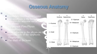
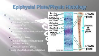
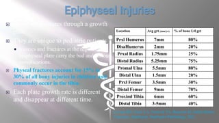
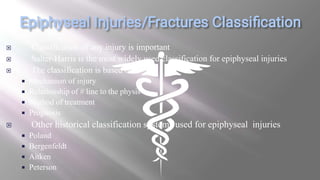
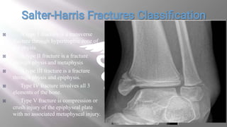
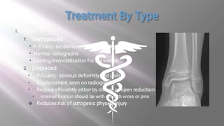
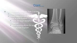
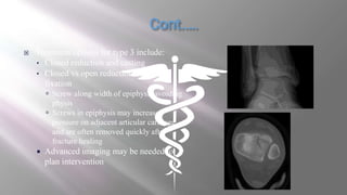
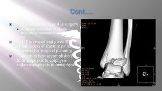
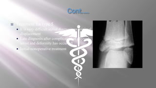
Morning five minute presentation
By:- Dr. Abdulshekur Abduljebar (MD, GSR3)
 Osseous Anatomy
 Growth Plate Histology
 Epiphyseal injuries
 Classification
 Management Principles
 Management By Type
 Complications
 Treatment Options for Complications
 References
 Epiphysis
 Secondary Ossification Center
 The epiphysis is the bone located
between the articular surface and
the physis
 Epiphyseal Plate/Physis
 Metaphysis
 Bone adjacent to the physis on the
opposite side of the epiphysis.
 Diaphysis
 The shaft of the bone
Zones of the Physis
 Germinal Zone
 Minimally active, scattered
chondrocytes
 Proliferative Zone
 Columns of chondrocytes actively
dividing
 Hypertrophic Zone
 Chondrocytes accumulate and
release calcium
 Weakest zone of physis
 Zone of endochondral ossification
 These are fractures through a growth
plate
 They are unique to pediatric patients
 Injuries and fractures at the region of
epiphyseal plate carry the bad and serious
complications.
 Physeal fractures account for 15% to
30% of all bony injuries in children and
commonly occur in the tibia.
 Each plate growth rate is different
and disappear at different time.
Location Avg grt (mm/yr) % of bone Ltl grt
Prxl Humerus 7mm 80%
DisaHumerus 2mm 20%
Prxal Radius 1.75mm 25%
Distal Radius 5.25mm 75%
Promal Ulna 5.5mm 80%
Distal Ulna 1.5mm 20%
Prxl Femur 3.5mm 30%
Distal Femur 9mm 70%
Proximl Tibia 6mm 60%
Distal Tibia 3-5mm 40%
Levine RH, Foris LA, Nezwek TA, Waseem M. Salter-Harris
Fractures. StatPearls. StatPearls Publishing; 2021.
 Classification of any injury is important
 Salter-Harris is the most widely used classification for epiphyseal injuries
 The classification is based on:-
 Mechanism of injury
 Relationship of # line to the physis
 Method of treatment
 Prognosis
 Other historical classification systems used for epiphyseal injuries
 Poland
 Bergenfeldt
 Aitken
 Peterson
 A type 1 fracture is a transverse
fracture through hypertrophic zone of
the physis
 A type II fracture is a fracture
through physis and metaphysis
 A type III fracture is a fracture
through physis and epiphysis.
 Type IV fracture involves all 3
elements of the bone.
 Type V fracture is compression or
crush injury of the epiphyseal plate
with no associated metaphyseal injury.
 X-ray:- It is difficult to assess as the physis is radiolucent and the
epiphysis is incompletely ossified.
 What to look for?
1. The physeal widening of the gap
2. Tilting of the epiphysis
3. Repeating X ray within few days
4. Comparing the injured side with the normal
 Sometimes advanced imaging is needed.
 CT Scan
 MRI
Achieve fracture healing with maintenance of growth potential
Achieve acceptable reduction and alignment
Maintenance of achieved reduction and alignment
Limit iatrogenic injury to physis
 Repeated, forceful reduction attempts
 Hardware across physis
 Treatment options for Type 2 include:
• Closed reduction and casting
• Closed reduction and percutaneous
screw or wire fixation
 Screw for larger metaphyseal
fragment
 Wires crossing physis for smaller
metaphyseal fragment
 Treatment options for type 3 include:
• Closed reduction and casting
• Closed vs open reduction, screw
fixation
 Screw along width of epiphysis avoiding
physis
 Screws in epiphysis may increase
pressure on adjacent articular cartilage
and are often removed quickly after
fracture healing
 Advanced imaging may be needed to
plan intervention
 Treatment for type 4 is surgery
 Anatomic reduction of physis
required to minimize risk of physeal
bar
 CT is crucial and gives 3D
visualization of fracture patterns;
Essential for surgical planning
 Fixation best accomplished
from epiphysis to epiphysis
and/or metaphysis to metaphysis
 Treatment for type 5
 It is very difficult initial diagnosis as minimal
displacement
 Late diagnosis after complication of physeal
arrest and deformity has occurred
 Initial nonoperative treatment
 When an entire physis arrests
 Longitudinal bone growth ceases completely at that physis
 When only part of physis arrests
 Angular deformity associated with shortening
 Often a much more difficult problem to address
 How to pick?
 Loss of abnormal physeal contour
 Sharply defined connection between epiphysis and metaphysis
 Tapering of Harris growth arrest line towards area of growth arrest
 Obvious angular deformity or segment shortening
 Surgical Physeal Arrest Resection
 Removal of arrest with continuation of physeal growth
 Complete Physis Arrest
 Ablation of growth in physis on one or both sides
 Hemi-ephiphysiodesis (angular) vs epiphysiodesis (growth correction of affected
and/or unaffected side)
 Treatment of angular or growth deformities
 Guided growth
 Osteotomies
 Fixators
Levine RH, Foris LA, Nezwek TA, Waseem M. Salter-Harris Fractures.
StatPearls. StatPearls Publishing; 2021.

1EPIPHYSEAL INJURIES (1).ppt

  • 1.
    Morning five minutepresentation By:- Dr. Abdulshekur Abduljebar (MD, GSR3)
  • 2.
     Osseous Anatomy Growth Plate Histology  Epiphyseal injuries  Classification  Management Principles  Management By Type  Complications  Treatment Options for Complications  References
  • 3.
     Epiphysis  SecondaryOssification Center  The epiphysis is the bone located between the articular surface and the physis  Epiphyseal Plate/Physis  Metaphysis  Bone adjacent to the physis on the opposite side of the epiphysis.  Diaphysis  The shaft of the bone
  • 4.
    Zones of thePhysis  Germinal Zone  Minimally active, scattered chondrocytes  Proliferative Zone  Columns of chondrocytes actively dividing  Hypertrophic Zone  Chondrocytes accumulate and release calcium  Weakest zone of physis  Zone of endochondral ossification
  • 5.
     These arefractures through a growth plate  They are unique to pediatric patients  Injuries and fractures at the region of epiphyseal plate carry the bad and serious complications.  Physeal fractures account for 15% to 30% of all bony injuries in children and commonly occur in the tibia.  Each plate growth rate is different and disappear at different time. Location Avg grt (mm/yr) % of bone Ltl grt Prxl Humerus 7mm 80% DisaHumerus 2mm 20% Prxal Radius 1.75mm 25% Distal Radius 5.25mm 75% Promal Ulna 5.5mm 80% Distal Ulna 1.5mm 20% Prxl Femur 3.5mm 30% Distal Femur 9mm 70% Proximl Tibia 6mm 60% Distal Tibia 3-5mm 40% Levine RH, Foris LA, Nezwek TA, Waseem M. Salter-Harris Fractures. StatPearls. StatPearls Publishing; 2021.
  • 6.
     Classification ofany injury is important  Salter-Harris is the most widely used classification for epiphyseal injuries  The classification is based on:-  Mechanism of injury  Relationship of # line to the physis  Method of treatment  Prognosis  Other historical classification systems used for epiphyseal injuries  Poland  Bergenfeldt  Aitken  Peterson
  • 7.
     A type1 fracture is a transverse fracture through hypertrophic zone of the physis  A type II fracture is a fracture through physis and metaphysis  A type III fracture is a fracture through physis and epiphysis.  Type IV fracture involves all 3 elements of the bone.  Type V fracture is compression or crush injury of the epiphyseal plate with no associated metaphyseal injury.
  • 8.
     X-ray:- Itis difficult to assess as the physis is radiolucent and the epiphysis is incompletely ossified.  What to look for? 1. The physeal widening of the gap 2. Tilting of the epiphysis 3. Repeating X ray within few days 4. Comparing the injured side with the normal  Sometimes advanced imaging is needed.  CT Scan  MRI
  • 9.
    Achieve fracture healingwith maintenance of growth potential Achieve acceptable reduction and alignment Maintenance of achieved reduction and alignment Limit iatrogenic injury to physis  Repeated, forceful reduction attempts  Hardware across physis
  • 11.
     Treatment optionsfor Type 2 include: • Closed reduction and casting • Closed reduction and percutaneous screw or wire fixation  Screw for larger metaphyseal fragment  Wires crossing physis for smaller metaphyseal fragment
  • 12.
     Treatment optionsfor type 3 include: • Closed reduction and casting • Closed vs open reduction, screw fixation  Screw along width of epiphysis avoiding physis  Screws in epiphysis may increase pressure on adjacent articular cartilage and are often removed quickly after fracture healing  Advanced imaging may be needed to plan intervention
  • 13.
     Treatment fortype 4 is surgery  Anatomic reduction of physis required to minimize risk of physeal bar  CT is crucial and gives 3D visualization of fracture patterns; Essential for surgical planning  Fixation best accomplished from epiphysis to epiphysis and/or metaphysis to metaphysis
  • 14.
     Treatment fortype 5  It is very difficult initial diagnosis as minimal displacement  Late diagnosis after complication of physeal arrest and deformity has occurred  Initial nonoperative treatment
  • 15.
     When anentire physis arrests  Longitudinal bone growth ceases completely at that physis  When only part of physis arrests  Angular deformity associated with shortening  Often a much more difficult problem to address  How to pick?  Loss of abnormal physeal contour  Sharply defined connection between epiphysis and metaphysis  Tapering of Harris growth arrest line towards area of growth arrest  Obvious angular deformity or segment shortening
  • 16.
     Surgical PhysealArrest Resection  Removal of arrest with continuation of physeal growth  Complete Physis Arrest  Ablation of growth in physis on one or both sides  Hemi-ephiphysiodesis (angular) vs epiphysiodesis (growth correction of affected and/or unaffected side)  Treatment of angular or growth deformities  Guided growth  Osteotomies  Fixators
  • 17.
    Levine RH, ForisLA, Nezwek TA, Waseem M. Salter-Harris Fractures. StatPearls. StatPearls Publishing; 2021.

Editor's Notes