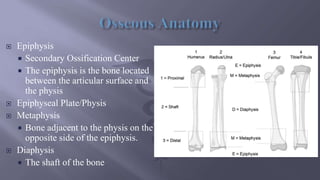
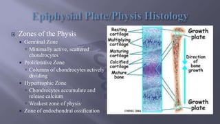
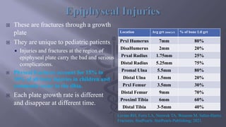
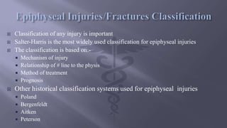
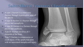
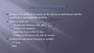
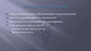
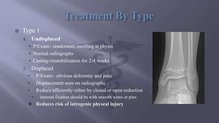
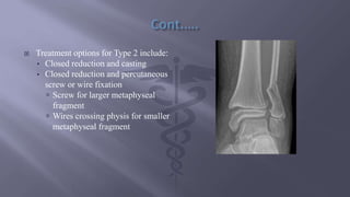
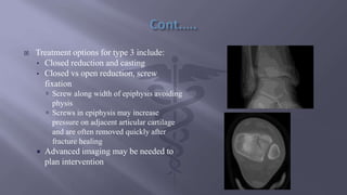
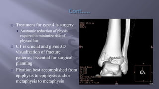
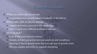
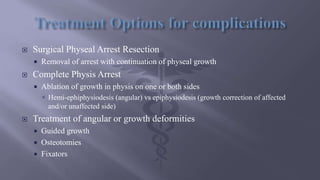
Morning five minute presentation
By:- Dr. Abdulshekur Abduljebar (MD, GSR3)
๏‚จ Osseous Anatomy
๏‚จ Growth Plate Histology
๏‚จ Epiphyseal injuries
๏‚จ Classification
๏‚จ Management Principles
๏‚จ Management By Type
๏‚จ Complications
๏‚จ Treatment Options for Complications
๏‚จ References
๏‚จ Epiphysis
๏‚ก Secondary Ossification Center
๏‚ก The epiphysis is the bone located
between the articular surface and
the physis
๏‚จ Epiphyseal Plate/Physis
๏‚จ Metaphysis
๏‚ก Bone adjacent to the physis on the
opposite side of the epiphysis.
๏‚จ Diaphysis
๏‚ก The shaft of the bone
๏‚จ Zones of the Physis
๏‚ก Germinal Zone
๏ƒบ Minimally active, scattered
chondrocytes
๏‚ก Proliferative Zone
๏ƒบ Columns of chondrocytes actively
dividing
๏‚ก Hypertrophic Zone
๏ƒบ Chondrocytes accumulate and
release calcium
๏ƒบ Weakest zone of physis
๏‚ก Zone of endochondral ossification
๏‚จ These are fractures through a growth
plate
๏‚จ They are unique to pediatric patients
๏‚ก Injuries and fractures at the region of
epiphyseal plate carry the bad and serious
complications.
๏‚จ Physeal fractures account for 15% to
30% of all bony injuries in children and
commonly occur in the tibia.
๏‚จ Each plate growth rate is different
and disappear at different time.
Location Avg grt (mm/yr) % of bone Ltl grt
Prxl Humerus 7mm 80%
DisaHumerus 2mm 20%
Prxal Radius 1.75mm 25%
Distal Radius 5.25mm 75%
Promal Ulna 5.5mm 80%
Distal Ulna 1.5mm 20%
Prxl Femur 3.5mm 30%
Distal Femur 9mm 70%
Proximl Tibia 6mm 60%
Distal Tibia 3-5mm 40%
Levine RH, Foris LA, Nezwek TA, Waseem M. Salter-Harris
Fractures. StatPearls. StatPearls Publishing; 2021.
๏‚จ Classification of any injury is important
๏‚จ Salter-Harris is the most widely used classification for epiphyseal injuries
๏‚จ The classification is based on:-
๏‚ก Mechanism of injury
๏‚ก Relationship of # line to the physis
๏‚ก Method of treatment
๏‚ก Prognosis
๏‚จ Other historical classification systems used for epiphyseal injuries
๏‚ก Poland
๏‚ก Bergenfeldt
๏‚ก Aitken
๏‚ก Peterson
๏‚จ A type 1 fracture is a transverse
fracture through hypertrophic zone of
the physis
๏‚จ A type II fracture is a fracture through
physis and metaphysis
๏‚จ A type III fracture is a fracture through
physis and epiphysis.
๏‚จ Type IV fracture involves all 3
elements of the bone.
๏‚จ Type V fracture is compression or
crush injury of the epiphyseal plate
with no associated metaphyseal injury.
๏‚จ X-ray:- It is difficult to assess as the physis is radiolucent and the
epiphysis is incompletely ossified.
๏‚จ What to look for?
1. The physeal widening of the gap
2. Tilting of the epiphysis
3. Repeating X ray within few days
4. Comparing the injured side with the normal
๏‚จ Sometimes advanced imaging is needed.
๏‚จ CT Scan
๏‚จ MRI
๏‚จ Achieve fracture healing with maintenance of growth potential
๏‚จ Achieve acceptable reduction and alignment
๏‚จ Maintenance of achieved reduction and alignment
๏‚จ Limit iatrogenic injury to physis
๏‚ก Repeated, forceful reduction attempts
๏‚ก Hardware across physis
๏‚จ Type 1
1. Undisplaced
โ€ข P/Exam:- tenderness, swelling at physis
โ€ข Normal radiographs
โ€ข Casting/immobilization for 2-4 weeks
2. Displaced
โ€ข P/Exam:- obvious deformity and pain
โ€ข Displacement seen on radiographs
โ€ข Reduce efficiently either by closed or open reduction
โ€ข internal fixation should be with smooth wires or pins
๏ถ Reduces risk of iatrogenic physeal injury
๏‚จ Treatment options for Type 2 include:
โ€ข Closed reduction and casting
โ€ข Closed reduction and percutaneous
screw or wire fixation
๏ƒบ Screw for larger metaphyseal
fragment
๏ƒบ Wires crossing physis for smaller
metaphyseal fragment
๏‚จ Treatment options for type 3 include:
โ€ข Closed reduction and casting
โ€ข Closed vs open reduction, screw
fixation
๏ƒบ Screw along width of epiphysis avoiding
physis
๏ƒบ Screws in epiphysis may increase
pressure on adjacent articular cartilage
and are often removed quickly after
fracture healing
๏‚ก Advanced imaging may be needed to
plan intervention
๏‚จ Treatment for type 4 is surgery
๏‚ก Anatomic reduction of physis
required to minimize risk of
physeal bar
๏‚จ CT is crucial and gives 3D
visualization of fracture
patterns; Essential for surgical
planning
๏‚จ Fixation best accomplished from
epiphysis to epiphysis and/or
metaphysis to metaphysis
๏‚จ Treatment for type 5
๏‚ก It is very difficult initial diagnosis as minimal displacement
๏‚ก Late diagnosis after complication of physeal arrest and deformity has occurred
๏‚ก Initial nonoperative treatment
๏‚จ When an entire physis arrests
๏‚ก Longitudinal bone growth ceases completely at that physis
๏‚จ When only part of physis arrests
๏‚ก Angular deformity associated with shortening
๏‚ก Often a much more difficult problem to address
๏‚จ How to pick?
๏‚ก Loss of abnormal physeal contour
๏‚ก Sharply defined connection between epiphysis and metaphysis
๏‚ก Tapering of Harris growth arrest line towards area of growth arrest
๏‚ก Obvious angular deformity or segment shortening
๏‚จ Surgical Physeal Arrest Resection
๏‚ก Removal of arrest with continuation of physeal growth
๏‚จ Complete Physis Arrest
๏‚ก Ablation of growth in physis on one or both sides
๏ƒบ Hemi-ephiphysiodesis (angular) vs epiphysiodesis (growth correction of affected
and/or unaffected side)
๏‚จ Treatment of angular or growth deformities
๏‚ก Guided growth
๏‚ก Osteotomies
๏‚ก Fixators
Levine RH, Foris LA, Nezwek TA, Waseem M. Salter-Harris Fractures.
StatPearls. StatPearls Publishing; 2021.

EPIPHYSEAL INJURIES .pptx

  • 1.
    Morning five minutepresentation By:- Dr. Abdulshekur Abduljebar (MD, GSR3)
  • 2.
    ๏‚จ Osseous Anatomy ๏‚จGrowth Plate Histology ๏‚จ Epiphyseal injuries ๏‚จ Classification ๏‚จ Management Principles ๏‚จ Management By Type ๏‚จ Complications ๏‚จ Treatment Options for Complications ๏‚จ References
  • 3.
    ๏‚จ Epiphysis ๏‚ก SecondaryOssification Center ๏‚ก The epiphysis is the bone located between the articular surface and the physis ๏‚จ Epiphyseal Plate/Physis ๏‚จ Metaphysis ๏‚ก Bone adjacent to the physis on the opposite side of the epiphysis. ๏‚จ Diaphysis ๏‚ก The shaft of the bone
  • 4.
    ๏‚จ Zones ofthe Physis ๏‚ก Germinal Zone ๏ƒบ Minimally active, scattered chondrocytes ๏‚ก Proliferative Zone ๏ƒบ Columns of chondrocytes actively dividing ๏‚ก Hypertrophic Zone ๏ƒบ Chondrocytes accumulate and release calcium ๏ƒบ Weakest zone of physis ๏‚ก Zone of endochondral ossification
  • 5.
    ๏‚จ These arefractures through a growth plate ๏‚จ They are unique to pediatric patients ๏‚ก Injuries and fractures at the region of epiphyseal plate carry the bad and serious complications. ๏‚จ Physeal fractures account for 15% to 30% of all bony injuries in children and commonly occur in the tibia. ๏‚จ Each plate growth rate is different and disappear at different time. Location Avg grt (mm/yr) % of bone Ltl grt Prxl Humerus 7mm 80% DisaHumerus 2mm 20% Prxal Radius 1.75mm 25% Distal Radius 5.25mm 75% Promal Ulna 5.5mm 80% Distal Ulna 1.5mm 20% Prxl Femur 3.5mm 30% Distal Femur 9mm 70% Proximl Tibia 6mm 60% Distal Tibia 3-5mm 40% Levine RH, Foris LA, Nezwek TA, Waseem M. Salter-Harris Fractures. StatPearls. StatPearls Publishing; 2021.
  • 6.
    ๏‚จ Classification ofany injury is important ๏‚จ Salter-Harris is the most widely used classification for epiphyseal injuries ๏‚จ The classification is based on:- ๏‚ก Mechanism of injury ๏‚ก Relationship of # line to the physis ๏‚ก Method of treatment ๏‚ก Prognosis ๏‚จ Other historical classification systems used for epiphyseal injuries ๏‚ก Poland ๏‚ก Bergenfeldt ๏‚ก Aitken ๏‚ก Peterson
  • 7.
    ๏‚จ A type1 fracture is a transverse fracture through hypertrophic zone of the physis ๏‚จ A type II fracture is a fracture through physis and metaphysis ๏‚จ A type III fracture is a fracture through physis and epiphysis. ๏‚จ Type IV fracture involves all 3 elements of the bone. ๏‚จ Type V fracture is compression or crush injury of the epiphyseal plate with no associated metaphyseal injury.
  • 8.
    ๏‚จ X-ray:- Itis difficult to assess as the physis is radiolucent and the epiphysis is incompletely ossified. ๏‚จ What to look for? 1. The physeal widening of the gap 2. Tilting of the epiphysis 3. Repeating X ray within few days 4. Comparing the injured side with the normal ๏‚จ Sometimes advanced imaging is needed. ๏‚จ CT Scan ๏‚จ MRI
  • 9.
    ๏‚จ Achieve fracturehealing with maintenance of growth potential ๏‚จ Achieve acceptable reduction and alignment ๏‚จ Maintenance of achieved reduction and alignment ๏‚จ Limit iatrogenic injury to physis ๏‚ก Repeated, forceful reduction attempts ๏‚ก Hardware across physis
  • 10.
    ๏‚จ Type 1 1.Undisplaced โ€ข P/Exam:- tenderness, swelling at physis โ€ข Normal radiographs โ€ข Casting/immobilization for 2-4 weeks 2. Displaced โ€ข P/Exam:- obvious deformity and pain โ€ข Displacement seen on radiographs โ€ข Reduce efficiently either by closed or open reduction โ€ข internal fixation should be with smooth wires or pins ๏ถ Reduces risk of iatrogenic physeal injury
  • 11.
    ๏‚จ Treatment optionsfor Type 2 include: โ€ข Closed reduction and casting โ€ข Closed reduction and percutaneous screw or wire fixation ๏ƒบ Screw for larger metaphyseal fragment ๏ƒบ Wires crossing physis for smaller metaphyseal fragment
  • 12.
    ๏‚จ Treatment optionsfor type 3 include: โ€ข Closed reduction and casting โ€ข Closed vs open reduction, screw fixation ๏ƒบ Screw along width of epiphysis avoiding physis ๏ƒบ Screws in epiphysis may increase pressure on adjacent articular cartilage and are often removed quickly after fracture healing ๏‚ก Advanced imaging may be needed to plan intervention
  • 13.
    ๏‚จ Treatment fortype 4 is surgery ๏‚ก Anatomic reduction of physis required to minimize risk of physeal bar ๏‚จ CT is crucial and gives 3D visualization of fracture patterns; Essential for surgical planning ๏‚จ Fixation best accomplished from epiphysis to epiphysis and/or metaphysis to metaphysis
  • 14.
    ๏‚จ Treatment fortype 5 ๏‚ก It is very difficult initial diagnosis as minimal displacement ๏‚ก Late diagnosis after complication of physeal arrest and deformity has occurred ๏‚ก Initial nonoperative treatment
  • 15.
    ๏‚จ When anentire physis arrests ๏‚ก Longitudinal bone growth ceases completely at that physis ๏‚จ When only part of physis arrests ๏‚ก Angular deformity associated with shortening ๏‚ก Often a much more difficult problem to address ๏‚จ How to pick? ๏‚ก Loss of abnormal physeal contour ๏‚ก Sharply defined connection between epiphysis and metaphysis ๏‚ก Tapering of Harris growth arrest line towards area of growth arrest ๏‚ก Obvious angular deformity or segment shortening
  • 16.
    ๏‚จ Surgical PhysealArrest Resection ๏‚ก Removal of arrest with continuation of physeal growth ๏‚จ Complete Physis Arrest ๏‚ก Ablation of growth in physis on one or both sides ๏ƒบ Hemi-ephiphysiodesis (angular) vs epiphysiodesis (growth correction of affected and/or unaffected side) ๏‚จ Treatment of angular or growth deformities ๏‚ก Guided growth ๏‚ก Osteotomies ๏‚ก Fixators
  • 17.
    Levine RH, ForisLA, Nezwek TA, Waseem M. Salter-Harris Fractures. StatPearls. StatPearls Publishing; 2021.

Editor's Notes

  • #5ย The children bones grow on their ends through an epiphysial plate (also called physis) that lay down new bone in both directions towards the joint and that part is called epiphysis, and toward the shaft and that part is called metaphysis. The shaft itself is called diaphysis.
  • #6ย The epiphysial plate appears radiolucent on x ray because they are mainly of cartilage. This growing plate will stop growing, gradually ossifies and disappears around the time of skeletal maturity. Each plate disappears at different time during life. Injuries and fractures at the region of epiphyseal plate carry the bad and serious complications of disturbing or stopping bone growth of all or part of the epiphysial plate giving rise to lateral shortness or deformities of the involved limb or joint.
  • #8ย A type 1 fracture is a transverse fracture Through the hypertrophic zone of the physis. In this injury, the width of the physis is increased.or there will be a transverse separation of the physis from the metaphysis .. The growing zone of the physis usually is not injured, and growth disturbance is uncommon. On clinical examination, the child has point tenderness at the which is suggestive of a type I fracture epiphyseal plate,