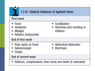
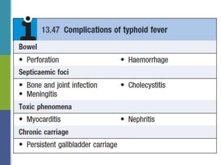
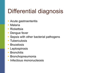
Enteric Fever
Sunil Kumar Daha
Introduction
 Over 17 million new cases of enteric fever occur
worldwide, mainly in India and Africa, causing 6
hundred thousand deaths per year.
 Enteric fever is an acute systemic disease
characterized by fever, headache & abdominal
discomfort
Geographical distribution
Typhoid fever – Salmonella enterica var Typhi
Paratyphoid fever - Salmonella enterica var
paratyphi A,B or C (20% of all cases of enteric fever)
Etiology
 Mode of transmission : Feco-oral route
 Infective dose : 10^5 – 10^9
 Incubation period: 10-14 days
Etiopathogenesis
Etiopathogenesis
Clinical features
 Insidious & non specific onset of illness
 Intermittent fever, headache, abdominal pain
 Hepatospleenomegaly, lymphadenopathy
 Maculopapular rash(red rose spots that fade on pressure),
bradycardia
 Diarrhoea, vomiting in children
 GI bleeding, Intestinal perforation, psychiatric symptoms
Differential diagnosis
 Acute gastroenteritis
 Malaria
 Rickettsia
 Dengue fever
 Sepsis with other bacterial pathogens
 Tuberculosis
 Brucelosis
 Leptospirosis
 Bronchitis
 Bronchopneumonia
 Infectious mononucleosis
Diagnosis
 Culture of S. typhi or S. paratyphi from the patient.
 Blood culture is positive in most cases in the first 2 weeks.
 Culture of intestinal secretions, faeces and urine.
 Bone marrow culture is more sensitive than blood culture
 Leucopenia
 Widal antigen test
How to start??
 Check ABC
 Send blood for culture and sensitivity
 Apply cold sponge
Fluid supplements if necessary
 Start empirical therapy
 Azithromycin 500mg PO BD or
 Ceftriaxone 1 gm IV BD
Prevention
 This is mainly through improved sanitation and clean water.
 Travelers should avoid drinking untreated water, ice in
drinks and eating ice creams.
 Typhoid vaccines are commercially available
 Dockrell DH, Sundar S, Angus BJ, Hobson RP. Infectious
disease,In: Walker BR, Colledge NR, Ralston SH, Penman I.
Davidson's principles and practice of medicine. 22nd ed.
Edinburgh : Churchill Livingstone/Elsevier; 2014.p.334-335.
References
Thank you!

Enteric fever and its management

  • 1.
  • 2.
    Introduction  Over 17million new cases of enteric fever occur worldwide, mainly in India and Africa, causing 6 hundred thousand deaths per year.  Enteric fever is an acute systemic disease characterized by fever, headache & abdominal discomfort
  • 3.
  • 4.
    Typhoid fever –Salmonella enterica var Typhi Paratyphoid fever - Salmonella enterica var paratyphi A,B or C (20% of all cases of enteric fever) Etiology
  • 5.
     Mode oftransmission : Feco-oral route  Infective dose : 10^5 – 10^9  Incubation period: 10-14 days Etiopathogenesis
  • 6.
  • 7.
    Clinical features  Insidious& non specific onset of illness  Intermittent fever, headache, abdominal pain  Hepatospleenomegaly, lymphadenopathy  Maculopapular rash(red rose spots that fade on pressure), bradycardia  Diarrhoea, vomiting in children  GI bleeding, Intestinal perforation, psychiatric symptoms
  • 10.
    Differential diagnosis  Acutegastroenteritis  Malaria  Rickettsia  Dengue fever  Sepsis with other bacterial pathogens  Tuberculosis  Brucelosis  Leptospirosis  Bronchitis  Bronchopneumonia  Infectious mononucleosis
  • 11.
    Diagnosis  Culture ofS. typhi or S. paratyphi from the patient.  Blood culture is positive in most cases in the first 2 weeks.  Culture of intestinal secretions, faeces and urine.  Bone marrow culture is more sensitive than blood culture  Leucopenia  Widal antigen test
  • 13.
    How to start?? Check ABC  Send blood for culture and sensitivity  Apply cold sponge Fluid supplements if necessary  Start empirical therapy  Azithromycin 500mg PO BD or  Ceftriaxone 1 gm IV BD
  • 14.
    Prevention  This ismainly through improved sanitation and clean water.  Travelers should avoid drinking untreated water, ice in drinks and eating ice creams.  Typhoid vaccines are commercially available
  • 15.
     Dockrell DH,Sundar S, Angus BJ, Hobson RP. Infectious disease,In: Walker BR, Colledge NR, Ralston SH, Penman I. Davidson's principles and practice of medicine. 22nd ed. Edinburgh : Churchill Livingstone/Elsevier; 2014.p.334-335. References
  • 16.

Editor's Notes

  • #8 Enteric fever occurs through the ingestion of the organism, and a variety of sources of fecal contamination have been reported, including street foods and contamination of water reservoirs. Human volunteer experiments established an infecting dose of about 10 5 -10 9 organisms, with an incubation period ranging from 4 to 14 days, depending on the inoculating dose of viable bacteria. After ingestion, S. Typhi organisms are thought to invade the body through the gut mucosa in the terminal ileum, possibly through specialized antigen-sampling cells known as M cells that overlie gut-associated lymphoid tissues, through enterocytes, or via a paracellular route. S. Typhi crosses the intestinal mucosal barrier after attachment to the microvilli by an intricate mechanism involving membrane ruffl ing, actin rearrangement, and internalization in an intracellular vacuole. In contrast to nontyphoidal Salmonella , S. Typhi expresses virulence factors that allow it to downregulate the pathogen recognition receptor – mediated host infl ammatory response. Within the Peyer patches in the terminal ileum, S. Typhi can traverse the intestinal barrier through several mechanisms, including the M cells in the follicleassociated epithelium, epithelial cells, and dendritic cells. At the villi, Salmonella can enter through the M cells or by passage through or between compromised epithelial cells. On contact with the epithelial cell, S. typhi assembles TTSS-1 and translocates effectors into the cytoplasm. These effectors activate host Rho guanosine triphosphatases (GTPases), resulting in the rearrangement of the actin cytoskeleton into membrane ruffl es, induction of mitogen-activated protein kinase (MAPK) pathways, and destabilization of tight junctions. Changes in the actin cytoskeleton are further modulated by the actin-binding proteins SipA and SipC and lead to bacterial uptake. MAPK signaling activates the transcription factors activator protein-1 (AP-1) and nuclear factor- κ B (NF- κ B), which turn on production of IL-8. The destabilization of tight junctions allows the transmigration of polymorphonuclear leukocytes (PMNs) from the basolateral surface to the apical surface, paracellular fl uid leakage, and access of bacteria to the basolateral surface. Shortly after internalization of S. Typhi by macropinocytosis, salmonellae are enclosed in a spacious phagosome that is formed by membrane ruffl es. Later, the phagosome fuses with lysosomes, acidifi es, and shrinks to become adherent around the bacterium, forming the Chapter 190 Salmonella ■ 955 Salmonella -containing vacuole (SCV). TTSS-2 is induced within the SCV and translocates effector proteins SifA and PipB2, which contribute to Salmonella -induced fi lament (Sif) formation along microtubules (see Fig. 190-2 ). After passing through the intestinal mucosa, S. Typhi organisms enter the mesenteric lymphoid system and then pass into the bloodstream via the lymphatics. This primary bacteremia is usually asymptomatic, and blood culture results are frequently negative at this stage of the disease. The blood-borne bacteria are disseminated throughout the body and are thought to colonize the organs of the RES, where they may replicate within macrophages. After a period of bacterial replication, S. Typhi organisms are shed back into the blood, causing a secondary bacteremia that coincides with the onset of clinical symptoms and marks the end of the incubation period ( Fig. 190-3 ). In vitro studies with human cell lines have shown qualitative and quantitative differences in the epithelial cell response to S. Typhi and S. Typhimurium with regard to cytokine and chemokine secretion. Thus, by avoiding the triggering of an early infl ammatory response in the gut, S. Typhi could instead colonize deeper tissues and organ systems. Infection with S. Typhi produces an infl ammatory response in the deeper mucosal layers and underlying lymphoid tissue, with hyperplasia of Peyer patches and subsequent necrosis and sloughing of overlying epithelium. The resulting ulcers can bleed but usually heal without scarring or stricture formation. The infl ammatory lesion may occasionally penetrate the muscularis and serosa of the intestine and produce perforation. The mesenteric lymph nodes, liver, and spleen are hyperemic and generally have areas of focal necrosis as well. A mononuclear response may be seen in the bone marrow in association with areas of focal necrosis. The morphologic changes of S. Typhi infection are less prominent in infants than in older children and adults. It is thought that several virulence factors, including TTSS-2, may be necessary for the virulence properties and ability to cause systemic infection. The surface Vi polysaccharide capsular antigen found in S. Typhi interferes with phagocytosis by preventing the binding of C3 to the surface of the bacterium. The ability of organisms to survive within macrophages after phagocytosis is an important virulence trait encoded by the PhoP regulon and may be related to metabolic effects on host cells. The occasional occurrence of diarrhea may be explained by the presence of a toxin related to cholera toxin and E. coli heat-labile enterotoxin. The clinical syndrome of fever and systemic symptoms is produced by a release of proinfl ammatory cytokines (IL-6, IL-1 β , and TNF- α ) from the infected cells. In addition to the virulence of the infecting organisms, host factors and immunity may also play an important role in predisposition to infection. There is an association between susceptibility to typhoid fever and human genes within the major histocompatibility complex class II and class III loci. Patients who are infected with HIV are at signifi cantly higher risk for clinical infection with S. Typhi and S. Paratyphi. Similarly, patients with Helicobacter pylori infection have an increased risk of acquiring typhoid fever.
  • #17 Airway, Breathing, circulation
  • #18 Ty21a, an oral live attenuated S. Typhi vaccine (given on days 1, 3, 5, and 7, with a booster every 5 years) Vi CPS, a parenteral vaccine consisting of purified Vi polysaccharide from the bacterial capsule (given in 1 dose, with a booster every 2 years).