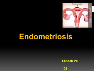
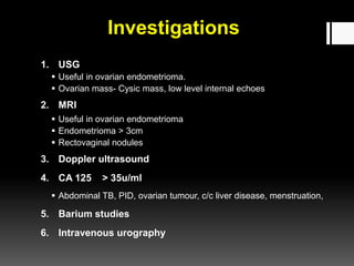
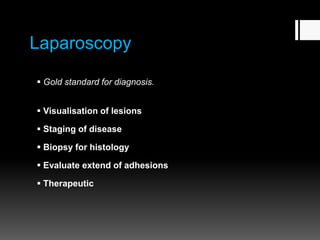
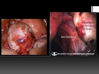
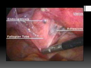
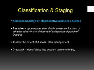
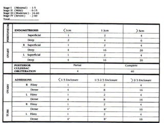
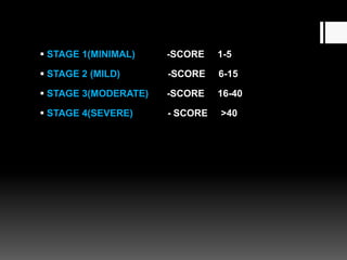
Endometriosis
Labeeb Pc
102
Topics to be dicussed…
 Introduction
 Pathogenesis
 Risk factors
 Classification
 Symptoms
 Physical examination
 Differential diagnosis
 Investigations
 Staging
Introduction
Presence of endometrial glands & stroma
outside the uterus
Benign
Incidence – 10%
Pathogenesis
1. Implantation Theory ( Sampson’s)
2. Coelomic metaplasia Theory ( Meyer )
3. Lymphatic & Vascular Metastatic Theory ( Halban )
4. Hormonal - estrogen
5. Genetic
6. Immunological
Implantation Theory
(Sampson’s)
 Retrograde menstruation
 Common in obstructive Mullerian anomalies,
cryptomenorrohea.
 Women with short & heavy menstrual cycles
 Scar endometriosis
 Dependant portion of pelvis
 Cytokines - Adhesion to peritoneal surface
 MMP - Invasion
 VEGF-A - Angiogenesis
 Growth factors - Growth
 Estrogen - Proliferative change
 Prostaglandin - inflammation, pain
Sites
Common – ovaries, pouch of Douglas,
uterosacral ligaments, Broad ligaments, fallopian
tubes, uterovesical fold, round ligament, appendix ,
vagina
Rectovaginal septum, sigmoid colon, cecum,
umbilicus, abdominal scars, tubal stumps
Risk Factors
1. Menstrual cycle
 Early menarche
 Heavy menstrual bleeding
 Short menstrual cycles
2. Delayed childbearing
3. Parity
 Low parity, nulliparous
4. High social class
5. First degree relatives
6. Low BMI
7. Obstructive Mullerian anomalies
8. Environmental
Classification
1. Superficial endometriosis
2. Ovarian endometriosis
3. Deep infiltrating endometriosis
Superficial Endometriosis
( Peritoneal)
 Dependent portion of pelvis.
 Most common – surface of ovaries.
 Pelvic peritoneum, pouch of Douglas, uterosacral ligaments,
Broad ligaments.
 Appearance –
 Early - Papular, vesicular
 Hemorrhagic - red, flame shaped
 Powder burn - puckered, blue- black - inactive old lesions
 Fibrotic - white
 Peritoneal cavity – yellowish brown fluid
 Cannot be palpated on clinical examination
 Difficult to visualise on imaging and diagnosis by laparoscopy
Ovarian Endometriosis
( Ovarian Endometrioma)
 Inversion & invagination of ovarian cortex , with superficial
endometriotic deposits.
 Adhesion of ovary to post. Peritoneum
 Chocolate Cysts.
 Cyst wall white or yellow.
 <12cm
 Histology- pseudoxanthoma cells - macrophages , are brown.
Deep Infiltrating Endometriosis
( Posterior Pelvic Endometriosis )
 Lesion extends >5mm beneath peritoneum.
 Usually in rectovaginal space,
also uterosacral ligaments, cervix , bowel or ureters.
 Can be felt on pelvic & per rectum examination – tender
induration & nodularity
 Can be visualised on imaging.
Extrapelvic Endometriosis
 Urinary tract
 GI tract
 Surgical scars
 Pulmonary
Symptoms
 Classic symptoms – Dysmenorrhoea, Dyspareunia, deep
seated pelvic pain.
 Menstrual – menorrhagia, Premenstrual spotting
 Infertility
 Cyclical bowel & bladder symptoms
 Scar endometriosis – cyclical pain
 Cyclical haemoptysis & haemothorax
Causes of infertility
 Ovulatory dysfunction
 Anovulation
 Luteal phase defect
 Luteinised unruptured follicle syndrome
 Immunological alteration
 Mechanical factors
 Dyspareunia
 Endometrial dysfunction
 Sperm inactivation
Physical Examination
1. Abdominal examination
 ovarian mass – tender, fixed, in iliac fossa
2. Per speculum examination
 Vaginal lesions – bluish puckered spots
3. Pelvic examination
 Fixed retroverted uterus
 Adnexal mass
 Tender uterosacral ligaments
4. Per rectal examination
Differential Diagnosis
1. Chronic PID
2. Uterine myomas
3. Ovarian malignant tumour
4. Rectal Ca
5. a/c abdominal catastrophe
6. c/c pelvic congestion syndrome
Investigations
1. USG
 Useful in ovarian endometrioma.
 Ovarian mass- Cysic mass, low level internal echoes
2. MRI
 Useful in ovarian endometrioma
 Endometrioma > 3cm
 Rectovaginal nodules
3. Doppler ultrasound
4. CA 125 > 35u/ml
 Abdominal TB, PID, ovarian tumour, c/c liver disease, menstruation,
5. Barium studies
6. Intravenous urography
Laparoscopy
 Gold standard for diagnosis.
 Visualisation of lesions
 Staging of disease
 Biopsy for histology
 Evaluate extend of adhesions
 Therapeutic
Classification & Staging
 American Society For Reproductive Medicine ( ASRM )
 Based on - appearance, size, depth, presence & extent of
adnexal adhesions and degree of obliteration of pouch of
Douglas
 To describe extent of disease, plan management.
 Drawback – doesn’t take into account pain or inferitlity
 STAGE 1(MINIMAL) -SCORE 1-5
 STAGE 2 (MILD) -SCORE 6-15
 STAGE 3(MODERATE) -SCORE 16-40
 STAGE 4(SEVERE) - SCORE >40
Endometriosis

Endometriosis

  • 1.
  • 2.
    Topics to bedicussed…  Introduction  Pathogenesis  Risk factors  Classification  Symptoms  Physical examination  Differential diagnosis  Investigations  Staging
  • 3.
    Introduction Presence of endometrialglands & stroma outside the uterus Benign Incidence – 10%
  • 4.
    Pathogenesis 1. Implantation Theory( Sampson’s) 2. Coelomic metaplasia Theory ( Meyer ) 3. Lymphatic & Vascular Metastatic Theory ( Halban ) 4. Hormonal - estrogen 5. Genetic 6. Immunological
  • 5.
    Implantation Theory (Sampson’s)  Retrogrademenstruation  Common in obstructive Mullerian anomalies, cryptomenorrohea.  Women with short & heavy menstrual cycles  Scar endometriosis  Dependant portion of pelvis
  • 6.
     Cytokines -Adhesion to peritoneal surface  MMP - Invasion  VEGF-A - Angiogenesis  Growth factors - Growth  Estrogen - Proliferative change  Prostaglandin - inflammation, pain
  • 7.
    Sites Common – ovaries,pouch of Douglas, uterosacral ligaments, Broad ligaments, fallopian tubes, uterovesical fold, round ligament, appendix , vagina Rectovaginal septum, sigmoid colon, cecum, umbilicus, abdominal scars, tubal stumps
  • 9.
    Risk Factors 1. Menstrualcycle  Early menarche  Heavy menstrual bleeding  Short menstrual cycles 2. Delayed childbearing 3. Parity  Low parity, nulliparous 4. High social class 5. First degree relatives 6. Low BMI 7. Obstructive Mullerian anomalies 8. Environmental
  • 10.
    Classification 1. Superficial endometriosis 2.Ovarian endometriosis 3. Deep infiltrating endometriosis
  • 11.
    Superficial Endometriosis ( Peritoneal) Dependent portion of pelvis.  Most common – surface of ovaries.  Pelvic peritoneum, pouch of Douglas, uterosacral ligaments, Broad ligaments.  Appearance –  Early - Papular, vesicular  Hemorrhagic - red, flame shaped  Powder burn - puckered, blue- black - inactive old lesions  Fibrotic - white  Peritoneal cavity – yellowish brown fluid
  • 12.
     Cannot bepalpated on clinical examination  Difficult to visualise on imaging and diagnosis by laparoscopy
  • 13.
    Ovarian Endometriosis ( OvarianEndometrioma)  Inversion & invagination of ovarian cortex , with superficial endometriotic deposits.  Adhesion of ovary to post. Peritoneum  Chocolate Cysts.  Cyst wall white or yellow.  <12cm  Histology- pseudoxanthoma cells - macrophages , are brown.
  • 16.
    Deep Infiltrating Endometriosis (Posterior Pelvic Endometriosis )  Lesion extends >5mm beneath peritoneum.  Usually in rectovaginal space, also uterosacral ligaments, cervix , bowel or ureters.  Can be felt on pelvic & per rectum examination – tender induration & nodularity  Can be visualised on imaging.
  • 17.
    Extrapelvic Endometriosis  Urinarytract  GI tract  Surgical scars  Pulmonary
  • 18.
    Symptoms  Classic symptoms– Dysmenorrhoea, Dyspareunia, deep seated pelvic pain.  Menstrual – menorrhagia, Premenstrual spotting  Infertility  Cyclical bowel & bladder symptoms  Scar endometriosis – cyclical pain  Cyclical haemoptysis & haemothorax
  • 19.
    Causes of infertility Ovulatory dysfunction  Anovulation  Luteal phase defect  Luteinised unruptured follicle syndrome  Immunological alteration  Mechanical factors  Dyspareunia  Endometrial dysfunction  Sperm inactivation
  • 20.
    Physical Examination 1. Abdominalexamination  ovarian mass – tender, fixed, in iliac fossa 2. Per speculum examination  Vaginal lesions – bluish puckered spots 3. Pelvic examination  Fixed retroverted uterus  Adnexal mass  Tender uterosacral ligaments 4. Per rectal examination
  • 21.
    Differential Diagnosis 1. ChronicPID 2. Uterine myomas 3. Ovarian malignant tumour 4. Rectal Ca 5. a/c abdominal catastrophe 6. c/c pelvic congestion syndrome
  • 22.
    Investigations 1. USG  Usefulin ovarian endometrioma.  Ovarian mass- Cysic mass, low level internal echoes 2. MRI  Useful in ovarian endometrioma  Endometrioma > 3cm  Rectovaginal nodules 3. Doppler ultrasound 4. CA 125 > 35u/ml  Abdominal TB, PID, ovarian tumour, c/c liver disease, menstruation, 5. Barium studies 6. Intravenous urography
  • 23.
    Laparoscopy  Gold standardfor diagnosis.  Visualisation of lesions  Staging of disease  Biopsy for histology  Evaluate extend of adhesions  Therapeutic
  • 26.
    Classification & Staging American Society For Reproductive Medicine ( ASRM )  Based on - appearance, size, depth, presence & extent of adnexal adhesions and degree of obliteration of pouch of Douglas  To describe extent of disease, plan management.  Drawback – doesn’t take into account pain or inferitlity
  • 28.
     STAGE 1(MINIMAL)-SCORE 1-5  STAGE 2 (MILD) -SCORE 6-15  STAGE 3(MODERATE) -SCORE 16-40  STAGE 4(SEVERE) - SCORE >40