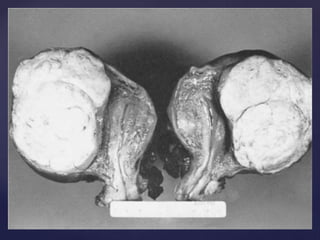
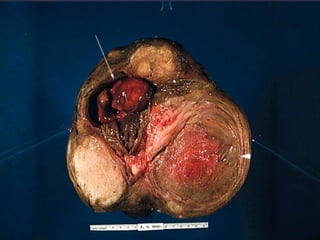
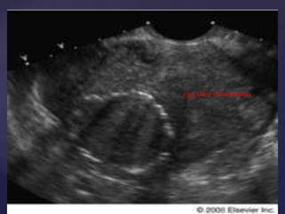
Fibroid uterus 
• Disease is prevalent in one among every 
four women as per studies 
 The commonest benign tumour of uterus 
 Commonest benign solid tumour in 
female
Terminology & Definition 
• “womb stone” 
• “scleromas” 
• “Fibroid” 
• “myoma” 
 Benign tumors Arising from the 
myometrium or muscles of its vessel walls 
Composed of smooth muscles 
interspersed with varying amounts of 
fibrous tissue 
 myoma, fibromyoma, leiofibromyoma, 
fibroleiomyoma, and fibroma
Aetiology & Pathogenesis 
 
 origin of uterine leiomyomas is incompletely 
understood 
 But cytogenetic studies have yielded some clues 
 Each tumor develops from a single muscle cell 
– a progenitor myocyte 
Cytogenetic analysis has demonstrated that myomas 
have multiple chromosomal abnormalities.
• Twenty percent of abnormalities involve translocations 
between chromosomes 12 and 14. 
• Seventeen percent involve a deletion of chromosome 7. 
• Twelve percent involve a deletion of chromosome 12. 
• oestrogen and progesterone receptors are found in higher 
concentrations in uterine myomas. 
• There also appear to be similarities between fibroids and 
keloid formation
Sites
CLASSIFICATION OF UTERINE FIBROIDS 
BODY(CORPOREAL) CERVICAL 
INTERSTITIAL(75%) SUBSEROUS (15%) SUBMUCOUS(5%) 
SESSILE PEDUNCULATED 
SUBSEROUS BROAD LIGAMENT WANDERING 
(PSEUDO) (PARASITIC) 
ANTERIOR POSTERIOR CENTRAL LATERAL
MORPHOLOGY
BODY/CORPOREAL FIBROIDS 
GROSS APPEARANCE 
 CIRCUMSCRIBED DISCRETE ROUND FIRM,GRAY WHITE TUMORS 
SIZE 
VISIBLE NODULES TO MASSIVE TUMOR 
CUT SECTION 
 SMOOTH AND WHITISH 
 WHORLED APPEARANCE
Secondary changes
Degenaration 
atrophy 
hyaline change 
calcification 
cystic degenaration 
red degenaration 
Torsion 
Infection 
Sarcomatous change – 0.2%
egg shell calcification 
tvs
 Atrophic 
 Hyaline  yellow, soft gelatinous areas 
 Cystic liquefaction follows extreme hyalinization 
 Calcific circulatory deprivation precipitation of ca 
carbonate & phosphate 
 Septic circulatory deprivation necrosis  infection 
 Myxomatous (fatty) uncommon, follows hyaline or cystic 
degenration
Red (carneous) degeneration 
 Commonly occurs during pregnancy 
 Edema & hypertrophy impede blood supply 
aseptic degeneration & infarction with venous 
thrombosis & hemorrhage 
 Painful but self-limiting 
 May result in preterm labor & rarely DIC 
2-MALIGNANT TRANSFORMATION 
 Transformation to leiomyosarcomas occurs in 0.1-0.5%
symptom 
 
 Asymptomatic 
 Abnormal uterine bleeding---- 30% 
 Pain abdomen --- 
 Mass per abdomen
Abnormal uterine bleeding 
• The most common symptom is menorrhagia 
• Heavy / prolonged bleeding (menorrhagia)  iron deficiency anemia 
• But intermenstrual spotting and disruption of a normal pattern are 
other frequent complaints 
• location of the myomas, submucous versus intramural, is not 
related to bleeding symptoms 
• symptoms of bleeding were related to the size of myomas 
• The older theory that the amount of menorrhagia is directly 
related to an increase of endometrial surface area has been 
disproven.
PAIN 
• Dull aching pain of Feeling a mass 
• RED DEGENERATION 
• TORSION HAEMORRHAGE, 
• ACUTE INFECTION 
• EXPULSION OF A SUBMUCOUS FIBROID 
• Back pain radiating to the lower extremities 
• Dysparunea
PRESSURE EFFECTS 
• If large may distort or obstruct other organs like ureters, bladder or 
rectum urinary symptoms, hydroureter, constipation, pelvic venous 
congestion & LL edema 
• Rarely a posterior fundal tumor extreme retroflexion of the uterus 
distorting the bladder base urinary retention 
• Parasitic tumor may cause bowel obstruction 
• Cervical tumors serosanguineous vaginal discharge, bleeding, 
dyspareunia or infertility
INFERTILITY 
“Woman postpones her pregnacy later fibroid postpones it” 
• The relationship is uncertain 
• Myomectomy is indicated only in long-standing 
infertility and recurrent abortion after all other potential 
factors have been investigated and treated. 
• submucous myomas that distort the uterine cavity are 
the myomas that may affect reproduction
VICTOR BONNEY 
INVENTOR: MYOMECTOMY CLAMP AND SCREW 
“ …in my early years as a 
gynaecological surgeon, a 
case occurred which 
profoundly affected my 
outlook. A lady, recently 
married, wishing above all 
things to have a child, 
underwent a subtotal 
hysterectomy on account of a 
single sub‐mucous fibroid.”
Master pelvic surgeon and pioneer of 
conservative surgery for the ovary and 
fibroids
Clinical 
Examination
 Clinically, the diagnosis of uterine myomas is 
usually confirmed by physical examination. Upon 
palpation, an enlarged, firm, irregular uterus may 
be felt. 
 The three conditions that commonly enter into the 
differential diagnosis include pregnancy, 
adenomyosis, and an ovarian neoplasm. 
 The discrimination between large ovarian tumors 
and myomatous uteri may be difficult on physical 
examination, because the extension of myomas 
laterally may make palpation of normal ovaries 
impossible during the pelvic examination. 
 The mobility of the pelvic mass and whether the 
mass moves independently or as part of the uterus 
may be helpful diagnostically.
INCIDENCE OF CLINICALLY DETECTABLE FIBROIDS IN 
PREGNANCY VARIES FROM 1 IN 500 TO 1 IN 1000.
 INCREASE IN SIZE– oestrogen and progestrone 
 RED DEGENERATION – charecterised by rapid 
enlargement of fibroid, acute onset of pain 
over the fibroid, mild pain and 
vomiting……..self limiting…… 
 INFECTION of the fibroid in peuperium 
 TORSION OF A PEDUNCULATED FIBROID
 Position size and type of fibroid determine 
their effect on pregnancy 
 Most complications occur when the fibroid is 
submucous and close to the placental 
implantation site.
 Miscarriage and preterm labour. 
 Both in first and second trimester
 Malpresentations 
 Non-engagement of head 
 Uterine inertia 
 Obstructed labour 
 PPH and retained placenta 
 Difficulty at CS
 Puerperal infection and morbid puerperium
Thanks to VISHNU H LAL & AL VAHSAB
Fibroid uterus in detail ..... odstetrics and gynecolgy

Fibroid uterus in detail ..... odstetrics and gynecolgy

  • 2.
    Fibroid uterus •Disease is prevalent in one among every four women as per studies  The commonest benign tumour of uterus  Commonest benign solid tumour in female
  • 3.
    Terminology & Definition • “womb stone” • “scleromas” • “Fibroid” • “myoma”  Benign tumors Arising from the myometrium or muscles of its vessel walls Composed of smooth muscles interspersed with varying amounts of fibrous tissue  myoma, fibromyoma, leiofibromyoma, fibroleiomyoma, and fibroma
  • 4.
    Aetiology & Pathogenesis   origin of uterine leiomyomas is incompletely understood  But cytogenetic studies have yielded some clues  Each tumor develops from a single muscle cell – a progenitor myocyte Cytogenetic analysis has demonstrated that myomas have multiple chromosomal abnormalities.
  • 5.
    • Twenty percentof abnormalities involve translocations between chromosomes 12 and 14. • Seventeen percent involve a deletion of chromosome 7. • Twelve percent involve a deletion of chromosome 12. • oestrogen and progesterone receptors are found in higher concentrations in uterine myomas. • There also appear to be similarities between fibroids and keloid formation
  • 7.
  • 9.
    CLASSIFICATION OF UTERINEFIBROIDS BODY(CORPOREAL) CERVICAL INTERSTITIAL(75%) SUBSEROUS (15%) SUBMUCOUS(5%) SESSILE PEDUNCULATED SUBSEROUS BROAD LIGAMENT WANDERING (PSEUDO) (PARASITIC) ANTERIOR POSTERIOR CENTRAL LATERAL
  • 12.
  • 15.
    BODY/CORPOREAL FIBROIDS GROSSAPPEARANCE  CIRCUMSCRIBED DISCRETE ROUND FIRM,GRAY WHITE TUMORS SIZE VISIBLE NODULES TO MASSIVE TUMOR CUT SECTION  SMOOTH AND WHITISH  WHORLED APPEARANCE
  • 16.
  • 17.
    Degenaration atrophy hyalinechange calcification cystic degenaration red degenaration Torsion Infection Sarcomatous change – 0.2%
  • 23.
  • 24.
     Atrophic Hyaline  yellow, soft gelatinous areas  Cystic liquefaction follows extreme hyalinization  Calcific circulatory deprivation precipitation of ca carbonate & phosphate  Septic circulatory deprivation necrosis  infection  Myxomatous (fatty) uncommon, follows hyaline or cystic degenration
  • 25.
    Red (carneous) degeneration  Commonly occurs during pregnancy  Edema & hypertrophy impede blood supply aseptic degeneration & infarction with venous thrombosis & hemorrhage  Painful but self-limiting  May result in preterm labor & rarely DIC 2-MALIGNANT TRANSFORMATION  Transformation to leiomyosarcomas occurs in 0.1-0.5%
  • 26.
    symptom  Asymptomatic  Abnormal uterine bleeding---- 30%  Pain abdomen ---  Mass per abdomen
  • 27.
    Abnormal uterine bleeding • The most common symptom is menorrhagia • Heavy / prolonged bleeding (menorrhagia)  iron deficiency anemia • But intermenstrual spotting and disruption of a normal pattern are other frequent complaints • location of the myomas, submucous versus intramural, is not related to bleeding symptoms • symptoms of bleeding were related to the size of myomas • The older theory that the amount of menorrhagia is directly related to an increase of endometrial surface area has been disproven.
  • 28.
    PAIN • Dullaching pain of Feeling a mass • RED DEGENERATION • TORSION HAEMORRHAGE, • ACUTE INFECTION • EXPULSION OF A SUBMUCOUS FIBROID • Back pain radiating to the lower extremities • Dysparunea
  • 29.
    PRESSURE EFFECTS •If large may distort or obstruct other organs like ureters, bladder or rectum urinary symptoms, hydroureter, constipation, pelvic venous congestion & LL edema • Rarely a posterior fundal tumor extreme retroflexion of the uterus distorting the bladder base urinary retention • Parasitic tumor may cause bowel obstruction • Cervical tumors serosanguineous vaginal discharge, bleeding, dyspareunia or infertility
  • 30.
    INFERTILITY “Woman postponesher pregnacy later fibroid postpones it” • The relationship is uncertain • Myomectomy is indicated only in long-standing infertility and recurrent abortion after all other potential factors have been investigated and treated. • submucous myomas that distort the uterine cavity are the myomas that may affect reproduction
  • 31.
    VICTOR BONNEY INVENTOR:MYOMECTOMY CLAMP AND SCREW “ …in my early years as a gynaecological surgeon, a case occurred which profoundly affected my outlook. A lady, recently married, wishing above all things to have a child, underwent a subtotal hysterectomy on account of a single sub‐mucous fibroid.”
  • 32.
    Master pelvic surgeonand pioneer of conservative surgery for the ovary and fibroids
  • 33.
  • 34.
     Clinically, thediagnosis of uterine myomas is usually confirmed by physical examination. Upon palpation, an enlarged, firm, irregular uterus may be felt.  The three conditions that commonly enter into the differential diagnosis include pregnancy, adenomyosis, and an ovarian neoplasm.  The discrimination between large ovarian tumors and myomatous uteri may be difficult on physical examination, because the extension of myomas laterally may make palpation of normal ovaries impossible during the pelvic examination.  The mobility of the pelvic mass and whether the mass moves independently or as part of the uterus may be helpful diagnostically.
  • 35.
    INCIDENCE OF CLINICALLYDETECTABLE FIBROIDS IN PREGNANCY VARIES FROM 1 IN 500 TO 1 IN 1000.
  • 36.
     INCREASE INSIZE– oestrogen and progestrone  RED DEGENERATION – charecterised by rapid enlargement of fibroid, acute onset of pain over the fibroid, mild pain and vomiting……..self limiting……  INFECTION of the fibroid in peuperium  TORSION OF A PEDUNCULATED FIBROID
  • 37.
     Position sizeand type of fibroid determine their effect on pregnancy  Most complications occur when the fibroid is submucous and close to the placental implantation site.
  • 38.
     Miscarriage andpreterm labour.  Both in first and second trimester
  • 39.
     Malpresentations Non-engagement of head  Uterine inertia  Obstructed labour  PPH and retained placenta  Difficulty at CS
  • 40.
     Puerperal infectionand morbid puerperium
  • 41.
    Thanks to VISHNUH LAL & AL VAHSAB

Editor's Notes

  • #3 Only 50% among them ever become symptomatic
  • #5 thus each myoma is monoclonal (Each myoma would have cells with the same abnormality.) The larger the myoma, the more an abnormal karyotype will be detected
  • #6 . The regions of chromosome 12 and 7 involve genes that may regulate growth-inducing proteins and cytokines, including transforming growth factor β (TGF-β) epidermal growth factor (EGF), insulin-like growth factors (IGF) 1 and 2, and platelet-derived growth factor (PDGF) ( Fig. 18-34 ).
  • #9 FOUND IN CERVIX AS WELL
  • #10 Subserosal myomas give the uterus its knobby contour during pelvic examination. Further growth of a subserosal myoma may lead to a pedunculated myoma wandering into the peritoneal cavity. This myoma may outgrow its uterine blood supply and obtain a secondary blood supply from another organ, such as the omentum, and become a parasitic myoma. Growth of a myoma in a lateral direction from the uterus may result in a broad ligament myoma ( Fig. 18-33 ). The clinical significance of broad ligament myomas is that they are difficult to differentiate on pelvic examination from a solid ovarian tumor. Large, broad ligament myomas may produce a hydroureter as they enlarge.
  • #14 Grossly, a myoma has a lighter color than the normal myometrium
  • #15 . On cut surface the tumor has a glistening, pearl-white appearance, with the smooth muscle arranged in a trabeculated or whorled configuration. smooth muscle cells are markedly elongated and have eosinophilic cytoplasm and elongated, cigar-shaped nuclei.
  • #21 Ground glass fibrous tissue proliferation
  • #22 Red degeneration; the ghosts of the muscle cells and their nuclei remain
  • #27 The most common symptoms related to myomas are pres-sure from an enlarging pelvic mass, pain including dysmenorrhea, and abnormal uterine bleeding. The severity of symptoms is usually related to the number, location, and size of the myomas. However, over two thirds of women with uterine myomas are asymptomatic.
  • #28 Symptoms depend on location, size, changes & pregnancy status One of three women with abnormal bleeding and submucous myomas also has endometrial hyperplasia, which may be the cause of the symptom. explanation is straightforward when there are areas of ulceration over submucous myomas. However, ulceration is a rare finding. The most popular theory is that myomas result in an abnormal microvascular growth pattern and function of the vessels in the adjacent endometrium.
  • #29 Vascular occlusion  necrosis, infection Torsion of a pedunculated fibroid acute pain Myometrial contractions to expel the myoma Red degenration acute pain Heaviness fullness in the pelvic area Feeling a mass If the tumor gets impacted in the pelvis pressure on nerves back pain radiating to the lower extremities Dysparunea if it is protruding to vagina
  • #31 Occasionally, myomas are the only identifiable abnormality after a detailed infertility investigation. NEXT SLIDE …. VICTOR BONNEY
  • #32 HE GOES ON TO DESCRIBE THE LATER ORDEAL IN HIS LIFE….
  • #33 WENT ON TO REMOVE 130 FIBROIDS FROM A UTERUS CONSIDERED A FEAT IN 1930
  • #37 TORSION is extremely rare ….. Only situation which necessitates a laprotomy and removal of myoma from the pregnant uterus….
  • #39 Submucous fibroids can lead to miscarriage both due to defective implantation and as there is less space for the foetus to grow