DIGESTIVE SYSTEM
•Download as PPTX, PDF•
0 likes•884 views

The digestive system breaks down food into nutrients that can be absorbed and used by the body. The mouth, stomach, and intestines make up the gastrointestinal tract, while accessory organs like the liver, pancreas and salivary glands aid digestion. In the mouth, chewing and saliva begin breaking down food. The stomach stores, churns, and breaks down food further using gastric juices and enzymes. The small intestine then absorbs nutrients before waste is eliminated in the large intestine and rectum.

Recommended
More Related Content
What's hot
What's hot (20)
Similar to DIGESTIVE SYSTEM
Similar to DIGESTIVE SYSTEM (20)
More from SURESH BABU EMANDI DEPARTMENT OF PHARMACOGNOSY Vikas Institute of Pharmaceutical scienes
More from SURESH BABU EMANDI DEPARTMENT OF PHARMACOGNOSY Vikas Institute of Pharmaceutical scienes (20)
Recently uploaded
Recently uploaded (20)
DIGESTIVE SYSTEM
- 2. Introduction • Digestion is defined as the process by which food is broken down into simple chemical substances that can be absorbed and used as nutrients by the body. • Most of the substances in the diet cannot be utilized as such. These substances must be broken into smaller particles, so that they can be absorbed into blood and distributed to various parts of the body for utilization. Digestive system is responsible for these functions.
- 3. Introduction • Digestive process is accomplished by mechanical and enzymatic breakdown of food into simpler chemical compounds. A normal young healthy adult consumes about 1 kg of solid diet and about 1 to 2 liter of liquid diet every day. All these food materials are subjected to digestive process, before being absorbed into blood and distributed to the tissues of the body. Digestive system plays the major role in the digestion and absorption of food substances.
- 4. Functions of digestive system • 1. Ingestion or consumption of food substances. • 2. Breaking them into small particles. • 3. Transport of small particles to different areas of the digestive tract. • 4. Secretion of necessary enzymes and other substances for digestion. • 5. Digestion of the food particles. • 6. Absorption of the digestive products (nutrients) • 7. Removal of unwanted substances from the body.
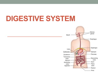
- 6. FUNCTIONALANATOMY OF DIGESTIVE SYSTEM • Digestive system is made up of gastrointestinal tract (GI tract) or alimentary canal and accessory organs, which help in the process of digestion and absorption. • GI tract is a tubular structure extending from the mouth up to anus, with a length of about 30 feet. • It opens to the external environment on both ends. • GI tract is formed by two types of organs: • 1. Primary digestive organs. • 2. Accessory digestive organs.
- 7. 1. Primary Digestive Organs • Primary digestive organs are the organs where actual digestion takes place. • Primary digestive organs are: • Mouth • Pharynx • Esophagus • Stomach • Duodenum • Small intestine • Large intestine. • Rectum and Anus.
- 8. 2. Accessory Digestive Organs • Accessory digestive organs are those which help primary digestive organs in the process of digestion. • Accessory digestive organs are: • i. Teeth • ii. Tongue • iii. Salivary glands • iv. Exocrine part of pancreas • v. Liver • vi. Gallbladder.
- 9. Mouth and Salivary Glands
- 10. FUNCTIONAL ANATOMY OF MOUTH • Mouth is otherwise known as oral cavity or buccal cavity. It is formed by cheeks, lips and palate. • It encloses the teeth, tongue and salivary glands. • Mouth opens anteriorly to the exterior through lips and posteriorly through faces into the pharynx. • Digestive juice present in the mouth is saliva, which is secreted by the salivary glands.
- 11. FUNCTIONS OF MOUTH • Primary function of mouth is eating and it has few other • important functions also. • Functions of mouth include: • 1. Ingestion of food materials • 2. Chewing the food and mixing it with saliva • 3. Appreciation of taste of the food • 4. Transfer of food (bolus) to the esophagus by • swallowing • 5. Role in speech • 6. Social functions such as smiling and other • expressions.
- 12. SALIVARY GLANDS • In humans, the saliva is secreted by three pairs of • Major (larger) salivary glands • Minor (small) salivary glands.
- 13. MAJOR SALIVARY GLANDS • Major glands are: • 1. Parotid glands • 2. Submaxillary or submandibular glands • 3. Sublingual glands.
- 14. 1. Parotid Glands • Parotid glands are the largest of all salivary glands, situated at the side of the face just below and in front of the ear. Each gland weighs about 20 to 30 g in adults. Secretions from these glands are emptied into the oral cavity by Stensen duct. This duct is about 35 mm to 40 mm long and opens inside the cheek against the upper second molar tooth.
- 15. 2. Submaxillary Glands • Submaxillary glands or submandibular glands are located in submaxillary triangle, medial to mandible. • Each gland weighs about 8 to 10 g. • Saliva from these glands is emptied into the oral cavity by Wharton duct, which is about 40 mm long. • The duct opens at the side of frenulum of tongue, by means of a small opening on the summit of papilla called caruncula sublingualis.
- 16. 3. Sublingual Glands • Sublingual glands are the smallest salivary glands situated in the mucosa at the floor of the mouth. • Each gland weighs about 2 to 3 g. • Saliva from these glands is poured into 5 to 15 small ducts called ducts of Rivinus. • These ducts open on small papillae beneath the tongue. One of the ducts is larger and it is called Bartholin duct. • It drains the anterior part of the gland and opens on caruncula sublingualis near the opening of submaxillary duct.
- 17. MINOR SALIVARY GLANDS • 1. Lingual Mucus Glands Lingual mucus glands are situated in posterior one third of the tongue, behind circumvallate papillae and at the tip and margins of tongue. • 2. Lingual Serous Glands Lingual serous glands are located near circumvallate papillae and filiform papillae. • 3. Buccal Glands Buccal glands or molar glands are present between the mucus membrane and buccinator muscle. Four to five of these are larger and situated outside buccinator, around the terminal part of parotid duct. • 4. Labial Glands Labial glands are situated beneath the mucus membrane around the orifice of mouth • 5. Palatal Glands Palatal glands are found beneath the mucus membrane of the soft palate.
- 18. STRUCTURE AND DUCT SYSTEM OF SALIVARY GLANDS • Salivary glands are formed by acini or alveoli. • Each acinus is formed by a small group of cells which surround a central globular cavity. Central cavity of each acinus is continuous with the lumen of the duct. The fine duct draining each acinus is called intercalated duct. Many intercalated ducts join together to form intralobular duct. • Few intralobular ducts join to form interlobular ducts,which unite to form the main duct of the gland. • A gland with this type of structure and duct system is called racemose type (racemose = bunch of grapes).
- 19. Diagram showing acini and duct system in salivary glands
- 20. Contribution by each major salivary gland is • i. Parotid glands : 25% • ii. Submaxillary glands : 70% • iii. Sublingual glands : 5%.
- 21. PROPERTIES OF SALIVA 1. Volume: 1000 mL to 1500 mL of saliva is secreted per day and it is approximately about 1 mL/minute. 2. Reaction: Mixed saliva from all the glands is slightly acidic with pH of 6.35 to 6.85. 3. Specific gravity: It ranges between 1.002 and 1.012 4. 4. Tonicity: Saliva is hypotonic to plasma.
- 22. COMPOSITION OF SALIVA • Mixed saliva contains 99.5% water and 0.5% solids.
- 23. FUNCTIONS OF SALIVA • Saliva is a very essential digestive juice. Since it has many functions, its absence leads to many inconveniences. Preparation of food for swallowing. Appreciation of taste. Digestive function Role in speech Excretory function. Regulation of body temperature. Regulation of water balance
- 24. PREPARATION OF FOOD FOR SWALLOWING When food is taken into the mouth, it is moistened and dissolved by saliva. The mucus membrane of mouth is also moistened by saliva. It facilitates chewing. By the movement of tongue, the moistened and masticated Food is rolled into a bolus. Mucin of saliva lubricates the bolus and facilitates swallowing.
- 25. Digestive function • Saliva has three digestive enzymes, • Salivary amylase, • Maltase • Lingual lipase
- 26. APPRECIATION OF TASTE • Taste is a chemical sensation. By its solvent action, • saliva dissolves the solid food substances, so that the dissolved substances can stimulate the taste buds. The stimulated taste buds recognize the taste.
- 27. Role in speech • By moistening and lubricating soft parts of mouth and lips, saliva helps in speech. • If the mouth becomes dry, articulation and pronunciation becomes difficult.
- 28. Cleaning & protective functions • i. Due to the constant secretion of saliva, the mouth and teeth are rinsed and kept free off food debris, shed epithelial cells and foreign particles. In this way, saliva prevents bacterial growth by removing materials, which may serve as culture media for the bacterial growth. • ii. Enzyme lysozyme of saliva kills some bacteria such as staphylococcus, streptococcus and brucella.
- 29. Excretory function • Many substances, both organic and inorganic, are • excreted in saliva. • It excretes substances like mercury, potassium iodide, lead, and thiocyanate. • Saliva also excretes some viruses such as those causing rabies and mumps.
- 30. Regulation of body temperature • In dogs and cattle, excessive dripping of saliva during panting helps in the loss of heat and regulation of body temperature. However, in human beings, sweat glands play a major role in temperature regulation and saliva does not play any role in this function.
- 31. Regulation of water balance • When the body water content decreases, salivary secretion also decreases. • This causes dryness of the mouth and induces thirst. When water is taken, it quenches the thirst and restores the body water content.
- 32. Stomach
- 33. Structure of stomach • The stomach is a J-shaped dilated portion of the alimentary tract situated in the epigastric, umbilical and left hypochondriac regions of the abdominal cavity. • Stomach is a hollow organ situated just below the diaphragm on the left side in the abdominal cavity. • Volume of empty stomach is 50 mL. • Under normal conditions, it can expand to accommodate 1 L to 1.5 L of solids and liquids. However, it is capable of expanding still further up to 4 L.
- 34. Parts of stomach stomach has four parts: • 1. Cardiac region • 2. Fundus • 3. Body or corpus • 4. Pyloric region. • 1. Cardiac Region • Cardiac region is the upper part of the stomach where esophagus opens. The opening is guarded by a sphincter called cardiac sphincter, which opens only towards stomach. This portion is also known as cardiac end.
- 35. 2. Fundus • Fundus is a small dome shaped structure. It is elevated above the level of esophageal opening 3. Body or Corpus Body is the largest part of stomach forming about 75% to 80% of the whole stomach. It extends from just below the fundus up to the pyloric region. 4. Pyloric Region Pyloric region has two parts, antrum and pyloric canal. The body of stomach ends in antrum.
- 36. • Junction between body and antrum is marked by an angular notch called incisura angularis. • Antrum is continued as the narrow canal, which is called pyloric canal or pyloric end. • Pyloric canal opens into first part of small intestine called duodenum. • The opening of pyloric canal is guarded by a sphincter called pyloric sphincter. It opens towards duodenum. Stomach has two curvatures. • One on the right side is lesser curvature and the other on left side is greater curvature.
- 37. STRUCTURE OF STOMACH WALL Stomach wall is formed by four layers of structures: 1. Outer serous layer: Formed by peritoneum. 2. Muscular layer: Made up of three layers of smooth muscle fibers, namely inner oblique, middle circular and outer longitudinal layers. 3. Submucus layer: Formed by areolar tissue, blood vessels, lymph vessels and Meissner nerve plexus. 4. Inner mucus layer: Lined by mucussecreting columnar epithelial cells. The gastric glands are situated in this layer. Under resting conditions, the mucosa of the stomach is thrown into many folds. These folds are called rugae. The rugae disappear when the stomach is distended after meals. Throughout the inner mucus layer, small depressions called gastric pits are present. Glands of the stomach open into these pits. Inner surface of mucus layer is covered by 2 mm thick mucus.
- 38. GLANDS OF STOMACH – GASTRIC GLANDS • Glands of the stomach or gastric glands are tubular structures made up of different types of cells. These glands open into the stomach cavity via gastric pits.
- 39. CLASSIFICATION OF GLANDS OF THE STOMACH • Gastric glands are classified into three types, on the basis of their location in the stomach: • 1. Fundic glands or main gastric glands or oxyntic glands: Situated in body and fundus of stomach • 2. Pyloric glands: Present in the pyloric part of the stomach • 3. Cardiac glands: Located in the cardiac region of the stomach
- 40. Parietal cells are different from other cells of the gland because of the presence of canaliculi (singular = canaliculus). Parietal cells empty their secretions into the lumen of the gland through the canaliculi. But, other cells empty their secretions directly into lumen of the gland.
- 41. STRUCTURE OF GASTRIC GLANDS 1. Fundic Glands Fundic glands are considered as the typical gastric glands. • These glands are long and tubular. • Each gland has three parts, viz. body, neck and isthmus. Cells of fundic glands • 1. Chief cells or pepsinogen cells • 2. Parietal cells or oxyntic cells • 3. Mucus neck cells • 4. Enterochromaffin (EC) cells or Kulchitsky cells • 5. Enterochromaffinlike (ECL) cells.
- 42. Pyloric Glands • Pyloric glands are short and tortuous in nature. • These glands are formed by • G cells, • Mucus cells, • EC cells and • ECL cells.
- 43. FUNCTIONS OF GASTRIC GLANDS • Function of the gastric gland is to secrete gastric juice. • Secretory activities of different cells of gastric glands and enteroendocrine cells.
- 44. Cardiac Glands Cardiac glands are also short and tortuous in structure, with many mucus cells. EC cells, ECL cells Chief cells are also present in the cardiac glands
- 45. 1. MECHANICAL FUNCTION i. Storage Function • Food is stored in the stomach for a long period, i.e. for 3 to 4 hours and emptied into the intestine slowly. • The maximum capacity of stomach is up to 1.5 L. Slow emptying of stomach provides enough time for proper digestion and absorption of food substances in the small intestine. ii. Formation of Chyme • Peristaltic movements of stomach mix the bolus with gastric juice and convert it into the semisolid material known as chyme
- 46. DIGESTIVE FUNCTION • „ DIGESTIVE FUNCTION Gastric juice acts mainly on proteins. Proteolytic enzymes of the gastric juice are pepsin and rennin.
- 47. • Gastric juice also contains some other enzymes like gastric lipase, gelatinase, urase and gastric amylase. Pepsin Pepsin is secreted as inactive pepsinogen. • Pepsinogen is converted into pepsin by hydrochloric acid. Optimum pH for activation of pepsinogen is below 6. • Action of pepsin Pepsin converts proteins into proteoses, peptones and polypeptides. • Pepsin also causes curdling and digestion of milk (casein). • Gastric Lipase Gastric lipase is a weak lipolytic enzyme when compared to pancreatic lipase. • It is active only when the pH is between 4 and 5 and becomes inactive at a pH below
- 48. PROTECTIVE FUNCTION FUNCTION OF MUCUS • Mucus is a mucoprotein, secreted by mucus neck cells of the gastric glands and surface mucus cells in fundus, body and other parts of stomach. It protects the gastric wall by the following ways: Mucus i. Protects the stomach wall from irritation or mechanical injury, by virtue of its high viscosity
- 49. HEMOPOIETIC FUNCTION • Intrinsic factor of Castle, secreted by parietal cells of gastric glands plays an important role in erythropoiesis. • It is necessary for the absorption of vitamin B12 (which is called extrinsic factor) from GI tract into the blood. • Vitamin B12 is an important maturation factor during erythropoiesis. • Absence of intrinsic factor in gastric juice causes deficiency of vitamin B12, leading to pernicious anemia
- 50. EXCRETORY FUNCTION • Many substances like toxins, alkaloids and metals are excreted through gastric juice
- 51. GASTRICOMPOSITIONOF C JUICEPROPERTIES Gastric juice is a mixture of secretions from different gastric glands. PROPERTIES OF GASTRIC JUICE • Volume : 1200 mL/day to 1500 mL/day. • Reaction : Gastric juice is highly acidic with a pH of 0.9 to 1.2. Acidity of gastric juice is due to the presence of hydrochloric acid. • Specific gravity : 1.002 to 1.004
- 52. COMPOSITION OF GASTRIC JUICE • Gastric juice contains 99.5% of water and 0.5% solids. Solids are organic and inorganic substances.for composition of gastric juice.
- 53. COMPOSITION OF GASTRIC JUICE
- 54. FUNCTIONS OF GASTRIC JUICE • FUNCTIONS OF HYDROCHLORIC ACID • Hydrochloric acid is present in the gastric juice: • i. Activates pepsinogen into pepsin • ii. Kills some of the bacteria entering the stomach along with food substances. This action is called bacteriolytic action • iii. Provides acid medium, which is necessary for the action of hormones.
- 55. GASTRIC JUICE SECRETION PHASES OF GASTRIC SECRETION • Secretion of gastric juice is a continuous process. But the quantity varies, depending upon time and stimulus. Accordingly, gastric secretion occurs in three different phases: I. Cephalic phase II. Gastric phase III. Intestinal phase. In human beings, a fourth phase called interdigestive phase exists. Each phase is regulated by neural mechanism or hormonal mechanism or both.
- 56. Regulationof gastric secretion CCKPZ = Cholecystokininpancreozymin,GIP= Gastric inhibitorypeptide,VIP= Vasoactive intestinalpeptide
- 57. CEPHALIC PHASE • Secretion of gastric juice by the stimuli arising from head region (cephalous) is called cephalic phase. • This phase of gastric secretion is regulated by nervous mechanism. The gastric juice secreted during this phase is called appetite juice. • During this phase, gastric secretion occurs even without the presence of food in stomach. The quantity of the juice is less but it is rich in enzymes and hydrochloric acid.
- 58. Nervous mechanism regulates cephalic phase Two types of reflexes occur: • 1. Unconditioned reflex • 2. Conditioned reflex. 1.Unconditioned Reflex • Unconditioned reflex is the inborn reflex. When food is placed in the mouth, salivary secretion is induced . Simultaneously, gastric secretion also occurs.
- 59. 2.Conditioned Reflex • Conditioned reflex is the reflex response acquired by previous experience. • Presence of food in the mouth is not necessary to elicit this reflex. • The sight, smell, hearing or thought of food, which induce salivary secretion induce gastric secretion also.
- 60. GASTRIC PHASE • Secretion of gastric juice when food enters the stomach is called gastric phase. This phase is regulated by both nervous and hormonal control. • Gastric juice secreted during this phase is rich in pepsinogen and hydrochloric acid. • Mechanisms involved in gastric phase are: 1. Nervous mechanism through local myenteric reflex and vagovagal reflex 2. Hormonal mechanism through gastrin Stimuli, which initiate these two mechanisms are: • 1. Distention of stomach • 2. Mechanical stimulation of gastric mucosa by bulk of food • 3. Chemical stimulation of gastric mucosa by the food contents.
- 61. INTESTINAL PHASE • Intestinal phase is the secretion of gastric juice when chyme enters the intestine. When chyme enters the • Intestine, initially, the gastric secretion increases but later it stops. Intestinal phase of gastric secretion is regulated by nervous and hormonal control. • Initial Stage of Intestinal Phase Chyme that enters the intestine stimulates the duodenal mucosa to release gastrin, which is transported to stomach by blood. • There it increases gastric secretion. Later Stage of Intestinal Phase After the initial increase, there is a decrease or complete stoppage of gastric secretion.
- 62. • Gastric secretion is inhibited by two factors: • 1. Enterogastric reflex • 2. Gastrointestinal (GI) hormones.
- 64. DISORDERS OF STOMACH • PEPTIC ULCER • Ulcer means the erosion of the surface of any organ due to shedding or sloughing of inflamed necrotic tissue that lines the organ. • Peptic ulcer means an ulcer in the wall of stomach or duodenum, caused by digestive action of gastric juice. • If peptic ulcer is found in stomach, it is called gastric ulcer • if found in duodenum, it is called duodenal ulcer.
- 70. Causes • i. Increased peptic activity due to excessive secretion of pepsin in gastric juice. • ii. Hyperacidity of gastric juice. • iii. Reduced alkalinity of duodenal content. • iv. Decreased mucin content in gastric juice or decreased protective activity in stomach or duodenum. • v. Constant physical or emotional stress. • vi. Food with excess spices or smoking (classical causes of ulcers). • vii. Long term use of NSAIDs (see above) such as Aspirin, Ibuprofen and Naproxen. • viii. Chronic inflammation due to Helicobacter pylori.
- 71. PANCREAS
- 73. FUNCTIONALANATOMYAND NERVE SUPPLY OF PANCREAS • Pancreas is a dual organ having two functions, namely endocrine function and exocrine function. • Endocrine function is concerned with the production of hormones. • The exocrine function is concerned with the secretion of digestive juice called pancreatic juice. „
- 74. • FUNCTIONAL ANATOMY OF EXOCRINE PART OF PANCREAS • Exocrine part of pancreas resembles salivary gland in structure. It is made up of acini or alveoli. • Each acinus has a single layer of acinar cells with a lumen in the center. Acinar cells contain zymogen granules, which possess digestive enzymes. • A small duct arises from lumen of each alveolus. Some of these ducts from neighboring alveoli unite to form intralobular duct. • All the intralobular ducts unite to form the main duct of pancreas called Wirsung duct. • Wirsung duct joins common bile duct to form ampulla of Vater, which opens into duodenum
- 75. PROPERTIESAND COMPOSITION OF PANCREATIC JUICE Properties of pancreatic juice • Volume : 500 to 800 mL/day • Reaction : Highly alkaline with a pH of 8 to 8.3 • Specific gravity : 1.010 to 1.018
- 76. COMPOSITION OF PANCREATIC JUICE • Pancreatic juice contains 99.5% of water and 0.5% of solids. The solids are the organic and inorganic substances. • Bicarbonate content is very high in pancreatic juice. • It is about 110 to 150 mEq/ L, against the plasma level of 24 mEq/L.
- 78. FUNCTIONS OF PANCREATIC JUICE • Pancreatic juice has digestive functions and neutralizing action. DIGESTIVE FUNCTIONS OF PANCREATIC JUICE • Pancreatic juice plays an important role in the digestion of proteins and lipids. It also has mild digestive action on carbohydrates.
- 79. DIGESTION OF PROTEINS • Major proteolytic enzymes of pancreatic juice are trypsin and chymotrypsin. Other proteolytic enzymes are carboxypeptidases, nuclease, elastase and collagenase. • 1. Trypsin • Trypsin is a single polypeptide with a molecular weight of 25,000. It contains 229 amino acids
- 80. • Chymotrypsin • Chymotrypsin is a polypeptide with a molecular weight of 25,700 and 246 amino acids. It is secreted as inactive chymotrypsinogen, which is activated into chymotrypsin by trypsin. • Actions of chymotrypsin • i. Digestion of proteins: Chymotrypsin is also an endopeptidase and it converts proteins into polypeptides
- 81. • Nucleases • Nucleases of pancreatic juice are ribonuclease and deoxyribonuclease, which are responsible for the digestion of nucleic acids. These enzymes convert the ribonucleic acid (RNA) and deoxyribonucleic acid (DNA) into mononucleotides.
- 82. • Elastase • Elastase is secreted as inactive proelastase, which is activated into elastase by trypsin. Elastase digests the elastic fibers. • Collagenase • Collagenase is secreted as inactive procollagenase, which is activated into collagenase by trypsin. It digests collagen.
- 83. DIGESTION OF LIPIDS • Lipolytic enzymes present in pancreatic juice are pancreatic lipase, cholesterol ester hydrolase, phospholipase A, phospholipase B, colipase and bilesalt- activated lipase
- 84. • DIGESTION OF CARBOHYDRATES • Pancreatic amylase is the amylolytic enzyme present in pancreatic juice. • Like salivary amylase, the pancreatic amylase also converts starch into dextrin and maltose.
- 85. LIVER Liver is a dual organ having both secretory and excretory functions. It is the largest gland in the body, weighing about 1.5 kg in man. It is located in the upper and right side of the abdominal cavity, immediately beneath diaphragm.
- 86. LIVER • Hepatic Lobes Liver is made up of many lobes called hepatic lobes Each lobe consists of many lobules called hepatic lobules. Hepatic Lobules • Hepatic lobule is the structural and functional unit of liver. • There are about 50,000 to 100,000 lobules in the liver. • The lobule is a honeycomb-like structure and it is made up of liver cells called hepatocytes.
- 87. Hepatocytes and Hepatic Plates • Hepatocytes are arranged in columns, which form the • hepatic plates. Each plate is made up of two columns of cells. In between the two columns of each plate lies a • bile canaliculus.
- 88. In between the neighboring plates, a blood space called sinusoid is present. Sinusoid is lined by the endothelial cells. In between the endothelial cells some special macrophages called Kupffer cells are present.
- 89. Portal Triads • Each lobule is surrounded by many portal triads. • Each portal triad consists of three vessels: • 1. A branch of Hepatic artery • 2. A branch of Portal vein • 3. A tributary of Bile duct. • Branches of hepatic artery and portal vein open into the sinusoid. • Sinusoid opens into the central vein. Central vein empties into hepatic vein. • Bile is secreted by hepatic cells and emptied into bile canaliculus. • From canaliculus, the bile enters the ributary of bile duct. Tributaries of bile duct from canaliculi of neighboring lobules unite to form small bile ducts. • These small bile ducts join together and finally form left and right hepatic ducts, which emerge out of liver
- 91. PROPERTIESAND COMPOSITION OF BILE PROPERTIES OF BILE • Volume : 800 to 1,200 mL/day • Reaction : Alkaline • pH : 8 to 8.6 • Specific gravity : 1.010 to 1.011 • Color : Golden yellow or green.
- 92. COMPOSITION OF BILE • Bile contains 97.6% of water and 2.4% of solids. • Solids include organic and inorganic substances.
- 93. SECRETION OF BILE • Bile is secreted by hepatocytes. The initial bile secreted by hepatocytes contains large quantity of bile acids, bile pigments, cholesterol, lecithin and fatty acids. • From hepatocytes, bile is released into canaliculi. From here, it passes through small ducts and hepatic ducts and reaches the common hepatic duct. From common hepatic duct, bile is diverted either directly into the intestine or into the gallbladder. • Sodium, bicarbonate and water are added to bile when it passes through the ducts. These substances are secreted by the epithelial cells of the ducts. Addition of sodium, bicarbonate and water increases the total quantity of bile.
- 94. STORAGE OF BILE • Most of the bile from liver enters the gallbladder, where it is stored. • It is released from gallbladder into the intestine whenever it is required.
- 95. FUNCTIONS OF BILE • Most of the functions of bile are due to the bile salts. „ DIGESTIVE FUNCTION • Bile salts are required for digestion and absorption of fats in the intestine. The functions of bile salts are: Emulsification of Fats Emulsification is the process by which the fat globules are broken down into minute droplets and made in the form of a milky fluid called emulsion in small intestine, by the action of bile salts.
- 96. ABSORPTIVE FUNCTIONS • Absorption of Fats Bile salts help in the absorption of digested fats from intestine into blood. Bile salts combine with fats and make complexes of fats called micelles. The fats in the form of micelles can be absorbed easily.
- 97. EXCRETORY FUNCTIONS Bile pigments are the major excretory products of the bile. Other substances excreted in bile are: i. Heavy metals like copper and iron ii. Some bacteria like typhoid bacteria iii. Some toxins iv. Cholesterol v. Lecithin vi. Alkaline phosphatase. „
- 98. LUBRICATION FUNCTION • The mucin in bile acts as a lubricant for the chyme in intestine.
- 99. LAXATIVE ACTION Laxative Action Laxative is an agent which induces defecation. Bile salts act as laxatives by stimulating peristaltic movements of the intestine Bile salts act as laxatives. ANTISEPTIC ACTION Bile inhibits the growth of certain bacteria in the lumen of intestine by its natural detergent action.
- 100. • Antiseptic action • Bile inhibits the growth of certain bacteria in the lumen of intestine by its natural detergent action. • Maintenance OF pH In Gastrointestinal Tract • As bile is highly alkaline, it neutralizes the acid chyme which enters the intestine from stomach. • Thus, an optimum pH is maintained for the action of digestive enzymes
- 101. FUNCTIONS OF LIVER • Liver is the largest gland and one of the vital organs of the body. • It performs many vital metabolic and homeostatic functions. „
- 102. • Metabolic function • Liver is the organ where maximum metabolic reactions such as metabolism of carbohydrates, proteins, fats, vitamins and many hormones are carried out. „ • Storage function • Many substances like glycogen, amino acids, iron, folic acid and vitamins A, B12 and D are stored in liver. „
- 103. Secretion of bile • Liver secretes bile which contains bile salts, bile pigments, cholesterol, fatty acids and lecithin. • The functions of bile are mainly due to bile salts. • Bile salts are required for digestion and absorption of fats in the intestine. • Bile helps to carry away waste products and breakdown fats, which are excreted through feces or urine.
- 104. Synthetic function • Liver produces glucose by gluconeogenesis. • It synthesizes all the plasma proteins and other proteins (except immunoglobulins) such as clotting factors, complement factors and hormonebinding proteins. • It also synthesizes steroids, somatomedin and heparin.
- 105. Excretory function • Liver excretes cholesterol, bile pigments, heavy metals (like lead, arsenic and bismuth), toxins, bacteria and virus (like that of yellow fever) through bile
- 106. Heat production • Enormous amount of heat is produced in the liver because of metabolic reactions. • Liver is the organ where maximum heat is produced.
- 107. HEMOPOIETIC FUNCTION • In fetus (hepatic stage), liver produces the blood cells . • It stores vitamin B12 necessary for erythropoiesis and iron necessary for synthesis • of hemoglobin. Liver produces thrombopoietin that promotes production of thrombocytes
- 108. HEMOLYTIC FUNCTION • The senile RBCs after a lifespan of 120 days are destroyed by reticuloendothelial cells (Kupffer cells) of liver
- 109. INACTIVATION OF HORMONESAND DRUGS • Liver catabolizes the hormones such as growth hormone, parathormone, cortisol, insulin, glucagon and estrogen. • It also inactivates the drugs, particularly the fat soluble drugs. • The fat soluble drugs are converted into water soluble substances, which are excreted through bile or urine.
- 110. DEFENSIVE AND DETOXIFICATION FUNCTIONS • Reticuloendothelial cells (Kupffer cells) of the liver play an important role in the defense of the body. • Liver is also involved in the detoxification of the foreign bodies. • i. Foreign bodies such as bacteria or antigens are swallowed and digested by reticuloendothelial cells of liver by means of phagocytosis. • ii. Reticuloendothelial cells of liver also produce substances like interleukins and tumor necrosis factors, which activate the immune system of the body. • iii. Liver cells are involved in the removal of toxic property of various harmful substances. • Removal of toxic property of the harmful agent is known as detoxification.
- 111. • Detoxification in liver occurs in two ways: • a. Total destruction of the substances by means of metabolic degradation. • b. Conversion of toxic substances into nontoxic materials by means of conjugation with glucuronic acid or sulfates
- 112. GALLBLADDER • Bile secreted from liver is stored in gallbladder. • The capacity of gallbladder is approximately 50 mL. Gallbladder is not essential for life and it is removed (cholecystectomy) in patients suffering from gallbladder dysfunction. • After cholecystectomy, patients do not suffer from any major disadvantage. • In some species, gallbladder is absent.
- 113. FUNCTIONS OF GALLBLADDER • Major functions of gallbladder are the storage and concentration of bile. • 1. Storage of Bile • Bile is continuously secreted from liver. But it is released into intestine only intermittently and most of the bile is stored in gallbladder till it is required.
- 114. • 2. Concentration of Bile • Bile is concentrated while it is stored in gallbladder. The mucosa of gallbladder rapidly reabsorbs water and electrolytes, except calcium and potassium. But the bile salts, bile pigments, cholesterol and lecithin are not reabsorbed. So, the concentration of these substances in bile increases 5 to 10 times
- 115. 3. Alteration of pH of Bile • The pH of bile decreases from 8 – 8.6 to 7 – 7.6 and it becomes less alkaline when it is stored in gallbladder.
- 116. 4. Secretion of Mucin Gallbladder secretes mucin and adds it to bile. When bile is released into the intestine, mucin acts as a lubricant for movement of chyme in the intestine.
- 117. 5. Maintenance of Pressure in Biliary System Due to the concentrating capacity, gallbladder maintains a pressure of about 7 cm H2 O in biliary system. This pressure in the biliary system is essential for the release of bile into the intestine
- 118. • Prevention of Gallstone Formation • Bile salts prevent the formation of gallstone by keeping the cholesterol and lecithin in solution. • In the absence of bile salts, cholesterol precipitates along with lecithin and forms gallstone.
- 119. Disorders of Liver- Jaundice or icterus • Jaundice or icterus is the condition characterized by yellow coloration of the skin, mucous membrane and deeper tissues due to increased bilirubin level in blood.
- 120. Jaundice or icterus • The word jaundice is derived from the French word ‘jaune’ meaning yellow. • The normal serum bilirubin level is 0.5 to 1.5 mg/dL. • Jaundice occurs when bilirubin level exceeds 2 mg/dL.
- 121. Types of Jaundice • Jaundice is classified into three types: • 1. Prehepatic or hemolytic jaundice • 2. Hepatic or hepatocellular jaundice • 3. Posthepatic or obstructive jaundice.
- 122. 1. Prehepatic or Hemolytic Jaundice • Hemolytic jaundice is the type of jaundice that occurs because of excessive destruction of RBCs resulting in increased blood level of free (unconjugated) bilirubin. • In this condition, the excretory function of liver is normal. But the quantity of bilirubin increases enormously. • The liver cells cannot excrete that much excess bilirubin rapidly.
- 123. • Unconjugated bilirubin is insoluble in water and is not excreted in urine. So, it accumulates in the blood resulting in jaundice. • Formation of urobilinogen also increases resulting in the excretion of more amount of urobilinogen in urine. Causes Any condition that causes hemolytic anemia can lead to hemolytic jaundice
- 124. Common causes of hemolytic jaundice • i. Renal disorder • ii. Hypersplenism • iii. Burns • iv. Infections such as malaria • v. Hemoglobin abnormalities such as sickle cell • anemia or thalassemia • vi. Drugs or chemical substances causing red cell damage • vii. Autoimmune diseases.
- 125. 2. Hepatic or Hepatocellular or Cholestatic Jaundice • Hepatic jaundice is the type of jaundice that occurs due to the damage of hepatic cells. Because of the damage, the conjugated bilirubin from liver cannot be excreted and it returns to blood. Causes • i. Infection (infective jaundice) by virus, resulting in hepatitis (viral hepatitis) • ii. Alcoholic hepatitis • iii. Cirrhosis of liver • iv. Exposure to toxic materials
- 126. Post hepatic or Obstructive or Extra hepatic Jaundice • Post hepatic or Obstructive or Extra hepatic • Jaundice Post hepatic type of jaundice occurs because of the obstruction of bile flow at any level of the biliary system. • The bile cannot be excreted into small intestine. So, bile salts and bile pigments enter the circulation. • The blood contains more amount of conjugated bilirubin • Causes • i. Gallstones • ii. Cancer of biliary system or pancreas.
- 127. CIRRHOSIS OF LIVER • Cirrhosis of liver refers to inflammation and damage of parenchyma of liver. It results in degeneration of hepatic cells and dysfunction of liver.
- 128. Causes • 1. Infection • 2. Retention of bile in liver due to obstruction of ducts of biliary system • 3. Enlargement of liver due to intoxication • 4. Inflammation around liver (perihepatitis) • 5. Infiltration of fat in hepatic cells.
- 129. Features • 1. Fever, nausea and vomiting • 2. Jaundice • 3. Increased heart rate and cardiac output • 4. Portal hypertension • 5. Muscular weakness and wasting of muscles • 6. Drowsiness • 7. Lack of concentration and confused state of mind • 8. Coma in advanced stages
- 130. DIARRHEA • Diarrhea is the frequent and profuse discharge of intestinal contents in loose and fluid form. It occurs due to the increased movement of intestine. It may be acute or chronic.
- 131. Causes • Normally, when digested food passes through colon, large portion of fluid is absorbed and only a semisolid stool remains. In diarrhea, the fluid is not absorbed sufficiently, resulting in watery bowel discharge. Acute diarrhea may be caused by temporary problems like infection and chronic diarrhea may be due to disorders of intestinal mucosa.
- 132. Features • Features Severe diarrhea results in loss of excess water and electrolytes. • This leads to dehydration and electrolyte imbalance. Chronic diarrhea results in hypokalemia and metabolic acidosis. • Other features of diarrhea are abdominal pain, nausea and bloating (a condition in which the subject feels the abdomen full and tight due to excess intestinal gas).
- 133. CONSTIPATION • Failure of voiding of feces, which produces discomfort is known as constipation. It is due to the lack of movements necessary for defecation. • Due to the absence of mass movement in colon, feces remain in the large intestine for a long time, resulting in absorption of fluid. So the feces become hard and dry.
- 134. Causes • 1. Dietary causes Lack of fiber or lack of liquids in diet causes constipation. • 2. Irregular bowel habit Irregular bowel habit is most common cause for constipation. • It causes constipation by inhibiting the normal defecation reflexes.
- 135. VOMITING • Vomiting or emesis is the abnormal emptying of stomach and upper part of intestine through esophagus and mouth. „
- 136. CAUSES OF VOMITING • 1. Presence of irritating contents in GI tract • 2. Mechanical stimulation of pharynx • 3. Pregnancy • 4. Excess intake of alcohol • 5. Nauseating sight, odor or taste • 6. Unusual stimulation of labyrinthine apparatus, as in the case of sea sickness, air sickness, car sickness or swinging • 7. Abnormal stimulation of sensory receptors in other organs like kidney, heart, semicircular canals or uterus • 8. Drugs like antibiotics, opiates, etc. • 9. Any GI disorder • 10. Acute infection like urinary tract infection, influenza, etc. • 11. Metabolic disturbances like carbohydrate starvation and ketosis (pregnancy), uremia, ketoacidosis (diabetes) and hypercalcemia.
- 137. SMALL INTESTIN E • Small intestine is the part of gastrointestinal (GI) tract, extending between the pyloric sphincter of stomach and ileocecal valve, which opens into large intestine. • It is called small intestine because of its small diameter, compared to that of the large intestine. But it is longer than large intestine. Its length is about 6 meter. • Important function of small intestine is absorption. Maximum absorption of digested food products takes place in small intestine.
- 138. Small intestine consists of three portions • 1. Proximal part known as duodenum • 2. Middle part known as jejunum • 3. Distal part known as ileum. Wall of the small intestine has all the four layers as in stomach
- 139. • INTESTINAL VILLI AND GLANDS OF SMALL INTESTINE „ INTESTINAL VILLI Mucous membrane of small intestine is covered by minute projections called villi. The height of villi is about 1 mm and the diameter is less than 1 mm. Villi are lined by columnar cells, which are called enterocytes. Each enterocyte gives rise to hair-like projections called microvilli. • Villi and microvilli increase the surface area of mucous membrane by many folds. Within each villus, there is a central channel called lacteal, which opens into lymphatic vessels. It contains blood vessels also
- 140. • Epithelial cells lining the intestinal glands undergo division by mitosis at a faster rate. Newly formed cells push the older cells upward over the lining of villi. These cells which move to villi are called enterocytes. Enterocytes secrete the enzymes. Old enterocytes are continuously shed into lumen along with enzymes. • Types of cells interposed between columnar cells of intestinal glands: • 1. Argentaffin cells or enterochromaffin cells, which secrete intrinsic factor of Castle • 2. Goblet cells, which secrete mucus • 3. Paneth cells, which secrete the cytokines called defensins
- 141. • CRYPTS OF LIEBERKÜHN OR INTESTINAL GLANDS Crypts of Lieberkühn or intestinal glands are simple tubular glands of intestine. Intestinal glands do not penetrate the muscularis mucosa of the intestinal wall, but open into the lumen of intestine between the villi. Intestinal glands are lined by columnar cells. Lining of each gland is continuous with epithelial lining of the villi BRUNNER GLANDS In addition to intestinal glands, the first part of duodenum contains some mucus glands, which are called Brunner glands. These glands penetrate muscularis mucosa and extend up to the submucus coat of the intestinal wall. Brunner glands open into the lumen of intestine directly. Brunner gland secretes mucus and traces of enzymes
- 142. PROPERTIESAND COMPOSITION OF SUCCUS ENTERICUS • Secretion from small intestine is called succus entericus. • „PROPERTIES OF SUCCUS ENTERICUS • Volume : 1800 mL/day • Reaction : Alkaline • pH : 8.3 • „COMPOSITION OF SUCCUS ENTERICUS • Succus entericus contains water (99.5%) and solids (0.5%). Solids include organic and inorganic substances. Bicarbonate concentration is slightly high in succus entericus.
- 143. COMPOSITION
- 144. FUNCTIONS OF SUCCUS ENTERICUS • „1. DIGESTIVE FUNCTION Enzymes of succus entericus act on the partially digested food and convert them into final digestive products. Enzymes are produced and released into succus entericus by enterocytes of the villi. • Proteolytic Enzymes Proteolytic enzymes present in succus entericus are the peptidases, which are given in. • These peptidases convert peptides into amino acids.
- 145. • Amylolytic Enzymes Amylolytic enzymes of succus entericus are listed in Lactase, sucrase and maltase convert the disaccharides (lactose, sucrose and maltose) into two molecules of monosaccharides • Dextrinase converts dextrin, maltose and maltriose into glucose. • Trehalase or trehalose glucohydrolase causes hydrolysis of trehalose (carbohydrate present in mushrooms and yeast) and converts it into glucose. • Lipolytic Enzyme Intestinal lipase acts on triglycerides and converts them into fatty acids
- 146. 2. PROTECTIVE FUNCTION • i. Mucus present in the succus entericus protects the intestinal wall from the acid chyme, which enters the intestine from stomach; thereby it prevents the intestinal ulcer. • ii. Defensins secreted by paneth cells of intestinal glands are the antimicrobial peptides. These peptides are called natural peptide antibiotics because of their role in killing the phagocytosed bacteria.
- 147. 3. ACTIVATOR FUNCTION • Enterokinase present in intestinal juice activates trypsinogen into trypsin. Trypsin, in turn activates other enzymes . 4. HEMOPOIETIC FUNCTION Intrinsic factor of Castle present in the intestine plays an important role in erythropoiesis . It is necessary for the absorption of vitamin B12. 5. HYDROLYTIC PROCESS Intestinal juice helps in all the enzymatic reactions of digestion.
- 148. FUNCTIONS OF SMALL INTESTINE • „. MECHANICAL FUNCTION Mixing movements of small intestine help in the thorough mixing of chyme with the digestive juices like succus entericus, pancreatic juice and bile. „ • 2. SECRETORY FUNCTION Small intestine secretes succus entericus, enterokinase and the GI hormones. „ • 3. HORMONAL FUNCTION Small intestine secretes many GI hormones such as secretin, cholecystokinin, etc. These hormones regulate the movement of GI tract and secretory activities of small intestine and pancreas . • 4. DIGESTIVE FUNCTION Refer functions of succus entericus.
- 150. Digestion,Absorption and Metabolism of Carbohydrates • Carbohydrates in diet • Human diet contains three types of carbohydrates: • „1. POLYSACCHARIDES • Large polysaccharides are glycogen, amylose and • amylopectin, which are in the form of starch (glucose • polymers). Glycogen is available in non-vegetarian diet. • Amylose and amylopectin are available in vegetarian • diet because of their plant origin. • „ 2. DISACCHARIDES • Two types of disaccharides are available in the diet. • i. Sucrose (Glucose + Fructose), which is called • table sugar or cane sugar • ii. Lactose (Glucose + Galactose), which is the • sugar available in milk.
- 151. MONOSACCHARIDES • Monosaccharides consumed in human diet are mostly glucose and fructose. • Other carbohydrates in the diet include • i. Alcohol • ii. Lactic acid • iii. Pyruvic acid • iv. Pectins • v. Dextrins • vi. Carbohydrates in meat. • Diet also contains large amount of cellulose, which cannot be digested in the human GI tract so it is not considered as a food for human beings
- 152. DIGESTION OF CARBOHYDRATES IN THE MOUTH • Enzymes involved in the digestion of carbohydrates are known as amylolytic enzymes. The only amylolytic enzyme present in saliva is the salivary amylase or ptyalin . • „IN THE STOMACH • Gastric juice contains a weak amylase, which plays a minor role in digestion of carbohydrates. • „INTHE INTESTINE • Amylolytic enzymes present in the small intestine are derived from pancreatic juice and succusentericus
- 153. CARBOHYDRATE DIGESTION • Final products of carbohydrate digestion are monosaccharides, which are glucose, fructose and galactose. • Glucose represents 80% of the final product of carbohydrate digestion. Galactose and fructose represent the remaining 20%
- 155. ABSORPTION OF GLUCOSE • Glucose is transported from the lumen of small intestine into the epithelial cells in the mucus membrane of small intestine, by means of sodium cotransport. • Energy for this is obtained by the binding process of sodium ion and glucose molecule to carrier protein. From the epithelial cell, glucose is absorbed into the portal vein by facilitated diffusion. • Sodium ion moves laterally into the intercellular space. From here, it is transported into blood by active transport, utilizing the energy liberated by breakdown of ATP
- 156. ABSORPTION OF GALACTOSE Galactose is also absorbed from the small intestine in the same mechanism as that of glucose. ABSORPTION OF FRUCTOSE Fructose is absorbed into blood by means of facilitated diffusion. Some molecules of fructose are converted into glucose. Glucose is absorbed as described above
- 157. METABOLISM OF CARBOHYDRATES • Metabolism is the process in which food substances undergo chemical and energy transformation. • After digestion and absorption, food substances must be utilized by the body. • The utilization occurs mainly by oxidative process in which the carbohydrates, proteins and lipids are burnt slowly to release energy. • This process is known as catabolism. Part of the released energy is utilized by tissues for physiological actions and rest of the energy is stored as rich energy phosphate bonds and in the form of proteins, carbohydrates and lipids in the tissues. • This process is called anabolism.
- 159. Digestion, Absorption and Metabolism of Proteins • PROTEINS IN DIET • Foodstuffs containing high protein content are meat, fish, egg and milk. Proteins are also available in wheat, soybeans, oats and various types of pulses. • Proteins present in common foodstuffs are: • 1. Wheat: Glutenin and gliadin, which constitute gluten • 2. Milk: Casein, lactalbumin, albumin and myosin • 3. Egg: Albumin and vitellin • 4. Meat: Collagen, albumin and myosin.
- 160. • Dietary proteins are formed by long chains of amino acids, bound together by peptide linkages. DIGESTION OF PROTEINS Enzymes responsible for the digestion of proteins are called proteolytic enzymes. „ IN THE MOUTH Digestion of proteins does not occur in mouth, since saliva does not contain any proteolytic enzymes. So, the digestion of proteins starts only in stomach IN THE STOMACH Pepsin is the only proteolytic enzyme in gastric juice. Rennin is also present in gastric juice. But it is absent in human. „ IN THE SMALL INTESTINE Most of the proteins are digested in the duodenum and jejunum by the proteolytic enzymes of the pancreatic juice and succus enter
- 162. FINAL PRODUCTS OF PROTEIN DIGESTION • Final products of protein digestion are the amino acids, which are absorbed into blood from intestine. ABSORPTION OF PROTEINS Proteins are absorbed in the form of amino acids from small intestine. The levoamino acids are actively absorbed by means of sodium cotransport, whereas, the dextro amino acids are absorbed by means of facilitated diffusion. Absorption of amino acids is faster in duodenum and jejunum and slower in ileum
- 163. LIPIDS IN DIET • Lipids are mostly consumed in the form of neutral fats, which are also known as triglycerides. Triglycerides are made up of glycerol nucleus and free fatty acids. Triglycerides form the major constituent in foods of animal origin and much less in foods of plant origin. Apart from triglycerides, usual diet also contains small quantities of cholesterol and cholesterol esters.
- 164. Source of lipids
- 165. Dietary fats are classified into two types • 1. Saturated fats • 2. Unsaturated fats. „ • SATURATED FATS • Saturated fats are the fats which contain triglycerides formed from only saturated fatty acids. The fatty acids having maximum amount of hydrogen ions without any double bonds between carbon atoms are called saturated fatty acids. „ • UNSATURATED FATS • Fats containing unsaturated fatty acids are known as unsaturated fats. Unsaturated fatty acids are fatty acids formed by dehydrogenation of saturated fatty acids. Unsaturated fats are classified into three types: • 1. Monounsaturated fats • 2. Polyunsaturated fats • 3. Trans fats.
- 166. DIGESTION OF LIPIDS • Lipids are digested by lipolytic enzymes. „ • IN THE MOUTH Saliva contains lingual lipase. This enzyme is secreted by lingual glands of mouth and swallowed along with saliva. So, the lipid digestion does not commence in the mouth. „ • IN THE STOMACH Gastric lipase or tributyrase is the lipolytic enzyme present in gastric juice. „ • IN THE INTESTINE Almost all the lipids are digested in the small intestine because of the availability of bile salts, pancreatic lipolytic enzymes and intestinal lipase. Role of Bile Salts Bile salts play an important role in the digestion of lipids.
- 167. Digestion of Lipids
- 168. FINAL PRODUCTS OF FAT DIGESTION • Fatty acids, cholesterol and monoglycerides are the final products of lipid digestion. „ • ABSORPTION OF LIPIDS • Monoglycerides, cholesterol and fatty acids from the micelles enter the cells of intestinal mucosa by simple diffusion. From here, further transport occurs as follows: In the mucosal cells, most of the monoglycerides are converted into triglycerides. Triglycerides are also formed by re-esterification of fatty acids with more than 10 to 12 carbon atoms. Some of the cholesterol is also esterified.