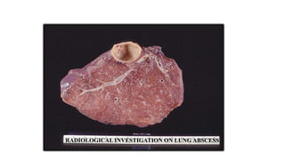
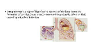
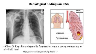
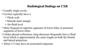
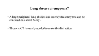
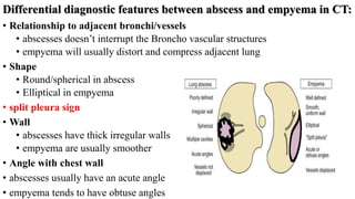
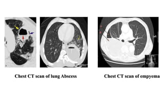
Welcome to my presentation
Presented by
RABEYA SULTANA MIM
Student ID :182408
Session: 2018-2019
Department of Physiotherapy & Rehabilitation
Jashore University of Science & Technology
RADIOLOGICAL INVESTIGATION ON LUNG ABSCESS
What is lung
abscess?
Pathophysiology
of pulmonary
abscess?
Radiological
Investigation of
it?
Clinical Features
of it?
Learning outcome
• Lung abscess is a type of liquefactive necrosis of the lung tissue and
formation of cavities (more than 2 cm) containing necrotic debris or fluid
caused by microbial infection.
Clinical features:
Early symptoms:
• Fever and chills
• Night sweats
• Persistent cough
• Later signs and symptoms :
• Pain with deep breathing
• Hemoptysis
• Shortness of breath
• Foul-smelling sputum
• Unintentional weight loss
• Fatigue
• Clubbing
Pathophysiology
Due to aspiration & Periodontal disease.
A bacterial inoculum from the gingival crevice reaches the lower
airways and infection is initiated
Aspiration pneumonitis
Progression to tissue necrosis 7-14 days later
Lung abscess.
Radiological Investigation by:
• Chest X-Ray(CXR)
• Chest CT Scan
• Chest X Ray: Parenchymal inflammation with a cavity containing an
air–fluid level
https://radiopaedia.org/cases/lung-abscess-17
Radiological findings on CXR
• Usually single cavity
• Cavities typically have a
• Thick-wall
• Smooth inner margin
• Air-fluid level
• More frequent in superior segments of lower lobes or posterior
segments of lower lobes
• Unlike pleural collections, lung abscesses frequently have a fluid
level which is approximately the same length on both the frontal
and lateral projection
• About 1/3 may have an associated empyema
Lung abscess or empyema?
• A large peripheral lung abscess and an encysted empyema can be
confused on a chest X-ray .
• Thoracic CT is usually needed to make the distinction.
Differential diagnostic features between abscess and empyema in CT:
• Relationship to adjacent bronchi/vessels
• abscesses doesn’t interrupt the Broncho vascular structures
• empyema will usually distort and compress adjacent lung
• Shape
• Round/spherical in abscess
• Elliptical in empyema
• split pleura sign
• Wall
• abscesses have thick irregular walls
• empyema are usually smoother
• Angle with chest wall
• abscesses usually have an acute angle
• empyema tends to have obtuse angles
Chest CT scan of lung Abscess Chest CT scan of empyema
Reference:
• En.wikipedia.org. 2022. Lung abscess - Wikipedia. [online] Available at:
<https://en.wikipedia.org/wiki/Lung_abscess> [Accessed 15 March 2022].
• Verywell Health. 2022. What Is a Lung Abscess?. [online] Available at:
<https://www.verywellhealth.com/lung-abscess-overview-4768089> [Accessed 15 March
2022].
• Emedicine.medscape.com. 2022. Lung Abscess: Background, Pathophysiology, Etiology.
[online] Available at: <https://emedicine.medscape.com/article/299425-overview#a5>
[Accessed 17 March 2022].
• Learningradiology.com. 2022. Learning Radiology - Lung, Abscess, pulmonary. [online]
Available at: <http://www.learningradiology.com/archives2008/COW%20306-
Lung%20abscess/lungabscesscorrect.html> [Accessed 17 March 2022].
• Learningradiology.com. 2022. Learning Radiology - Lung, Abscess, pulmonary. [online]
Available at: <http://www.learningradiology.com/archives2008/COW%20306-
Lung%20abscess/lungabscesscorrect.html> [Accessed 17 March 2022].
THANK YOU

MIM (Lung abscess).pptx

  • 1.
    Welcome to mypresentation Presented by RABEYA SULTANA MIM Student ID :182408 Session: 2018-2019 Department of Physiotherapy & Rehabilitation Jashore University of Science & Technology
  • 2.
  • 3.
    What is lung abscess? Pathophysiology ofpulmonary abscess? Radiological Investigation of it? Clinical Features of it? Learning outcome
  • 4.
    • Lung abscessis a type of liquefactive necrosis of the lung tissue and formation of cavities (more than 2 cm) containing necrotic debris or fluid caused by microbial infection.
  • 5.
    Clinical features: Early symptoms: •Fever and chills • Night sweats • Persistent cough • Later signs and symptoms : • Pain with deep breathing • Hemoptysis • Shortness of breath • Foul-smelling sputum • Unintentional weight loss • Fatigue • Clubbing
  • 6.
    Pathophysiology Due to aspiration& Periodontal disease. A bacterial inoculum from the gingival crevice reaches the lower airways and infection is initiated Aspiration pneumonitis Progression to tissue necrosis 7-14 days later Lung abscess.
  • 7.
    Radiological Investigation by: •Chest X-Ray(CXR) • Chest CT Scan
  • 8.
    • Chest XRay: Parenchymal inflammation with a cavity containing an air–fluid level https://radiopaedia.org/cases/lung-abscess-17 Radiological findings on CXR
  • 9.
    Radiological findings onCXR • Usually single cavity • Cavities typically have a • Thick-wall • Smooth inner margin • Air-fluid level • More frequent in superior segments of lower lobes or posterior segments of lower lobes • Unlike pleural collections, lung abscesses frequently have a fluid level which is approximately the same length on both the frontal and lateral projection • About 1/3 may have an associated empyema
  • 10.
    Lung abscess orempyema? • A large peripheral lung abscess and an encysted empyema can be confused on a chest X-ray . • Thoracic CT is usually needed to make the distinction.
  • 11.
    Differential diagnostic featuresbetween abscess and empyema in CT: • Relationship to adjacent bronchi/vessels • abscesses doesn’t interrupt the Broncho vascular structures • empyema will usually distort and compress adjacent lung • Shape • Round/spherical in abscess • Elliptical in empyema • split pleura sign • Wall • abscesses have thick irregular walls • empyema are usually smoother • Angle with chest wall • abscesses usually have an acute angle • empyema tends to have obtuse angles
  • 12.
    Chest CT scanof lung Abscess Chest CT scan of empyema
  • 14.
    Reference: • En.wikipedia.org. 2022.Lung abscess - Wikipedia. [online] Available at: <https://en.wikipedia.org/wiki/Lung_abscess> [Accessed 15 March 2022]. • Verywell Health. 2022. What Is a Lung Abscess?. [online] Available at: <https://www.verywellhealth.com/lung-abscess-overview-4768089> [Accessed 15 March 2022]. • Emedicine.medscape.com. 2022. Lung Abscess: Background, Pathophysiology, Etiology. [online] Available at: <https://emedicine.medscape.com/article/299425-overview#a5> [Accessed 17 March 2022]. • Learningradiology.com. 2022. Learning Radiology - Lung, Abscess, pulmonary. [online] Available at: <http://www.learningradiology.com/archives2008/COW%20306- Lung%20abscess/lungabscesscorrect.html> [Accessed 17 March 2022]. • Learningradiology.com. 2022. Learning Radiology - Lung, Abscess, pulmonary. [online] Available at: <http://www.learningradiology.com/archives2008/COW%20306- Lung%20abscess/lungabscesscorrect.html> [Accessed 17 March 2022].
  • 15.