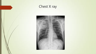
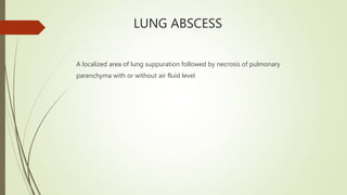
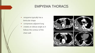
Dr Jamal Umar
TMO PULMONOLOGY
MTI KTH PESHAWAR
CASE PRESENTATION
HISTORY
 A 35 years old male patient
 2 mo history of Productive Cough (copious purulent sputum with no hemoptysis),
 Right sided chest pain( in 4th ICS, gradually increased in intensity, aggravated with
deep breathing and coughing, relieved with pain killers, radiating to the back and
ipsilateral shoulder)
 Fever (low grade, continuous with rigors and chills)
 Shortness of breath( MMRC Grade 2, gradually progressive, exertional, no
orthopnea, PND)
 Nausea and vomiting
HISTORY
 Scrap picker by profession
 No History of travel abroad
 No history of IV drug abuse
 No History of leg swelling
 No significant drug history
 No past history of HTN, DM, Hep B, Hep C, TB, asthma or any other respiratory
illness
EXAMINATION
 BP 110/80 Pulse 112/min SaO2 94 % RA, RR 20/min Temp 100 F
 GPE
Anemic, Clubbed,
 CVS - S1 + S2 no added sounds or murmurs
 Resp - R sided decreased breath sounds with inspiratory crackles on auscultation
 GIT - Not significant
 CNS - Not significant
LABORATORY
 Hb 11g/dl TLC 22.2 Pt 415 CRP 285
 Sputum AFB/ Xpert MTB Neg - 2 months back ( Started on ATT for 2 months
without evidence)
 Sputum bacterial C/S Neg
 Urine RE Normal
 Urea 40 Cr 1.0 RBS 85 HbA1C 5.03 LFT normal
 HBsAg /Anti HCV/Anti HIV Neg
Chest X ray
Chest X ray
DIAGNOSIS
LUNG ABSCESS
A localized area of lung suppuration followed by necrosis of pulmonary
parenchyma with or without air fluid level
CLASSIFICATION
ACUTE vs CHRONIC
PRIMARY vs SECONDARY
SINGLE vs MULTIPLE
PATHOPHYSIOLOGY
 The bacterial inoculum reaches the lung parenchyma, often in a dependent lung
area.
 Pneumonitis, followed by necrosis, occurs over 7– 14 days.
 Cavitation occurs when parenchymal necrosis leads to communication
with the bronchus
 entry of air and expectoration of necrotic material leading to the
formation of an air-fluid level.
 Bronchial obstruction leads to atelectasis with stasis and subsequent
infection, which can predispose to abscess formation.
RISK FACTORS
Aspiration
Impaired
consciousness
1. Post anesthesia
2. CVA
3. Seizure disorders
4. Alcoholism
Periodontal disease and pyorrhea
RISK FACTORS
 Dysphagia or pharyngeal
dysfunction
 Endobronchial obstruction
( foreign body, tumour)
 IV drug abuse( R heart
endocarditis caused by
Staph aureus)
RISK FACTORS
 Pulmonary embolism
 Septic embolism
 Bronchiectasis
 Immunocompromised states
CKD, DM, HIV
CLINICAL PRESENTATION
 SIGNS
Tachypnea
Tachycardia
Clubbing
Weight loss
Pyrexia
 SYMPTOMS
Fever with chills
Productive Cough
Dyspnea
Pleurisy
Chest heaviness
Malaise
Night sweats
Shoulder pain
MICROBIOLOGY
 Anaerobes (93%)
Fusobacterium nucleatum and
necrophorum
Bacteroides fragilis
Pigmented and non pigmented Prevotella
Peptococcus and peptostreptococcus
 Aerobes
Streptococcus ‘milleri’ group
Staphylococcus aureus
Klebsiella spp
Pseudomonas aeruginosa
Streptococcus pyogenes
Haemophilus influenza
Nocardia
DIFFERENTIAL DIAGNOSIS
 Cavitating carcinoma- Primary or metastatic
 Cavitatory TB
 GPA (Wegener’s)
 Infected pulmonary cyst or bulla (can produce a fluid level, usually thinner-walled)
 Aspergilloma
 Pulmonary infarct
 Rheumatoid nodule
 Sarcoidosis
 Bronchiectasis.
INVESTIGATIONS
MICROBIOLOGICAL CULTURES
 Blood cultures
 Sputum or bronchoscopic specimen (BAL or brushings rarely needed)
 Transthoracic percutaneous needle aspiration (CT- or US-guided)
(Risk of bleeding, pneumothorax, and seeding of infection to pleural space,
if abscess not adjacent to the pleura)
IMAGING STUDIES
 Useful to exclude aspirated foreign body, underlying neoplasm, or bronchial
stenosis and obstruction
 These include CXR and CT scan chest
CHEST X RAY
 consolidation
 cavitation
 air-fluid level (if the patient is unwell, the CXR is likely to be taken in a semi-
recumbent position, so an air-fluid level may not be visible).
 50% of abscesses are in the posterior segment of the right upper lobe or the apical
segments of either lower lobe
Chest X ray
Chest X ray
COMPUTED TOMOGRAPHY
CHEST( CT Chest)
 if the diagnosis is in doubt and cannot be confirmed from the CXR appearance
 if the clinical response to treatment is inadequate
 to define the exact position of the abscess (which may be useful for physiotherapy
or if surgery is being considered—rarely needed)
 can determine the presence of obstructing endobronchial disease eg due to
malignancy or foreign body,
 useful in defining the extent of disease in a very sick patient who has had
significant haemoptysis.
COMPUTED TOMOGRAPHY
CHEST( CT Chest)
CT Chest
LUNG ABSCESS vs EMPYEMA THORACIS
• Lung abscess appears as a
rounded intrapulmonary mass
• no compression of adjacent lung
• with a thickened irregular wall
• making an acute angle at its
contact with the chest wall.
EMPYEMA THORACIS
 empyema typically has a
lenticular shape
 compresses adjacent lung
 creates an obtuse angle as it
follows the contour of the
chest wall.
MANAGEMENT
ANTIBIOTICS
 to cover aerobic and anaerobic infection including β-lactam/ β-lactamase
inhibitors, e.g. co-amoxiclav and clindamycin.
 Long courses are needed
 Risk of Clostridium difficile diarrhea
 Infections are usually mixed, therefore antibiotics to cover these
 Metronidazole to cover anaerobes
 Common practice would be 1 – 2 weeks IV treatment with a further 2–6 weeks oral
antibiotics, often until outpatient clinic review.
DRAINAGE
 Spontaneous drainage is common with the production of purulent sputum
 increased with postural drainage and physiotherapy
 No data to support use of bronchoscopic drainage
 Percutaneous drainage with radiologically placed small percutaneous drains for
peripheral abscesses may be useful in those failing to respond to antibiotic and
supportive treatment
 usually placed under US guidance
SURGERY
 Surgery is rarely required if appropriate antibiotic treatment is given
 It is usually reserved for complicated infections failing to respond to standard
treatment after at least 6 weeks of treatment
 May be needed if
a. Very large abscess (>6cm diameter)
b. Resistant organisms
c. Haemorrhage
d. Recurrent disease
Lobectomy or pneumonectomy is occasionally needed if severe infection with an
abscess leaves a large volume of damaged lung that is hard to sterilize.
FAILED RESPONSE TO TREATMENT
 If slow to respond, consider
1. Underlying malignancy
2. Unusual microbiology, e.g. mycobacterium, fungi
3. Immunosuppression
4. Large cavity (>6cm)
5. Non-bacterial cause, e.g. cavitating malignancy, GPA (Wegener’s)
6. Other cause of persistent fever, e.g. Clostridium difficile diarrhoea, antibiotic-
associated fever.
PROGNOSIS
 85% cure rate in the absence of underlying disease.
 Mortality is reported as high as 75% in immunocompromised patients.
 Poor prognostic factors
• presence of underlying lung disease
• increasing age
• large abscesses (>6cm)
• Staphylococcus aureus infection
• Immunocompromised patients
REFERENCES
Oxford Handbook of Respiratory Medicine Third edition
Clinical respiratory medicine Fourth edition
Fishman's Pulmonary Diseases and Disorders Fifth edition
QUESTION 1
All of the following are risk factors of lung abscess except
a) Hemorrhagic stroke
b) Seizure disorders
c) Local anesthesia
d) Poor dentition
e) Chronic kidney disease
f) Alcoholism
QUESTION 2
Causes of cavitory lung disease include which of the following
a. Pulmonary embolism
b. Sarcoidosis
c. Adenocarcinoma lung
d. Rheumatoid arthritis
e. Eosinophilic pneumonia
f. Pneumoconiosis
g. Friedlanders pneumonia
h. MAC
i. Cryptogenic fibrosing alveolitis
QUESTION 3
All of the following organisms can cause cavitating pneumonia except
a. Entemoeba histolytica
b. Pseudomonas aeruginosa
c. Nocardia asteroids
d. Mycoplasma pneumonia
e. Staph aureus
f. Aspergillus spp
QUESTION 4
 A 70 yrs old male patient known case of COPD presented to us with Shortness of
breath, Productive cough, High grade fever with rigors and chills and chest
heaviness for 1 month. The patient had a history of 30 years of water pipe use
(Chilam). OE BP 130/90 Pulse 112 SaO2 90% RA and Temp 101 F.
A CXR was done.
 What is your diagnosis
 How will you classify the disease
 What will be your diagnostic approach in this patient
 How will you manage this patient

Lung abscess

  • 1.
    Dr Jamal Umar TMOPULMONOLOGY MTI KTH PESHAWAR
  • 2.
    CASE PRESENTATION HISTORY  A35 years old male patient  2 mo history of Productive Cough (copious purulent sputum with no hemoptysis),  Right sided chest pain( in 4th ICS, gradually increased in intensity, aggravated with deep breathing and coughing, relieved with pain killers, radiating to the back and ipsilateral shoulder)  Fever (low grade, continuous with rigors and chills)  Shortness of breath( MMRC Grade 2, gradually progressive, exertional, no orthopnea, PND)  Nausea and vomiting
  • 3.
    HISTORY  Scrap pickerby profession  No History of travel abroad  No history of IV drug abuse  No History of leg swelling  No significant drug history  No past history of HTN, DM, Hep B, Hep C, TB, asthma or any other respiratory illness
  • 4.
    EXAMINATION  BP 110/80Pulse 112/min SaO2 94 % RA, RR 20/min Temp 100 F  GPE Anemic, Clubbed,  CVS - S1 + S2 no added sounds or murmurs  Resp - R sided decreased breath sounds with inspiratory crackles on auscultation  GIT - Not significant  CNS - Not significant
  • 5.
    LABORATORY  Hb 11g/dlTLC 22.2 Pt 415 CRP 285  Sputum AFB/ Xpert MTB Neg - 2 months back ( Started on ATT for 2 months without evidence)  Sputum bacterial C/S Neg  Urine RE Normal  Urea 40 Cr 1.0 RBS 85 HbA1C 5.03 LFT normal  HBsAg /Anti HCV/Anti HIV Neg
  • 6.
  • 7.
  • 8.
  • 9.
    LUNG ABSCESS A localizedarea of lung suppuration followed by necrosis of pulmonary parenchyma with or without air fluid level
  • 10.
    CLASSIFICATION ACUTE vs CHRONIC PRIMARYvs SECONDARY SINGLE vs MULTIPLE
  • 11.
    PATHOPHYSIOLOGY  The bacterialinoculum reaches the lung parenchyma, often in a dependent lung area.  Pneumonitis, followed by necrosis, occurs over 7– 14 days.  Cavitation occurs when parenchymal necrosis leads to communication with the bronchus  entry of air and expectoration of necrotic material leading to the formation of an air-fluid level.  Bronchial obstruction leads to atelectasis with stasis and subsequent infection, which can predispose to abscess formation.
  • 12.
    RISK FACTORS Aspiration Impaired consciousness 1. Postanesthesia 2. CVA 3. Seizure disorders 4. Alcoholism Periodontal disease and pyorrhea
  • 13.
    RISK FACTORS  Dysphagiaor pharyngeal dysfunction  Endobronchial obstruction ( foreign body, tumour)  IV drug abuse( R heart endocarditis caused by Staph aureus)
  • 14.
    RISK FACTORS  Pulmonaryembolism  Septic embolism  Bronchiectasis  Immunocompromised states CKD, DM, HIV
  • 15.
    CLINICAL PRESENTATION  SIGNS Tachypnea Tachycardia Clubbing Weightloss Pyrexia  SYMPTOMS Fever with chills Productive Cough Dyspnea Pleurisy Chest heaviness Malaise Night sweats Shoulder pain
  • 16.
    MICROBIOLOGY  Anaerobes (93%) Fusobacteriumnucleatum and necrophorum Bacteroides fragilis Pigmented and non pigmented Prevotella Peptococcus and peptostreptococcus  Aerobes Streptococcus ‘milleri’ group Staphylococcus aureus Klebsiella spp Pseudomonas aeruginosa Streptococcus pyogenes Haemophilus influenza Nocardia
  • 17.
    DIFFERENTIAL DIAGNOSIS  Cavitatingcarcinoma- Primary or metastatic  Cavitatory TB  GPA (Wegener’s)  Infected pulmonary cyst or bulla (can produce a fluid level, usually thinner-walled)  Aspergilloma  Pulmonary infarct  Rheumatoid nodule  Sarcoidosis  Bronchiectasis.
  • 18.
    INVESTIGATIONS MICROBIOLOGICAL CULTURES  Bloodcultures  Sputum or bronchoscopic specimen (BAL or brushings rarely needed)  Transthoracic percutaneous needle aspiration (CT- or US-guided) (Risk of bleeding, pneumothorax, and seeding of infection to pleural space, if abscess not adjacent to the pleura)
  • 19.
    IMAGING STUDIES  Usefulto exclude aspirated foreign body, underlying neoplasm, or bronchial stenosis and obstruction  These include CXR and CT scan chest
  • 20.
    CHEST X RAY consolidation  cavitation  air-fluid level (if the patient is unwell, the CXR is likely to be taken in a semi- recumbent position, so an air-fluid level may not be visible).  50% of abscesses are in the posterior segment of the right upper lobe or the apical segments of either lower lobe
  • 21.
  • 22.
  • 23.
    COMPUTED TOMOGRAPHY CHEST( CTChest)  if the diagnosis is in doubt and cannot be confirmed from the CXR appearance  if the clinical response to treatment is inadequate  to define the exact position of the abscess (which may be useful for physiotherapy or if surgery is being considered—rarely needed)  can determine the presence of obstructing endobronchial disease eg due to malignancy or foreign body,  useful in defining the extent of disease in a very sick patient who has had significant haemoptysis.
  • 24.
  • 25.
    CT Chest LUNG ABSCESSvs EMPYEMA THORACIS • Lung abscess appears as a rounded intrapulmonary mass • no compression of adjacent lung • with a thickened irregular wall • making an acute angle at its contact with the chest wall.
  • 26.
    EMPYEMA THORACIS  empyematypically has a lenticular shape  compresses adjacent lung  creates an obtuse angle as it follows the contour of the chest wall.
  • 27.
    MANAGEMENT ANTIBIOTICS  to coveraerobic and anaerobic infection including β-lactam/ β-lactamase inhibitors, e.g. co-amoxiclav and clindamycin.  Long courses are needed  Risk of Clostridium difficile diarrhea  Infections are usually mixed, therefore antibiotics to cover these  Metronidazole to cover anaerobes  Common practice would be 1 – 2 weeks IV treatment with a further 2–6 weeks oral antibiotics, often until outpatient clinic review.
  • 28.
    DRAINAGE  Spontaneous drainageis common with the production of purulent sputum  increased with postural drainage and physiotherapy  No data to support use of bronchoscopic drainage  Percutaneous drainage with radiologically placed small percutaneous drains for peripheral abscesses may be useful in those failing to respond to antibiotic and supportive treatment  usually placed under US guidance
  • 29.
    SURGERY  Surgery israrely required if appropriate antibiotic treatment is given  It is usually reserved for complicated infections failing to respond to standard treatment after at least 6 weeks of treatment  May be needed if a. Very large abscess (>6cm diameter) b. Resistant organisms c. Haemorrhage d. Recurrent disease Lobectomy or pneumonectomy is occasionally needed if severe infection with an abscess leaves a large volume of damaged lung that is hard to sterilize.
  • 30.
    FAILED RESPONSE TOTREATMENT  If slow to respond, consider 1. Underlying malignancy 2. Unusual microbiology, e.g. mycobacterium, fungi 3. Immunosuppression 4. Large cavity (>6cm) 5. Non-bacterial cause, e.g. cavitating malignancy, GPA (Wegener’s) 6. Other cause of persistent fever, e.g. Clostridium difficile diarrhoea, antibiotic- associated fever.
  • 31.
    PROGNOSIS  85% curerate in the absence of underlying disease.  Mortality is reported as high as 75% in immunocompromised patients.  Poor prognostic factors • presence of underlying lung disease • increasing age • large abscesses (>6cm) • Staphylococcus aureus infection • Immunocompromised patients
  • 32.
    REFERENCES Oxford Handbook ofRespiratory Medicine Third edition Clinical respiratory medicine Fourth edition Fishman's Pulmonary Diseases and Disorders Fifth edition
  • 34.
    QUESTION 1 All ofthe following are risk factors of lung abscess except a) Hemorrhagic stroke b) Seizure disorders c) Local anesthesia d) Poor dentition e) Chronic kidney disease f) Alcoholism
  • 35.
    QUESTION 2 Causes ofcavitory lung disease include which of the following a. Pulmonary embolism b. Sarcoidosis c. Adenocarcinoma lung d. Rheumatoid arthritis e. Eosinophilic pneumonia f. Pneumoconiosis g. Friedlanders pneumonia h. MAC i. Cryptogenic fibrosing alveolitis
  • 36.
    QUESTION 3 All ofthe following organisms can cause cavitating pneumonia except a. Entemoeba histolytica b. Pseudomonas aeruginosa c. Nocardia asteroids d. Mycoplasma pneumonia e. Staph aureus f. Aspergillus spp
  • 37.
    QUESTION 4  A70 yrs old male patient known case of COPD presented to us with Shortness of breath, Productive cough, High grade fever with rigors and chills and chest heaviness for 1 month. The patient had a history of 30 years of water pipe use (Chilam). OE BP 130/90 Pulse 112 SaO2 90% RA and Temp 101 F. A CXR was done.
  • 39.
     What isyour diagnosis  How will you classify the disease  What will be your diagnostic approach in this patient  How will you manage this patient