Downloaded 37 times

![Daftar Pustaka
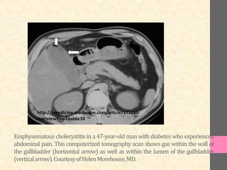
• Emphysematous Cholecystitis. April 9, 2015 [cited May 12, 2015];
Retrieved from: http://emedicine.medscape.com/article/173885-
overview
• Sunnapwar A, Raut AA, Nagar AM, Katre R. Emphysematous
cholecystitis: Imaging findings in nine patients. Indian J Radiol
Imaging. 2011;21(2):142–6.
• http://radiopaedia.org/articles/emphysematous-cholecystitis](https://image.slidesharecdn.com/emphysematouscholecystitisdryopi-150529115556-lva1-app6892/85/Emphysematous-cholecystitis-11-320.jpg)

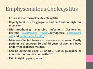
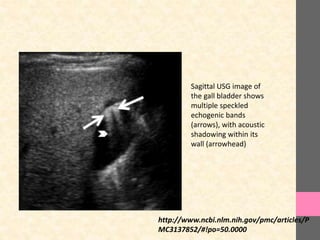
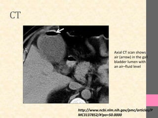
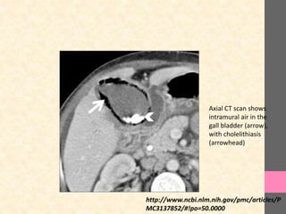
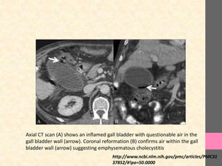
Emphysematous cholecystitis is a severe form of acute cholecystitis caused by gas-forming bacterial infections that can lead to gallbladder gangrene or perforation. It commonly affects men between 50-70 years old with underlying conditions like diabetes. Diagnosis can be made using imaging modalities like CT, USG, or X-ray that detect air within the gallbladder walls or lumen. Patients experience right upper quadrant pain. Without prompt treatment, emphysematous cholecystitis carries a high risk of mortality.


![PERI-PROSTHETIC FRACTURE NAIL-PLATE CONSTRUCT [NPC].pptx](https://cdn.slidesharecdn.com/ss_thumbnails/drarunkumardrmohamedashrafperiprostheticfrasturenail-plateconstructnpc-260209164459-7e9d15a1-thumbnail.jpg?width=640&height=640&fit=bounds)


