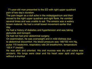
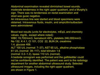
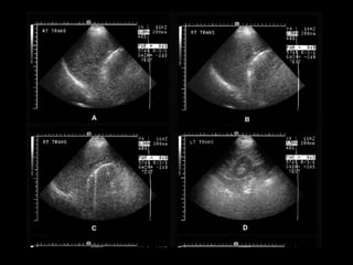
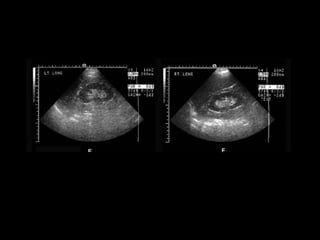
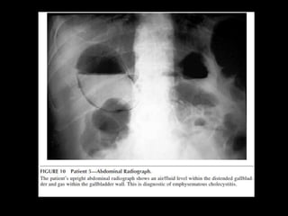
CHOLECYSTITIS
CASE REVIEW
A 71-year-old man presented to the ED with right upper quadrant
pain of two day’s duration.
The pain began as a dull ache in the midepigastrium and then
moved to the right upper quadrant and right flank. He vomited
several times and was unable to eat. The emesis was a watery
brown material. He had a small bowel movement earlier that
day.
He had a history of diabetes and hypertension and was taking
glyburide and lisinopril.
He had not had prior abdominal surgery.
On examination, he was overweight and in mild distress due
to abdominal discomfort. His blood pressure was 148/100 mm Hg,
pulse 110 beats/min, respiratory rate 24 breaths/min, temperature
100.4 F (rectal).
He was alert and oriented. His oral mucosa was dry and sclera was
anicteric. His lungs were clear and his heart wasr apid and regular
without a murmur
Abdominal examination revealed diminished bowel sounds,
moderate tenderness in the right upper quadrant, and a Murphy’s
sign. There was no tenderness on rectal examination and
stool was guiac negative.
An intravenous line was started and blood specimens were
obtained. Intravenous fluids, insulin, and ampicillin/sulbactam
were administered

Blood test results (units for electrolytes, mEq/L and chemistry
values, mg/dL, except where noted):
WBC 19,700/mm3, hematocrit 49%, platelets 246,000/mm3.
Na 132, K 4.1, Cl 101, CO2 22, BUN 24, creatinine
1.4, glucose 406.
ALT 100 U/L (normal: 7–37), AST 65 U/L, alkaline phosphatase
61 U/L (normal: 39–117), total bilirubin 1.6
(normal: 0.2–1.2), lipase 110 U/L (normal).
A bedside sonogram was performed and the gallbladder could
not be confidently identified. The patient was sent to the radiology
department for another abdominal ultrasound study. Selected
ultrasound images, including the right upper quadrant,
are shown in Figure 1.
TYPICAL SONOGRAPHIC
   APPEARANCE OF
     GALLSTONE
UNDER WHAT CIRCUMSTANCES
WOULD THE GALLBLADDER NOT
HAVE ITS TYPICAL APPEARANCE?
• When patient has recently eaten.
• Multiple episodes of cholecystitis (scarred and
  shrunken)
• Filled w/stones or contracted around
  gallstones
• Air filled gallblader (empysematouse
  cholecystitis)
WALL-ECHO-SHADOW (WES) OR DOUBLE ARC
EMPHYSEMATOUS CHOLECYSTITIS
•   Emphysematous cholecystitis < 1%
•   ATC: elderly, male, dbt
•   Mortality rate: 15% (1.4 cholecystitis)
•   TTO: CX because of perforation
•   Test of choice: Abdominal RX
• Air in the biliary system also occurs w/ enteric-
  biliary fistula (gallstone ileus or surgical
  anastomosis)
  – Gallbladder is collpased rather than distended
GAS IN THE GALLBLADDER WALL IS
PATHOGNOMONIC FOR
EMPHYSEMATOUS CHOLECYSTITIS
BEST INITIAL TEST?
• ULTRASONOGRAPHY : bright echogenic
  crescent in the gallbladder fossa with dirty
  shadowing and ring down artifacts.
  – Similar:
     • Contracted stone-filled gallbladder (WES)
     • Porcelain gallbladder w/ calcified wall due to chronic
       cholecystitis
• CONFIRMATION:
  – Abdominal rx or CT
IMAGING DIAGNOSIS OF ACUTE
          CHOLECYSTITIS
• Abdominal rx: 15% stones calcified
• Ultrasound : > sensitive
• Symptomatic:
  – 80% without
DIAGNOSIS OF ACUTE CHOLECYSTITIS
• CLINICAL PRESENTATION
• SIGNS OF GALLBLADDER INFLAMATION ON
  SONOGRAPHY OR CT
  – GALLBLADDER WALL THICKENING (>3 TO 5MM)
  – PERICHOLECYSTIC FLUID
  – SONOGRPHIC MURPHY’S SIGN
CLINICAL PRESENTATION!
Persitant pain
Focal tenderness
Murphy sign
Leukocytosis
Fever

Colecistitis

  • 3.
  • 4.
  • 5.
    A 71-year-old manpresented to the ED with right upper quadrant pain of two day’s duration. The pain began as a dull ache in the midepigastrium and then moved to the right upper quadrant and right flank. He vomited several times and was unable to eat. The emesis was a watery brown material. He had a small bowel movement earlier that day. He had a history of diabetes and hypertension and was taking glyburide and lisinopril. He had not had prior abdominal surgery. On examination, he was overweight and in mild distress due to abdominal discomfort. His blood pressure was 148/100 mm Hg, pulse 110 beats/min, respiratory rate 24 breaths/min, temperature 100.4 F (rectal). He was alert and oriented. His oral mucosa was dry and sclera was anicteric. His lungs were clear and his heart wasr apid and regular without a murmur
  • 6.
    Abdominal examination revealeddiminished bowel sounds, moderate tenderness in the right upper quadrant, and a Murphy’s sign. There was no tenderness on rectal examination and stool was guiac negative. An intravenous line was started and blood specimens were obtained. Intravenous fluids, insulin, and ampicillin/sulbactam were administered Blood test results (units for electrolytes, mEq/L and chemistry values, mg/dL, except where noted): WBC 19,700/mm3, hematocrit 49%, platelets 246,000/mm3. Na 132, K 4.1, Cl 101, CO2 22, BUN 24, creatinine 1.4, glucose 406. ALT 100 U/L (normal: 7–37), AST 65 U/L, alkaline phosphatase 61 U/L (normal: 39–117), total bilirubin 1.6 (normal: 0.2–1.2), lipase 110 U/L (normal). A bedside sonogram was performed and the gallbladder could not be confidently identified. The patient was sent to the radiology department for another abdominal ultrasound study. Selected ultrasound images, including the right upper quadrant, are shown in Figure 1.
  • 9.
    TYPICAL SONOGRAPHIC APPEARANCE OF GALLSTONE
  • 12.
    UNDER WHAT CIRCUMSTANCES WOULDTHE GALLBLADDER NOT HAVE ITS TYPICAL APPEARANCE?
  • 13.
    • When patienthas recently eaten. • Multiple episodes of cholecystitis (scarred and shrunken) • Filled w/stones or contracted around gallstones • Air filled gallblader (empysematouse cholecystitis)
  • 14.
  • 18.
  • 19.
    Emphysematous cholecystitis < 1% • ATC: elderly, male, dbt • Mortality rate: 15% (1.4 cholecystitis) • TTO: CX because of perforation • Test of choice: Abdominal RX
  • 21.
    • Air inthe biliary system also occurs w/ enteric- biliary fistula (gallstone ileus or surgical anastomosis) – Gallbladder is collpased rather than distended
  • 22.
    GAS IN THEGALLBLADDER WALL IS PATHOGNOMONIC FOR EMPHYSEMATOUS CHOLECYSTITIS
  • 23.
    BEST INITIAL TEST? •ULTRASONOGRAPHY : bright echogenic crescent in the gallbladder fossa with dirty shadowing and ring down artifacts. – Similar: • Contracted stone-filled gallbladder (WES) • Porcelain gallbladder w/ calcified wall due to chronic cholecystitis • CONFIRMATION: – Abdominal rx or CT
  • 25.
    IMAGING DIAGNOSIS OFACUTE CHOLECYSTITIS • Abdominal rx: 15% stones calcified • Ultrasound : > sensitive • Symptomatic: – 80% without
  • 27.
    DIAGNOSIS OF ACUTECHOLECYSTITIS • CLINICAL PRESENTATION • SIGNS OF GALLBLADDER INFLAMATION ON SONOGRAPHY OR CT – GALLBLADDER WALL THICKENING (>3 TO 5MM) – PERICHOLECYSTIC FLUID – SONOGRPHIC MURPHY’S SIGN
  • 29.
    CLINICAL PRESENTATION! Persitant pain Focaltenderness Murphy sign Leukocytosis Fever