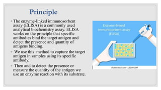
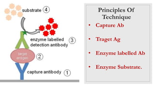
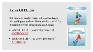
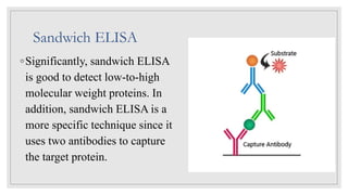
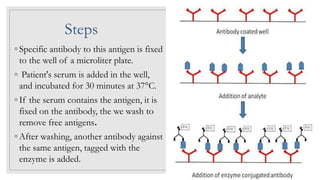
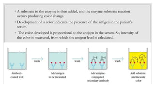
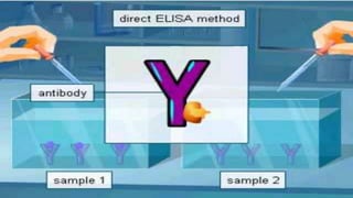
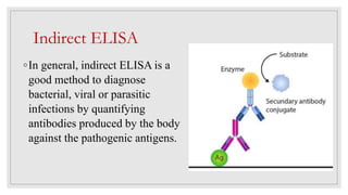
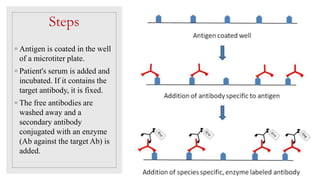
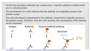
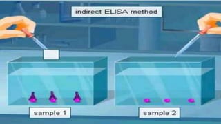
ELISA
Enzyme Linked
Immunosorbant Assay
◦ The enzyme-linked immunosorbent
assay (ELISA) is a commonly used
analytical biochemistry assay. ELISA
works on the principle that specific
antibodies bind the target antigen and
detect the presence and quantity of
antigens binding.
◦ We use this method to capture the target
antigen in samples using its specific
antibody.
◦ Then and to detect the presence or
measure the quantity of the antigen we
use an enzyme reaction with its substrate.
Principle
Principles Of
Technique
• Capture Ab
• Traget Ag
• Enzyme labelled Ab
• Enzyme Substrate.
Enzymes used in
ELISA
◦Alkaline phosphatase
(ALP).
◦β-galactosidase.
◦Horse radish
peroxidase (HRP).
Use of ELISA in medical field
◦ Medical professionals frequently use ELISA tests as blood tests to
detect antigens that may be present in the blood. The substances
detected by ELISA tests can include hormones, an allergen, viral
antigens (dengue fever, for example), bacterial antigens (TB, for
example), and antibodies that the body has made in response to
infection (antibodies to hepatitis B or HIV for example) or
vaccination.
Types Of ELISA
◦ ELISA tests can be classified into two types
depending upon the different methods used for
binding between antigen and antibodies:
 Indirect ELISA – to detect presence of
ANTIBODIES.
 Sandwich ELISA – to detect presence of
ANTIGENS.
Sandwich ELISA
◦Significantly, sandwich ELISA
is good to detect low-to-high
molecular weight proteins. In
addition, sandwich ELISA is a
more specific technique since it
uses two antibodies to capture
the target protein.
Steps
◦ Specific antibody to this antigen is fixed
to the well of a microliter plate.
◦ Patient's serum is added in the well,
and incubated for 30 minutes at 37°C.
◦ If the serum contains the antigen, it is
fixed on the antibody, the we wash to
remove free antigens.
◦ After washing, another antibody against
the same antigen, tagged with the
enzyme is added.
◦ A substrate to the enzyme is then added, and the enzyme substrate reaction
occurs producing color change.
◦ Development of a color indicates the presence of the antigen in the patient's
serum.
◦ The color developed is proportional to the antigen in the serum. So, intensity of
the color is measured, from which the antigen level is calculated.
Indirect ELISA
◦In general, indirect ELISA is a
good method to diagnose
bacterial, viral or parasitic
infections by quantifying
antibodies produced by the body
against the pathogenic antigens.
Steps
◦ Antigen is coated in the well
of a microtiter plate.
◦ Patient's serum is added and
incubated. If it contains the
target antibody, it is fixed.
◦ The free antibodies are
washed away and a
secondary antibody
conjugated with an enzyme
(Ab against the target Ab) is
added.
◦ All the free secondary antibodies are washed away. A specific substrate is added which
gives a colored product.
◦ The development of a color indicates that the antibody was originally present in the
patient's serum.
◦ The color developed is proportional to the antibody concentration originally present in
the patient's serum. Therefore, from the color intensity, the concentration of the antibody
can be calculated.
Components of
ELISA Kit
◦ Test sample
◦ Antibody /Antigen
◦ microtiter plates
◦ Enzyme labelled Ab
◦ Chromogenic
Substrate
◦ Washing buffer
◦ Standard solutions.
Advantages of ELISA Disadvantages of ELISA
Fast—90 samples tested in 2-3 hr
Measurement of enzyme activity can be
more complex than measurement of
activity of some type of radioisotopes
Many samples can be processed at once Enzyme activity may be affected by
plasma constituentsñ
Small sample size required (10 μL~
100μL)
Kits are commercially available, but not
cheap
Colorimetric results –easily observed and
measured (spectrophotometer)
Easy to learn, simple procedure.
Highly sensitive and specific .
https://youtu.be/o1vRMFvq8sU
Sandwich ELISA protocol
ELISA test procedure
More information for those who are interested :

ELISA

  • 1.
  • 2.
    ◦ The enzyme-linkedimmunosorbent assay (ELISA) is a commonly used analytical biochemistry assay. ELISA works on the principle that specific antibodies bind the target antigen and detect the presence and quantity of antigens binding. ◦ We use this method to capture the target antigen in samples using its specific antibody. ◦ Then and to detect the presence or measure the quantity of the antigen we use an enzyme reaction with its substrate. Principle
  • 3.
    Principles Of Technique • CaptureAb • Traget Ag • Enzyme labelled Ab • Enzyme Substrate.
  • 4.
    Enzymes used in ELISA ◦Alkalinephosphatase (ALP). ◦β-galactosidase. ◦Horse radish peroxidase (HRP).
  • 5.
    Use of ELISAin medical field ◦ Medical professionals frequently use ELISA tests as blood tests to detect antigens that may be present in the blood. The substances detected by ELISA tests can include hormones, an allergen, viral antigens (dengue fever, for example), bacterial antigens (TB, for example), and antibodies that the body has made in response to infection (antibodies to hepatitis B or HIV for example) or vaccination.
  • 6.
    Types Of ELISA ◦ELISA tests can be classified into two types depending upon the different methods used for binding between antigen and antibodies:  Indirect ELISA – to detect presence of ANTIBODIES.  Sandwich ELISA – to detect presence of ANTIGENS.
  • 7.
    Sandwich ELISA ◦Significantly, sandwichELISA is good to detect low-to-high molecular weight proteins. In addition, sandwich ELISA is a more specific technique since it uses two antibodies to capture the target protein.
  • 8.
    Steps ◦ Specific antibodyto this antigen is fixed to the well of a microliter plate. ◦ Patient's serum is added in the well, and incubated for 30 minutes at 37°C. ◦ If the serum contains the antigen, it is fixed on the antibody, the we wash to remove free antigens. ◦ After washing, another antibody against the same antigen, tagged with the enzyme is added.
  • 9.
    ◦ A substrateto the enzyme is then added, and the enzyme substrate reaction occurs producing color change. ◦ Development of a color indicates the presence of the antigen in the patient's serum. ◦ The color developed is proportional to the antigen in the serum. So, intensity of the color is measured, from which the antigen level is calculated.
  • 11.
    Indirect ELISA ◦In general,indirect ELISA is a good method to diagnose bacterial, viral or parasitic infections by quantifying antibodies produced by the body against the pathogenic antigens.
  • 12.
    Steps ◦ Antigen iscoated in the well of a microtiter plate. ◦ Patient's serum is added and incubated. If it contains the target antibody, it is fixed. ◦ The free antibodies are washed away and a secondary antibody conjugated with an enzyme (Ab against the target Ab) is added.
  • 13.
    ◦ All thefree secondary antibodies are washed away. A specific substrate is added which gives a colored product. ◦ The development of a color indicates that the antibody was originally present in the patient's serum. ◦ The color developed is proportional to the antibody concentration originally present in the patient's serum. Therefore, from the color intensity, the concentration of the antibody can be calculated.
  • 15.
    Components of ELISA Kit ◦Test sample ◦ Antibody /Antigen ◦ microtiter plates ◦ Enzyme labelled Ab ◦ Chromogenic Substrate ◦ Washing buffer ◦ Standard solutions.
  • 16.
    Advantages of ELISADisadvantages of ELISA Fast—90 samples tested in 2-3 hr Measurement of enzyme activity can be more complex than measurement of activity of some type of radioisotopes Many samples can be processed at once Enzyme activity may be affected by plasma constituentsñ Small sample size required (10 μL~ 100μL) Kits are commercially available, but not cheap Colorimetric results –easily observed and measured (spectrophotometer) Easy to learn, simple procedure. Highly sensitive and specific .
  • 17.
    https://youtu.be/o1vRMFvq8sU Sandwich ELISA protocol ELISAtest procedure More information for those who are interested :