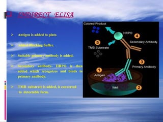
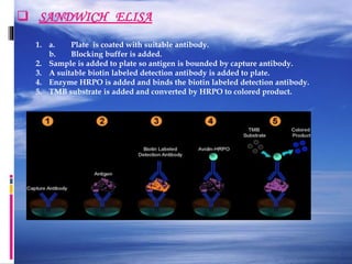
What is ELISA.
Principle.
History.
Types of ELISA method.
1.Direct ELISA.
2.Indirect ELISA.
3.Sandwhich ELISA.
Conclusion.
References.
Antibodies (also known as immunoglobulins abbreviated Ig) are gamma globulin proteins that are found in blood and are used by the immune system to identify and neutralize foreign objects, such as bacteria and viruses.
The dark energy paradox leads to a new structure of spacetime.pptx
ELISA, principle and method by kk sahu
1. Method of ELISA
By
KAUSHAL KUMAR SAHU
Assistant Professor (Ad Hoc)
Department of Biotechnology
Govt. Digvijay Autonomous P. G. College
Raj-Nandgaon ( C. G. )
2. Synopsis
What is ELISA.
Principle.
History.
Types of ELISA method.
1.Direct ELISA.
2.Indirect ELISA.
3.Sandwhich ELISA.
Conclusion.
References.
3. Definitions
Antibodies (also known as immunoglobulins abbreviated
Ig) are gamma globulin proteins that are found in blood
and are used by the immune system to identify and
neutralize foreign objects, such as bacteria and viruses.
4. Antigens
A substance that when introduced into the body
stimulates the production of an antibody
Immunoassay
A laboratory technique that makes use of the binding
between an antigen and its homologous antibody in
order to identify and quantify the specific antigen or
antibody in a sample
5. INTRODUCTION
ELISA, or enzyme-linked immunosorbent assay, is an
immunoassay technique involving the reaction of
antigen and antibody in vitro.
ELISA is a sensitive and specific assay for the detection
and quantification of antigens or antibodies.
ELISA tests are usually performed in microwell
plates.
The ELISA test, or the enzyme immunoassay (EIA),
was the first screening test commonly employed for
HIV. It has a high sensitivity.
7. What is ELISA
It is enzyme- linked immunosorbent assay.
It is also called enzyme immunoassay.
Is a biochemical technique used mainly in immunology
to detect the presence of an antibody or an antigen in a
sample.
The technique is divided into 3 types.
1-Direct ELISA.
2- Sandwich ELISA.
3- Indirect ELISA .
8. Principle of ELISA
ELISA is a sensitive method for quantification of
protein, which is suitable for various application. The
basic principle of an ELISA is to use an enzyme to
detect the Ag-Ab binding (antigen- antibody binding).
The enzyme converts a colorless substrate (chromogen)
to a colored product, indicating the presence of Ag:Ab
binding.
9. History
Antigen- antibody binding hypothesis was given by John
marrack 1938.
A technique to prepare immunosorbent to fix ag or ab to
surface of a container was published by Wide & Porath in
1966.
In 1971 Perlmann & engvall invented ELISA.
In 1975 George kohler & Milstein invented 1st monoclonal
antibody.
10. Different antigen in sample
substrate
Colored
product
Primary antibody
Secondary antibody
11. HOW DOES ELISA WORK ?
There are variations of the ELISA test but the most basic type
consist of an antibody attached to the solid surface. This antibody
has affinity for (will latch onto) the substance of interest.
For example… Human Chorionic Gonadotropin (HCG), the
commonly measured protein which indicates pregnancy.
A mixture of purified HCG linked to an enzyme and sample
(blood, urine, etc.) under test are added to the test system. If no
HCG is present in the test sample the only HCG with linked
enzyme will bind. The more HCG which is present in the test
sample the less enzyme linked HCG will bind.
12. Reagents
Substrate: TMB (3,3',5,5', tetramethyl benzidine) The
enzyme acts as a catalyst to oxidize substrate in the
presence of Hydrogen peroxide to produce a blue color.
Reaction stopped with dilute acid to cause complex to
turn yellow.
Enzyme: Horse Radish Peroxidase (HRPO)
Antibody: IgG fraction of serum purified by afinity
chrometography.
Solid phase: poly vinyl chloride.
Dilute buffer: phosphate buffer, neutral ph.
14. DIRECT ELISA
The direct ELISA uses the method of directly labeling the
antibody itself.
Micro well plates are coated with a sample containing the
target antigen,
and the binding of labeled antibody is quantitated by a
colorimetric, chemiluminescent, or fluorescent end-point.
15. DIRECT ELISA
1. Apply a sample of known
antigen to a surface.
2. Enzyme linked primary
antibody is applied to the plate.
3. Washed, After this wash, only
the antibody-antigen
complexes remain attached.
4. Apply a substrate which is
converted by the enzyme to
elicit a chromogenic signal.
16. Advantages of Direct Detection
Quick methodology since only one antibody is used.
Cross-reactivity of secondary antibody is eliminated.
Disadvantages of Direct Detection
Immunoreactivity of the primary antibody may be reduced
as a result of labeling.
Labeling of every primary antibody is time-consuming
and expensive.
17. INDIRECT ELISA
The indirect ELISA utilizes an unlabeled primary
antibody in conjunction with a labeled secondary
antibody. Since the labeled secondary antibody is
directed against all antibodies of a given species (e.g.
anti-mouse), it can be used with a wide variety of
primary antibodies (e.g. all mouse monoclonal
antibodies).
18. INDIRECT ELISA
Antigen is added to plate.
Added Blocking buffer.
Suitable primary antibody is added.
Secondary antibody- HRPO is then
added which recognizes and binds to
primary antibody.
TMB substrate is added, is converted
to detectable form.
19. Advantages of indirect detection
Wide variety of labeled secondary antibodies are available
commercially.
Versatile, since many primary antibodies can be made in
one species and the same labeled secondary antibody can
be used for detection.
Immuno reactivity of the primary antibody is not affected
by labeling.
Sensitivity is increased because each primary antibody
contains several epitopes that can be bound by the labeled
secondary antibody, allowing for signal amplification.
20. Disadvantages of indirect detection
Cross-reactivity may occur with the secondary antibody,
resulting in nonspecific signal.
An extra incubation step is required in the procedure.
21. SANDWICH ELISA
1. a. Plate is coated with suitable antibody.
b. Blocking buffer is added.
2. Sample is added to plate so antigen is bounded by capture antibody.
3. A suitable biotin labeled detection antibody is added to plate.
4. Enzyme HRPO is added and binds the biotin labeled detection antibody.
5. TMB substrate is added and converted by HRPO to colored product.
23. ELISA data is typically graphed with Optical density Vs Log concentration to produce a
sigmoidal curve.
Known concentrations of antigen are used to produce a standard curve and then this
data is used to measure the concentration of unknown samples by comparison to the
linear portion of the standard curve.
This can be done directly on the graph or with curve fitting software which is typically
found on ELISA plate readers. Which measure the colour density .
24. APPLICATIONS
Screening donated blood for evidence of viral contamination by
HIV-1 and HIV-2 (presence of anti-HIV antibodies)
Hepatitis C (presence of antibodies)
Hepatitis B (testing for both antibodies and a viral antigen)
Measuring hormone levels
HCG (as a test for pregnancy)
LH (determining the time of ovulation)
TSH, T3 and T4 (for thyroid function)
Detecting infections
Sexually-transmitted agents like HIV, syphilis and chlamydia
Hepatitis B and C
Toxoplasma gondii
Detecting allergens in food and house dust
25. CONCLUSION
ELISA is a sensitive and specific assay for the
detection and quantification of antigens or
antibodies.
ELISA tests are usually performed in
microwell plates.