SlideShare a Scribd company logo
Dr. NikitDr. Nikit
Moderator : Dr. SachinModerator : Dr. Sachin
ELECTROPHYSIOLOGICAL TESTSELECTROPHYSIOLOGICAL TESTS
 Clinical electrophysiological tests are objective testsClinical electrophysiological tests are objective tests
which allow assessment of nearly the entire length ofwhich allow assessment of nearly the entire length of
visual pathway.visual pathway.
 Electrophysiological tests :Electrophysiological tests :
 Electroretinogram (ERG)Electroretinogram (ERG)
 Electrooculogram (EOG)Electrooculogram (EOG)
 Visually Evoked Potentials (VEP)Visually Evoked Potentials (VEP)
 Objective assessment of retinal function is importantObjective assessment of retinal function is important
for following reasons :for following reasons :
 Helps in ‘localization of site of lesion’ in the visualHelps in ‘localization of site of lesion’ in the visual
pathway. Eg. Macula or entire retina.pathway. Eg. Macula or entire retina.
 Allows ‘quantitative assessment of degree ofAllows ‘quantitative assessment of degree of
malfunction’ which can be followed up over time tomalfunction’ which can be followed up over time to
project long term prognosis.project long term prognosis.
 ERG is an electric potential generated by retina inERG is an electric potential generated by retina in
response to brief stimulus of light.response to brief stimulus of light.
 The ‘amplitude of ERG’ (amount of electric potentialThe ‘amplitude of ERG’ (amount of electric potential
generated) is directly proportional to area ofgenerated) is directly proportional to area of
functioning retina stimulated.functioning retina stimulated.
ELECRORETINOGRAM (ERG)ELECRORETINOGRAM (ERG)
 BASIC PRINCIPLE OF ERG :BASIC PRINCIPLE OF ERG :
 Sudden illumination of retina.Sudden illumination of retina.
 Simultaneous activation of all the retinal cells toSimultaneous activation of all the retinal cells to
generate the current.generate the current.
 Currents generated by all the retinal cells mix, thenCurrents generated by all the retinal cells mix, then
pass through vitreous & extra cellular spaces.pass through vitreous & extra cellular spaces.
 High RPE resistance prevents summated current fromHigh RPE resistance prevents summated current from
passing posteriorly.passing posteriorly.
 The small portion of the summated current whichThe small portion of the summated current which
escapes through the cornea is recorded as ERG.escapes through the cornea is recorded as ERG.
 ‘‘a wave’a wave’ : It’s a ‘negative’: It’s a ‘negative’
(downward) wave &(downward) wave &
reflectsreflects photoreceptor
function..
 ‘‘b wave’b wave’ : It is a ‘positive’: It is a ‘positive’
(upward) wave & reflects(upward) wave & reflects
bipolar cell activitybipolar cell activity..
 ‘‘Oscillatory potentials ‘:Oscillatory potentials ‘:
Small rippling currentsSmall rippling currents
produced byproduced by inner plexiforminner plexiform
layer.layer.
ERG WAVEFORMSERG WAVEFORMS
 A normal ERG has 5 distinct responses :A normal ERG has 5 distinct responses :
 Rod responseRod response
 Maximal combined responseMaximal combined response Dark adaptedDark adapted
 Oscillatory potentialsOscillatory potentials
 Single flash cone responseSingle flash cone response
Light adaptedLight adapted
 30 Hz flicker response30 Hz flicker response
ERG RESPONSESERG RESPONSES
 ROD RESPONSE (Photopic ERG) :ROD RESPONSE (Photopic ERG) :
 Produced by dark adapting patientProduced by dark adapting patient
for 20 min. & then stimulating retinafor 20 min. & then stimulating retina
with dim light flash which is belowwith dim light flash which is below
cone threshold.cone threshold.
 The resultant waveform hasThe resultant waveform has
‘prominent b (positive) wave ‘& no‘prominent b (positive) wave ‘& no
detectable ‘a (negative) wave’.detectable ‘a (negative) wave’.
 MAXIMAL COMBINEDMAXIMAL COMBINED
RESPONSE :RESPONSE :
 It is a larger waveform generatedIt is a larger waveform generated
by using bright flash in darkby using bright flash in dark
adapted state which maximallyadapted state which maximally
stimulates both rods & cones.stimulates both rods & cones.
 It results in prominent ‘aIt results in prominent ‘a (negative)(negative)
wave & ‘bwave & ‘b (positive)(positive) wave’ withwave’ with
‘oscillatory potentials’ which are‘oscillatory potentials’ which are
superimposed on ‘b wave’.superimposed on ‘b wave’.
 CONE RESPONSES :CONE RESPONSES :
 ‘‘Single flash responseSingle flash response’ is obtained by’ is obtained by
maintaining the patient in lightmaintaining the patient in light
adapted state & stimulating theadapted state & stimulating the
retina with bright white flash.retina with bright white flash.
 The rods are suppressed by lightThe rods are suppressed by light
adaptation & donot contribute to theadaptation & donot contribute to the
waveform.waveform.
 With patient in light adapted state, aWith patient in light adapted state, a
flickering stimulus at 30 Hz can alsoflickering stimulus at 30 Hz can also
be used to filter rod response &be used to filter rod response &
measure cone responsemeasure cone response (30 Hz flicker(30 Hz flicker
response)response)
ELECTRODES USED IN ERGELECTRODES USED IN ERG
Jet Electrode Gold Plated Electrode Skin ElectrodeJet Electrode Gold Plated Electrode Skin Electrode
DTL Electrode HK Loops Burian Allen ElectrodeDTL Electrode HK Loops Burian Allen Electrode
A) BRIGHT FLASH ERG :A) BRIGHT FLASH ERG :
UsedUsed for assessment of retinal function in ‘for assessment of retinal function in ‘severelyseverely
traumatized eyetraumatized eye‘ or ‘‘ or ‘eye with dense media opacity’eye with dense media opacity’ likelike
dense VH, corneal opacity or advanced cataract.dense VH, corneal opacity or advanced cataract.
The flash used is aboutThe flash used is about 10.000 times brighter10.000 times brighter than thatthan that
used in standardused in standard ERG.ERG.
SPECIALISED FORMS OF ERGSPECIALISED FORMS OF ERG
 In this procedure successive responses are obtainedIn this procedure successive responses are obtained
with flashes of increasing intensity, allowing the timewith flashes of increasing intensity, allowing the time
for re-adaptation in between flashes.for re-adaptation in between flashes.
 AA non recordable flash ERGnon recordable flash ERG is an ominous sign foris an ominous sign for
visual prognosis.visual prognosis.
B) FOCAL ERG (FERG):B) FOCAL ERG (FERG):
UsedUsed for detecting small focal lesionsfor detecting small focal lesions or pathologiesor pathologies
which are missed by standard full field ERG.which are missed by standard full field ERG.
A small stimulus of 4A small stimulus of 4oo
size is projected on area of retinasize is projected on area of retina
to be tested.to be tested.
Due to light scattering & poor signal to noise ratio, thisDue to light scattering & poor signal to noise ratio, this
technique is mostly used in research setting than intechnique is mostly used in research setting than in
clinical setting.clinical setting.
 Clinical uses of FERG :Clinical uses of FERG :
 Early detection of cone dystrophy or macular diseaseEarly detection of cone dystrophy or macular disease
before the fundus changes are evident.before the fundus changes are evident.
 Can differentiate between early macular & optic nerveCan differentiate between early macular & optic nerve
pathology.pathology.
 Can be used for evaluation of any type focal macularCan be used for evaluation of any type focal macular
pathology.pathology.
C)C) MULTIFOCAL ERG (mfERG) :MULTIFOCAL ERG (mfERG) :
The stimuli consists denselyThe stimuli consists densely
arranged black or white hexagonalarranged black or white hexagonal
elements displayed on CRT monitor.elements displayed on CRT monitor.
These hexagonal elements changeThese hexagonal elements change
from light to dark independently &from light to dark independently &
this change results into recording ofthis change results into recording of
mfERG.mfERG.
 Based on retinal activity, the recorded mfERG appearsBased on retinal activity, the recorded mfERG appears
in ‘topographic map form’ & also in ‘small ERGin ‘topographic map form’ & also in ‘small ERG
waveforms’ from various parts of retina.waveforms’ from various parts of retina.
 Both cone & rod mfERG forms can be recorded.Both cone & rod mfERG forms can be recorded.
 mfERG findings also correlate with visual field defectsmfERG findings also correlate with visual field defects
but very small scotomas can be missed by this method.but very small scotomas can be missed by this method.
2D Map 3D Map2D Map 3D Map
Normal MFERGNormal MFERG
Retinitis Pigmentosa Stargardt’s DiseaseRetinitis Pigmentosa Stargardt’s Disease
Macular Degenearation Chloroquine RetinopathyMacular Degenearation Chloroquine Retinopathy
D) PATTERN ERG (PERG)D) PATTERN ERG (PERG) ::
It mainly represents inner retinal activityIt mainly represents inner retinal activity (especially(especially
ganglion cell activity)ganglion cell activity)
Useful inUseful in differentiating optic nerve disorders fromdifferentiating optic nerve disorders from
macular disorders.macular disorders.
Unlike flash ERG, pattern ERG is a very small response.Unlike flash ERG, pattern ERG is a very small response.
Recorded with full correction of refractive errors asRecorded with full correction of refractive errors as
visualization of stimulus for extended time is essentialvisualization of stimulus for extended time is essential
for recording.for recording.
 PERG WAVEFORMS :PERG WAVEFORMS :
 P1 or P50P1 or P50 : Initial corneal positive response.: Initial corneal positive response.
 N1 or N95N1 or N95 : Immediate cornea negative response.: Immediate cornea negative response.
 This 50 & 95 represents the time in milliseconds from theThis 50 & 95 represents the time in milliseconds from the
onset of stimulus to peak of positive or negative response.onset of stimulus to peak of positive or negative response.
 Evaluation of visual function in infants & children.Evaluation of visual function in infants & children.
 To determine presence or absence of retinal function.To determine presence or absence of retinal function.
 To evaluate progression of retinal degeneration.To evaluate progression of retinal degeneration.
 To confirm diagnosis of a particular diseaseTo confirm diagnosis of a particular disease
(dystrophies).(dystrophies).
 For early detection of toxic retinopathies.For early detection of toxic retinopathies.
 To give visual prognosis.To give visual prognosis.
 Assisting in diagnosing the retinal conditions in whichAssisting in diagnosing the retinal conditions in which
clinical findings don't match with visual complaintsclinical findings don't match with visual complaints
(unexplained visual loss).(unexplained visual loss).
Indications & Clinical Uses of ERGIndications & Clinical Uses of ERG
ERG IN CLINICAL CASESERG IN CLINICAL CASES
 DIABETIC RETINOPATHY :DIABETIC RETINOPATHY :
 In DR there isIn DR there is reduction in amplitudereduction in amplitude
&& delay of peak implicit timesdelay of peak implicit times..
 These changes are directlyThese changes are directly
proportional to severity ofproportional to severity of
retinopathy.retinopathy.
 Amplitude of oscillatory potentialsAmplitude of oscillatory potentials
(OP)(OP) is a good predictor ofis a good predictor of
progression of retinopathy fromprogression of retinopathy from
NPDR to PDR.NPDR to PDR.
 Abnormal amplitude of OP indicateAbnormal amplitude of OP indicate
high risk of developing PDR.high risk of developing PDR.
 RETINAL DETACHMENT (RD)RETINAL DETACHMENT (RD)
& CENTRAL SEROUS& CENTRAL SEROUS
RETINOPATHY (CSR) :RETINOPATHY (CSR) :
 In RD & CSR there isIn RD & CSR there is significantsignificant
reduction in ERG amplitudereduction in ERG amplitude..
 However there is no significantHowever there is no significant
change seen in waveforms of ERG.change seen in waveforms of ERG.
 RETINOSCHISIS :RETINOSCHISIS :
 ERG in retinoschisis is typically characterized byERG in retinoschisis is typically characterized by
marked decrease amplitudemarked decrease amplitude oror absence of b wave.absence of b wave.
 RETINITIS PIGMENTOSA :RETINITIS PIGMENTOSA :
 A full field ERG in RP showsA full field ERG in RP shows
marked reduction in both rodmarked reduction in both rod
& cone signals although& cone signals although lossloss
of rod signals isof rod signals is
predominant.predominant.
 There is significantThere is significant reductionreduction
in amplitude of both a & bin amplitude of both a & b
waves of ERG.waves of ERG.
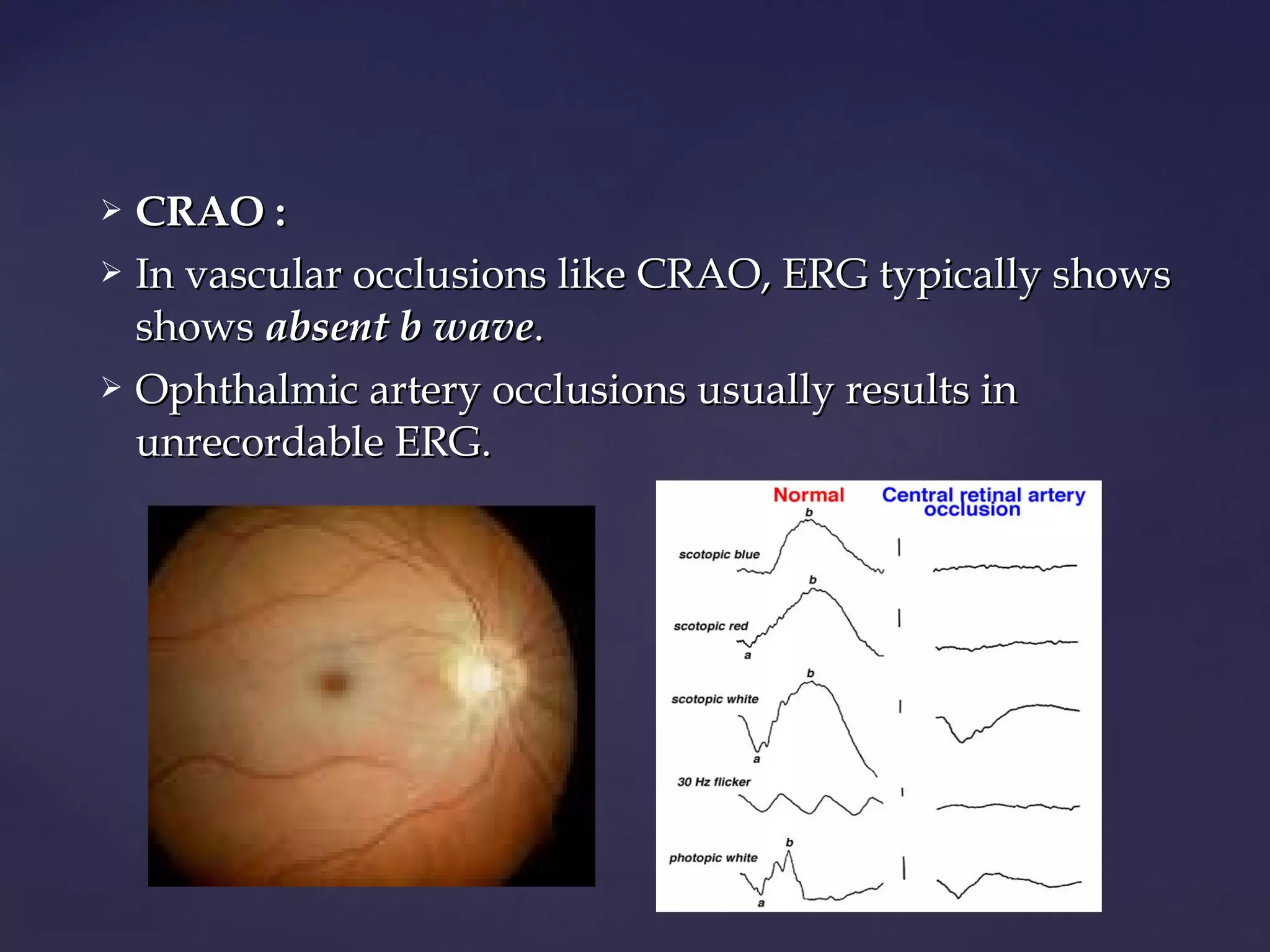
 CRAO :CRAO :
 In vascular occlusions like CRAO, ERG typically showsIn vascular occlusions like CRAO, ERG typically shows
showsshows absent b waveabsent b wave..
 Ophthalmic artery occlusions usually results inOphthalmic artery occlusions usually results in
unrecordable ERG.unrecordable ERG.
 CONE DYSTROPHY :CONE DYSTROPHY :
 ERG in cone dystrophy showsERG in cone dystrophy shows goodgood
rod b-waves that are just slower.rod b-waves that are just slower.
 The early cone response of theThe early cone response of the
scotopic red flash ERG is missing.scotopic red flash ERG is missing.
 The scotopic bright white ERG isThe scotopic bright white ERG is
fairly normal in appearance but withfairly normal in appearance but with
slow implicit times.slow implicit times.
 The 30 Hz flicker & photopic whiteThe 30 Hz flicker & photopic white
ERGs which are dependent uponERGs which are dependent upon
cones are very poor.cones are very poor.
 RETAINED IOFB :RETAINED IOFB :
 A retained metallic FB like iron &A retained metallic FB like iron &
copper shows changes in ERG earlycopper shows changes in ERG early
as well as late stages.as well as late stages.
 A characteristic change isA characteristic change is b-waveb-wave
amplitude is reduced by 50% oramplitude is reduced by 50% or
moremore as compared with normal eye.as compared with normal eye.
 No intervention finally results intoNo intervention finally results into
an unrecordable ERG (an unrecordable ERG (Zero ERGZero ERG))
 It is recording of standing potential of the eyeIt is recording of standing potential of the eye
 The electrodes are placed at inner & outer canthus ofThe electrodes are placed at inner & outer canthus of
the eye with reference electrode placed on forehead.the eye with reference electrode placed on forehead.
 The patient is asked to look back & forth between aThe patient is asked to look back & forth between a
pair of fixation lights separated by 30pair of fixation lights separated by 30oo
of visual anglesof visual angles
on Ganzfeld globe.on Ganzfeld globe.
ELECTRO-OCULOGRAMELECTRO-OCULOGRAM
 Like ERG, EOG reflects activity of entire retina & usedLike ERG, EOG reflects activity of entire retina & used
to evaluateto evaluate combined photoreceptor-RPE activity.combined photoreceptor-RPE activity.
 As validity of results depends upon consistent trackingAs validity of results depends upon consistent tracking
of fixation target over 30 min., this test is not suitableof fixation target over 30 min., this test is not suitable
in unco-operative patients & children.in unco-operative patients & children.
 Also EOG depends upon a minimum degree of lightAlso EOG depends upon a minimum degree of light
adaptation so it is not reliable in patients with denseadaptation so it is not reliable in patients with dense
cataracts.cataracts.
 CORNEOFUNDAL POTENTIAL :CORNEOFUNDAL POTENTIAL :
 It is the source of voltage obtained in EOG & it rendersIt is the source of voltage obtained in EOG & it renders
the cornea positive by 0.006 to 0.010 V as comparedthe cornea positive by 0.006 to 0.010 V as compared
with the back of the eye.with the back of the eye.
 The corneofundal potential results from metabolicThe corneofundal potential results from metabolic
activity of RPE (mainly) as well as corneal & lensactivity of RPE (mainly) as well as corneal & lens
epithelium.epithelium.
 Contributions of corneal & lens epithelium are notContributions of corneal & lens epithelium are not
photosensitive but that of RPE is, which is substantialyphotosensitive but that of RPE is, which is substantialy
increased during light adaptationincreased during light adaptation && decreased during darkdecreased during dark
adaptation.adaptation.
 For EOG to be normal, it requires as little as 20-25 % ofFor EOG to be normal, it requires as little as 20-25 % of
normal functioning retina.normal functioning retina.
 Thus abnormal EOG indicates a dense pathologyThus abnormal EOG indicates a dense pathology
involving entire retina.involving entire retina.
 ARDEN’S RATIO :ARDEN’S RATIO :
 It is the ratio of ‘largest EOG amplitude during lightIt is the ratio of ‘largest EOG amplitude during light
adaptation’adaptation’ (light peak)(light peak) to ‘least amplitude duringto ‘least amplitude during
dark adaptation’dark adaptation’ (dark trough).(dark trough).
 Clinically normal value of this ratio is 1.85 or higher.Clinically normal value of this ratio is 1.85 or higher.
 Values below 1.85 are considered subnormal & thoseValues below 1.85 are considered subnormal & those
below 1.30 are considered severely subnormal orbelow 1.30 are considered severely subnormal or
extinguished.extinguished.
 BEST’S DISEASE :BEST’S DISEASE :
 Abnormal EOG with normal ERGAbnormal EOG with normal ERG is ais a
hallmark.hallmark.
 Other examples ofOther examples of ERG to EOGERG to EOG
dissociationdissociation are :are :
 Diffuse fundus flavimaculatousDiffuse fundus flavimaculatous
 Pattern dystrophy of RPEPattern dystrophy of RPE
eg. Butterfly Macular Dystrophy.eg. Butterfly Macular Dystrophy.
 Chloroquine retinopathyChloroquine retinopathy
 Metallosis bulbiMetallosis bulbi
EOG IN CLINICAL CASESEOG IN CLINICAL CASES
 Also called as ‘visually evoked response (VER)’ orAlso called as ‘visually evoked response (VER)’ or
‘cortical potentials’.‘cortical potentials’.
 It is the electrical response of the brain to suddenIt is the electrical response of the brain to sudden
appearance / disappearance / change of visualappearance / disappearance / change of visual
stimulus.stimulus.
 Like EEG, VEP is detected by placing surface electrodesLike EEG, VEP is detected by placing surface electrodes
at scalp which can be placed anywhere, but shouldat scalp which can be placed anywhere, but should
always include posterior occipital area.always include posterior occipital area.
VISUALLY EVOKED POTENTIALS (VEP)VISUALLY EVOKED POTENTIALS (VEP)
 The occipital electrode (Inion) lies near visual area thusThe occipital electrode (Inion) lies near visual area thus
called ascalled as reference electrodereference electrode..
 The vertex electrode is placed over non visual areaThe vertex electrode is placed over non visual area
which detects minimum activity in response to visualwhich detects minimum activity in response to visual
stimulation is called asstimulation is called as active electactive electroderode..
 The 3The 3rdrd
electrode is placed over forehead is calledelectrode is placed over forehead is called
ground electrode.ground electrode.
VEP ELECTRODESVEP ELECTRODES
 The stimulus shown is aThe stimulus shown is a flash of lightflash of light
(diffuse light spot, annulus ) or(diffuse light spot, annulus ) or
patterned stimuluspatterned stimulus (illuminated(illuminated
checkerboard)checkerboard)
 The stimuli are repetitivelyThe stimuli are repetitively
presented at random within a shortpresented at random within a short
period of time. Eg. 1 cycle/second forperiod of time. Eg. 1 cycle/second for
100 seconds.100 seconds.
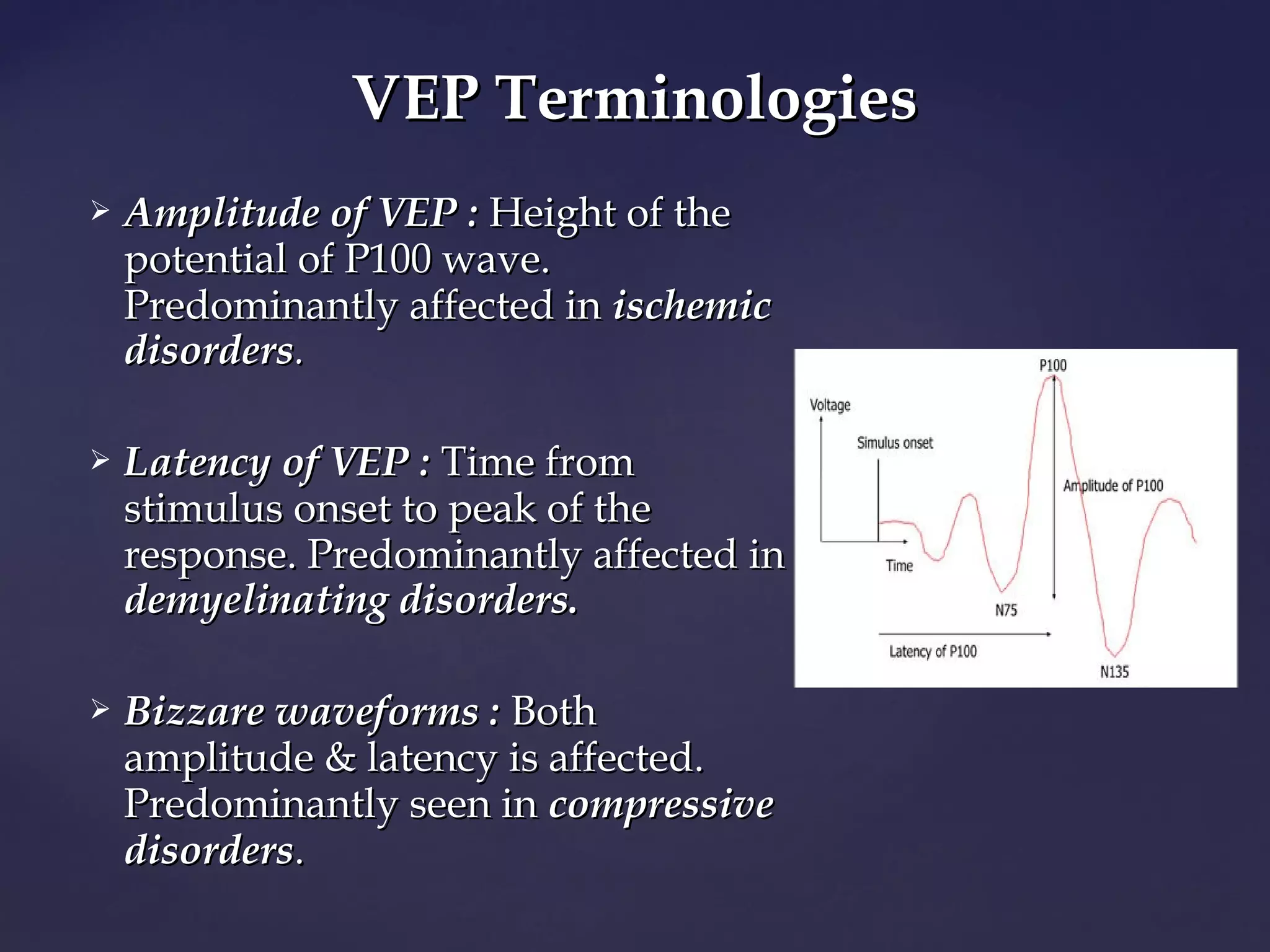
 The standard flash VEP is characterized by positiveThe standard flash VEP is characterized by positive
wave (Pwave (P11 or P100) which most commonly studiedor P100) which most commonly studied
clinically & 2 negative waves (Nclinically & 2 negative waves (N11 or Nor N7575 & N& N22 or Nor N135135))..
 Amplitude of VEP :Amplitude of VEP : Height of theHeight of the
potential of P100 wave.potential of P100 wave.
Predominantly affected inPredominantly affected in ischemicischemic
disordersdisorders..
 LatencyLatency of VEP :of VEP : Time fromTime from
stimulus onset to peak of thestimulus onset to peak of the
response. Predominantly affected inresponse. Predominantly affected in
demyelinating disorders.demyelinating disorders.
 Bizzare waveforms :Bizzare waveforms : BothBoth
amplitude & latency is affected.amplitude & latency is affected.
Predominantly seen inPredominantly seen in compressivecompressive
disordersdisorders..
VEP TerminologiesVEP Terminologies
 Recording visual acuity in nonverbal patients.Recording visual acuity in nonverbal patients.
 Macular function test.Macular function test.
 Screening and early diagnosis of Multiple Sclerosis.Screening and early diagnosis of Multiple Sclerosis.
 To identify optic nerve diseases, visual pathwayTo identify optic nerve diseases, visual pathway
abnormalities.abnormalities.
 Amblyopia : latency relatively spared, so VEP can beAmblyopia : latency relatively spared, so VEP can be
used to monitor response to occlusion therapy.used to monitor response to occlusion therapy.
 Detection of a malingerer.Detection of a malingerer.
 To detect color blindness : Using chromatic patternedTo detect color blindness : Using chromatic patterned
light stimuli.light stimuli.
APPLICATIONS OF VEPAPPLICATIONS OF VEP
 TOXIC & COMPRESSIVE OPTIC NEUROPATHY :TOXIC & COMPRESSIVE OPTIC NEUROPATHY :
 Following 2 changes are seen :Following 2 changes are seen :
 Decreased amplitude of P100 wave.Decreased amplitude of P100 wave.
 Increase in latency period.Increase in latency period.
 Decreased amplitude of P100 is more predominant
than increased latency period.than increased latency period.
VEP IN CLINICAL CASESVEP IN CLINICAL CASES
 MULTIPLE SCLEROSIS :MULTIPLE SCLEROSIS :
 Abnormalities in VEP are bilateral & seen 90 % of casesAbnormalities in VEP are bilateral & seen 90 % of cases
irrespective of visual symptoms.irrespective of visual symptoms.
 In MS,In MS, increase in latency period is more predominant
than decrease in P100 amplitude.than decrease in P100 amplitude.
 OPTIC NEURITIS :OPTIC NEURITIS :
 In optic neuritis, VEP showsIn optic neuritis, VEP shows increasedincreased
latency periodlatency period &/or&/or decreaseddecreased
amplitudeamplitude as compared to normal eye.as compared to normal eye.
 These findingsThese findings develop even beforedevelop even before
occurrence of visual symptoms & coloroccurrence of visual symptoms & color
defects.defects.
 In recovery stage, amplitude mayIn recovery stage, amplitude may
return to normal but latency periodreturn to normal but latency period
continues to be decreased.continues to be decreased.
NORMAL OPTIC NEURITISNORMAL OPTIC NEURITIS
VEP IN OPTIC NEURITISVEP IN OPTIC NEURITIS
 LESIONS OF VISUAL PATHWAY :LESIONS OF VISUAL PATHWAY :
 ‘‘Asymmetric amplitudesAsymmetric amplitudes’ of VEP :’ of VEP :
 Recorded over both hemispheres,Recorded over both hemispheres,
 When both eyes are stimulated separately, impliesWhen both eyes are stimulated separately, implies
‘‘hemianopic pattern of visual loss’.hemianopic pattern of visual loss’.
 ‘‘Decreased amplitudesDecreased amplitudes’ of VEP ,’ of VEP ,
 Recorded over contralateral hemisphere,Recorded over contralateral hemisphere,
 when both eyes are stimulated separately indicatewhen both eyes are stimulated separately indicate
‘‘bitemporal visual loss’bitemporal visual loss’ & may localize site of& may localize site of opticoptic
chiasmal pathology.chiasmal pathology.
Electrophysiology

More Related Content

What's hot

Electrophysiology of retina
Electrophysiology of retinaElectrophysiology of retina
Electrophysiology of retinaGanesh Gaikwad
 
Saccadic Eye Movement
Saccadic Eye Movement Saccadic Eye Movement
Saccadic Eye Movement
Ade Wijaya
 
Vep
VepVep
Electroretinogram and Clinical Applications
Electroretinogram and Clinical ApplicationsElectroretinogram and Clinical Applications
Electroretinogram and Clinical Applications
Vikas Khatri
 
Electrophysiology VEP, ERG, EOG
Electrophysiology VEP, ERG, EOGElectrophysiology VEP, ERG, EOG
Electrophysiology VEP, ERG, EOG
Mero Eye
 
Electrophysiology: ERG & VEP
Electrophysiology: ERG & VEPElectrophysiology: ERG & VEP
Electrophysiology: ERG & VEP
eadvisor
 
Oct
OctOct
Aberrometry
AberrometryAberrometry
Aberrometry
Herojit Asem
 
Eye movements - Anatomy, Physiology, Clinical Applications
Eye movements - Anatomy, Physiology, Clinical ApplicationsEye movements - Anatomy, Physiology, Clinical Applications
Eye movements - Anatomy, Physiology, Clinical Applications
Rahul Kumar
 
Autorefractometer
AutorefractometerAutorefractometer
Autorefractometer
Devanshu Arora
 
Visual field evaluation
Visual field evaluation Visual field evaluation
Visual field evaluation
poornima sewwandi
 
Supranuclear eye movement control (1)
Supranuclear eye movement control (1)Supranuclear eye movement control (1)
Supranuclear eye movement control (1)
drnaveent
 
Sherrington’s Law
Sherrington’s LawSherrington’s Law
Sherrington’s Law
Tala't Khan
 
ELECTROOCULOGRAM (EOG) - Basics
ELECTROOCULOGRAM (EOG) - BasicsELECTROOCULOGRAM (EOG) - Basics
ELECTROOCULOGRAM (EOG) - Basics
AtheenaPandian Enterprises
 
Cover tests
Cover testsCover tests
Cover tests
Dr Samarth Mishra
 
Accommodation
AccommodationAccommodation
Accommodation
dhara sorathiya
 

What's hot (20)

Electrophysiology of retina
Electrophysiology of retinaElectrophysiology of retina
Electrophysiology of retina
 
Saccadic Eye Movement
Saccadic Eye Movement Saccadic Eye Movement
Saccadic Eye Movement
 
Vep
VepVep
Vep
 
Erg
ErgErg
Erg
 
Electroretinogram and Clinical Applications
Electroretinogram and Clinical ApplicationsElectroretinogram and Clinical Applications
Electroretinogram and Clinical Applications
 
Electrophysiology VEP, ERG, EOG
Electrophysiology VEP, ERG, EOGElectrophysiology VEP, ERG, EOG
Electrophysiology VEP, ERG, EOG
 
Electrophysiology: ERG & VEP
Electrophysiology: ERG & VEPElectrophysiology: ERG & VEP
Electrophysiology: ERG & VEP
 
Oct
OctOct
Oct
 
Aberrometry
AberrometryAberrometry
Aberrometry
 
Eye movements - Anatomy, Physiology, Clinical Applications
Eye movements - Anatomy, Physiology, Clinical ApplicationsEye movements - Anatomy, Physiology, Clinical Applications
Eye movements - Anatomy, Physiology, Clinical Applications
 
Autorefractometer
AutorefractometerAutorefractometer
Autorefractometer
 
Light and Dark Adaptation
Light and Dark AdaptationLight and Dark Adaptation
Light and Dark Adaptation
 
Visual field evaluation
Visual field evaluation Visual field evaluation
Visual field evaluation
 
Supranuclear eye movement control (1)
Supranuclear eye movement control (1)Supranuclear eye movement control (1)
Supranuclear eye movement control (1)
 
Sherrington’s Law
Sherrington’s LawSherrington’s Law
Sherrington’s Law
 
ELECTROOCULOGRAM (EOG) - Basics
ELECTROOCULOGRAM (EOG) - BasicsELECTROOCULOGRAM (EOG) - Basics
ELECTROOCULOGRAM (EOG) - Basics
 
Nystagmus
NystagmusNystagmus
Nystagmus
 
Cover tests
Cover testsCover tests
Cover tests
 
Accommodation
AccommodationAccommodation
Accommodation
 
Retinoscopy
RetinoscopyRetinoscopy
Retinoscopy
 

Viewers also liked

Electrophysiology in Ophthalmology
Electrophysiology in OphthalmologyElectrophysiology in Ophthalmology
Electrophysiology in Ophthalmology
Vignesh Shenoy
 
Electroretinography
ElectroretinographyElectroretinography
ElectroretinographyFrank FAMOSE
 
Electrophysiological tests in ophthalmology by Dr.Vaibhav.k postgraduate dept...
Electrophysiological tests in ophthalmology by Dr.Vaibhav.k postgraduate dept...Electrophysiological tests in ophthalmology by Dr.Vaibhav.k postgraduate dept...
Electrophysiological tests in ophthalmology by Dr.Vaibhav.k postgraduate dept...
Vaibhav Kanduri
 
Multifcoal ERG in Hydroxychloroquine Retinopathy
Multifcoal ERG in Hydroxychloroquine RetinopathyMultifcoal ERG in Hydroxychloroquine Retinopathy
Multifcoal ERG in Hydroxychloroquine Retinopathyneurophq8
 
ELECTROOCULOGRAPY
ELECTROOCULOGRAPYELECTROOCULOGRAPY
ELECTROOCULOGRAPY
shakil2604
 
ERG and VEP Lecture sept 20, 2015
ERG and VEP Lecture sept 20, 2015ERG and VEP Lecture sept 20, 2015
ERG and VEP Lecture sept 20, 2015
Visionary Ophthamology
 
Vep and its practical importance
Vep and its practical importanceVep and its practical importance
Vep and its practical importance
nikhilzandu
 
Electrooculography
ElectrooculographyElectrooculography
Electrooculography
Towfeeq Umar
 
Evoked potential - An overview
Evoked potential - An overviewEvoked potential - An overview
Evoked potential - An overviewAnbarasi rajkumar
 
Brain computer interface
Brain computer interfaceBrain computer interface
Brain computer interface
ANISHA KUMAR
 
Toxicidad ocular por antimalaricos
Toxicidad ocular por antimalaricosToxicidad ocular por antimalaricos
Toxicidad ocular por antimalaricosrotreuma6
 
Electrophysiology
ElectrophysiologyElectrophysiology
Electrophysiology
siddiqui 786
 
Plaquenil and retinopathy by dr. michael duplessie ophthalmologist
Plaquenil and retinopathy by dr. michael duplessie ophthalmologist Plaquenil and retinopathy by dr. michael duplessie ophthalmologist
Plaquenil and retinopathy by dr. michael duplessie ophthalmologist
Michael Duplessie
 
Maculopatia por cloroquina
Maculopatia por cloroquinaMaculopatia por cloroquina
Maculopatia por cloroquinaclaudialpheratz
 
andy's EOG Resources
andy's EOG Resourcesandy's EOG Resources
andy's EOG ResourcesAndy Lim
 
45 drug related retinopathies
45 drug related retinopathies45 drug related retinopathies
45 drug related retinopathies
Mohamad Jeffrey Ismail
 
Paediatric ophthalmology
Paediatric ophthalmologyPaediatric ophthalmology
Paediatric ophthalmology
OphthalmicDocs Chiong
 
cloroquina toxicidad
cloroquina toxicidadcloroquina toxicidad
cloroquina toxicidad
javier torres
 

Viewers also liked (19)

Electrophysiology in Ophthalmology
Electrophysiology in OphthalmologyElectrophysiology in Ophthalmology
Electrophysiology in Ophthalmology
 
Electroretinography
ElectroretinographyElectroretinography
Electroretinography
 
Electrophysiological tests in ophthalmology by Dr.Vaibhav.k postgraduate dept...
Electrophysiological tests in ophthalmology by Dr.Vaibhav.k postgraduate dept...Electrophysiological tests in ophthalmology by Dr.Vaibhav.k postgraduate dept...
Electrophysiological tests in ophthalmology by Dr.Vaibhav.k postgraduate dept...
 
Multifcoal ERG in Hydroxychloroquine Retinopathy
Multifcoal ERG in Hydroxychloroquine RetinopathyMultifcoal ERG in Hydroxychloroquine Retinopathy
Multifcoal ERG in Hydroxychloroquine Retinopathy
 
ELECTROOCULOGRAPY
ELECTROOCULOGRAPYELECTROOCULOGRAPY
ELECTROOCULOGRAPY
 
ERG and VEP Lecture sept 20, 2015
ERG and VEP Lecture sept 20, 2015ERG and VEP Lecture sept 20, 2015
ERG and VEP Lecture sept 20, 2015
 
Vep and its practical importance
Vep and its practical importanceVep and its practical importance
Vep and its practical importance
 
Electrooculography
ElectrooculographyElectrooculography
Electrooculography
 
Evoked potential - An overview
Evoked potential - An overviewEvoked potential - An overview
Evoked potential - An overview
 
Brain computer interface
Brain computer interfaceBrain computer interface
Brain computer interface
 
Toxicidad ocular por antimalaricos
Toxicidad ocular por antimalaricosToxicidad ocular por antimalaricos
Toxicidad ocular por antimalaricos
 
Electrophysiology
ElectrophysiologyElectrophysiology
Electrophysiology
 
Plaquenil and retinopathy by dr. michael duplessie ophthalmologist
Plaquenil and retinopathy by dr. michael duplessie ophthalmologist Plaquenil and retinopathy by dr. michael duplessie ophthalmologist
Plaquenil and retinopathy by dr. michael duplessie ophthalmologist
 
Maculopatia por cloroquina
Maculopatia por cloroquinaMaculopatia por cloroquina
Maculopatia por cloroquina
 
andy's EOG Resources
andy's EOG Resourcesandy's EOG Resources
andy's EOG Resources
 
45 drug related retinopathies
45 drug related retinopathies45 drug related retinopathies
45 drug related retinopathies
 
Paediatric ophthalmology
Paediatric ophthalmologyPaediatric ophthalmology
Paediatric ophthalmology
 
cloroquina toxicidad
cloroquina toxicidadcloroquina toxicidad
cloroquina toxicidad
 
Eog ppt
Eog pptEog ppt
Eog ppt
 

Similar to Electrophysiology

Electrophysiology of retina
Electrophysiology of retinaElectrophysiology of retina
Electrophysiology of retina
slidenka
 
Electrodiadnostic tests ERG and VEP
Electrodiadnostic tests ERG and VEPElectrodiadnostic tests ERG and VEP
Electrodiadnostic tests ERG and VEP
Pravda Chaturvedi
 
Electrophysiology in retinitis pigmentosa
Electrophysiology in retinitis pigmentosaElectrophysiology in retinitis pigmentosa
Electrophysiology in retinitis pigmentosa
Hind Safwat
 
Electrophysiological tests in ophthalmology
Electrophysiological tests in ophthalmologyElectrophysiological tests in ophthalmology
Electrophysiological tests in ophthalmology
SAMEEKSHA AGRAWAL
 
electrophysiology-2936-2936.docx
electrophysiology-2936-2936.docxelectrophysiology-2936-2936.docx
electrophysiology-2936-2936.docx
KanekiSSS
 
Electrophysiology of the Eye
Electrophysiology of the EyeElectrophysiology of the Eye
Electrophysiology of the Eye
Vedant Murkey
 
ELECTRORETINOGRAPHY (in veterinary).pptx
ELECTRORETINOGRAPHY (in veterinary).pptxELECTRORETINOGRAPHY (in veterinary).pptx
ELECTRORETINOGRAPHY (in veterinary).pptx
Urfeya Mirza
 
Clinical Visual Electrophysiology
 Clinical  Visual  Electrophysiology  Clinical  Visual  Electrophysiology
Clinical Visual Electrophysiology
Manish Dahal
 
Electrophysiological vision(erg eog vep).ppt
Electrophysiological vision(erg eog vep).pptElectrophysiological vision(erg eog vep).ppt
Electrophysiological vision(erg eog vep).ppt
thakurbimal2
 
Electrophysiology
ElectrophysiologyElectrophysiology
Electrophysiology
Hira Dahal
 
Electroretinography basics
Electroretinography   basicsElectroretinography   basics
Electroretinography basics
AtheenaPandian Enterprises
 
Optical coherence tomography
Optical coherence tomographyOptical coherence tomography
Optical coherence tomographySamuel Ponraj
 
Sensory perceptionexaminationandaids fin
Sensory perceptionexaminationandaids finSensory perceptionexaminationandaids fin
Sensory perceptionexaminationandaids finMUBOSScz
 
Electrophysiological tests for vareious occular disorder and interpretation
Electrophysiological tests for vareious occular  disorder and interpretationElectrophysiological tests for vareious occular  disorder and interpretation
Electrophysiological tests for vareious occular disorder and interpretationpragyarai53
 
Erg and eog
Erg and eogErg and eog
Erg and eog
dr.gyanendra gupta
 
Evaluation of visual function with opaque media
Evaluation of visual function with opaque media Evaluation of visual function with opaque media
Evaluation of visual function with opaque media
Kunal Shinde
 
Intraoperative Flash Visual Evoked Potentials
Intraoperative Flash Visual Evoked PotentialsIntraoperative Flash Visual Evoked Potentials
Intraoperative Flash Visual Evoked Potentials
Anurag Tewari MD
 
Electrophysiological tests for vareious occular disorder and interpretation
Electrophysiological tests for vareious occular  disorder and interpretationElectrophysiological tests for vareious occular  disorder and interpretation
Electrophysiological tests for vareious occular disorder and interpretationpragyarai53
 
Electrophysiological tests of retina
Electrophysiological tests of retina Electrophysiological tests of retina
Electrophysiological tests of retina
Piyushi Sao
 

Similar to Electrophysiology (20)

Electrophysiology of retina
Electrophysiology of retinaElectrophysiology of retina
Electrophysiology of retina
 
Electrodiadnostic tests ERG and VEP
Electrodiadnostic tests ERG and VEPElectrodiadnostic tests ERG and VEP
Electrodiadnostic tests ERG and VEP
 
Electrophysiology in retinitis pigmentosa
Electrophysiology in retinitis pigmentosaElectrophysiology in retinitis pigmentosa
Electrophysiology in retinitis pigmentosa
 
Electrophysiological tests in ophthalmology
Electrophysiological tests in ophthalmologyElectrophysiological tests in ophthalmology
Electrophysiological tests in ophthalmology
 
electrophysiology-2936-2936.docx
electrophysiology-2936-2936.docxelectrophysiology-2936-2936.docx
electrophysiology-2936-2936.docx
 
Electrophysiology of the Eye
Electrophysiology of the EyeElectrophysiology of the Eye
Electrophysiology of the Eye
 
ELECTRORETINOGRAPHY (in veterinary).pptx
ELECTRORETINOGRAPHY (in veterinary).pptxELECTRORETINOGRAPHY (in veterinary).pptx
ELECTRORETINOGRAPHY (in veterinary).pptx
 
Clinical Visual Electrophysiology
 Clinical  Visual  Electrophysiology  Clinical  Visual  Electrophysiology
Clinical Visual Electrophysiology
 
Electrophysiological vision(erg eog vep).ppt
Electrophysiological vision(erg eog vep).pptElectrophysiological vision(erg eog vep).ppt
Electrophysiological vision(erg eog vep).ppt
 
Electrophysiology
ElectrophysiologyElectrophysiology
Electrophysiology
 
Electroretinography basics
Electroretinography   basicsElectroretinography   basics
Electroretinography basics
 
Optical coherence tomography
Optical coherence tomographyOptical coherence tomography
Optical coherence tomography
 
Sensory perceptionexaminationandaids fin
Sensory perceptionexaminationandaids finSensory perceptionexaminationandaids fin
Sensory perceptionexaminationandaids fin
 
Fields glaucoma
Fields glaucomaFields glaucoma
Fields glaucoma
 
Electrophysiological tests for vareious occular disorder and interpretation
Electrophysiological tests for vareious occular  disorder and interpretationElectrophysiological tests for vareious occular  disorder and interpretation
Electrophysiological tests for vareious occular disorder and interpretation
 
Erg and eog
Erg and eogErg and eog
Erg and eog
 
Evaluation of visual function with opaque media
Evaluation of visual function with opaque media Evaluation of visual function with opaque media
Evaluation of visual function with opaque media
 
Intraoperative Flash Visual Evoked Potentials
Intraoperative Flash Visual Evoked PotentialsIntraoperative Flash Visual Evoked Potentials
Intraoperative Flash Visual Evoked Potentials
 
Electrophysiological tests for vareious occular disorder and interpretation
Electrophysiological tests for vareious occular  disorder and interpretationElectrophysiological tests for vareious occular  disorder and interpretation
Electrophysiological tests for vareious occular disorder and interpretation
 
Electrophysiological tests of retina
Electrophysiological tests of retina Electrophysiological tests of retina
Electrophysiological tests of retina
 

More from Laxmi Eye Institute

Important trials in Glaucoma
Important trials in GlaucomaImportant trials in Glaucoma
Important trials in Glaucoma
Laxmi Eye Institute
 
Congenital glaucoma
Congenital glaucomaCongenital glaucoma
Congenital glaucoma
Laxmi Eye Institute
 
Ocular pharmacology
Ocular pharmacologyOcular pharmacology
Ocular pharmacology
Laxmi Eye Institute
 
Supranuclear pathways and lesions
Supranuclear pathways and lesionsSupranuclear pathways and lesions
Supranuclear pathways and lesions
Laxmi Eye Institute
 
IOL power calculation special situations
IOL power calculation special situations IOL power calculation special situations
IOL power calculation special situations
Laxmi Eye Institute
 
Corneal dystrophy
Corneal dystrophy Corneal dystrophy
Corneal dystrophy
Laxmi Eye Institute
 
Ice syndrome
Ice syndromeIce syndrome
Ice syndrome
Laxmi Eye Institute
 
Scleritis a case presentation
Scleritis a case presentationScleritis a case presentation
Scleritis a case presentation
Laxmi Eye Institute
 
Visual pathway
Visual pathway Visual pathway
Visual pathway
Laxmi Eye Institute
 
CCP
CCPCCP
Ocular tb
Ocular tbOcular tb
Causes of low vision in adult
Causes of low vision in adultCauses of low vision in adult
Causes of low vision in adult
Laxmi Eye Institute
 
Macular hole
Macular holeMacular hole
Macular hole
Laxmi Eye Institute
 
Trial set
Trial setTrial set
ASSESMENT OF VISUAL ACUITY IN CHILDREN
ASSESMENT OF VISUAL ACUITY IN CHILDRENASSESMENT OF VISUAL ACUITY IN CHILDREN
ASSESMENT OF VISUAL ACUITY IN CHILDREN
Laxmi Eye Institute
 
INTRAOCULAR FOREIGN BODY
INTRAOCULAR FOREIGN BODYINTRAOCULAR FOREIGN BODY
INTRAOCULAR FOREIGN BODY
Laxmi Eye Institute
 
VITAMIN A & VISUAL CYCLE
VITAMIN A & VISUAL CYCLEVITAMIN A & VISUAL CYCLE
VITAMIN A & VISUAL CYCLE
Laxmi Eye Institute
 
Malignant glaucoma
Malignant glaucomaMalignant glaucoma
Malignant glaucoma
Laxmi Eye Institute
 
Lasers in Glaucoma
Lasers in GlaucomaLasers in Glaucoma
Lasers in Glaucoma
Laxmi Eye Institute
 
Uveitic Glaucoma
Uveitic GlaucomaUveitic Glaucoma
Uveitic Glaucoma
Laxmi Eye Institute
 

More from Laxmi Eye Institute (20)

Important trials in Glaucoma
Important trials in GlaucomaImportant trials in Glaucoma
Important trials in Glaucoma
 
Congenital glaucoma
Congenital glaucomaCongenital glaucoma
Congenital glaucoma
 
Ocular pharmacology
Ocular pharmacologyOcular pharmacology
Ocular pharmacology
 
Supranuclear pathways and lesions
Supranuclear pathways and lesionsSupranuclear pathways and lesions
Supranuclear pathways and lesions
 
IOL power calculation special situations
IOL power calculation special situations IOL power calculation special situations
IOL power calculation special situations
 
Corneal dystrophy
Corneal dystrophy Corneal dystrophy
Corneal dystrophy
 
Ice syndrome
Ice syndromeIce syndrome
Ice syndrome
 
Scleritis a case presentation
Scleritis a case presentationScleritis a case presentation
Scleritis a case presentation
 
Visual pathway
Visual pathway Visual pathway
Visual pathway
 
CCP
CCPCCP
CCP
 
Ocular tb
Ocular tbOcular tb
Ocular tb
 
Causes of low vision in adult
Causes of low vision in adultCauses of low vision in adult
Causes of low vision in adult
 
Macular hole
Macular holeMacular hole
Macular hole
 
Trial set
Trial setTrial set
Trial set
 
ASSESMENT OF VISUAL ACUITY IN CHILDREN
ASSESMENT OF VISUAL ACUITY IN CHILDRENASSESMENT OF VISUAL ACUITY IN CHILDREN
ASSESMENT OF VISUAL ACUITY IN CHILDREN
 
INTRAOCULAR FOREIGN BODY
INTRAOCULAR FOREIGN BODYINTRAOCULAR FOREIGN BODY
INTRAOCULAR FOREIGN BODY
 
VITAMIN A & VISUAL CYCLE
VITAMIN A & VISUAL CYCLEVITAMIN A & VISUAL CYCLE
VITAMIN A & VISUAL CYCLE
 
Malignant glaucoma
Malignant glaucomaMalignant glaucoma
Malignant glaucoma
 
Lasers in Glaucoma
Lasers in GlaucomaLasers in Glaucoma
Lasers in Glaucoma
 
Uveitic Glaucoma
Uveitic GlaucomaUveitic Glaucoma
Uveitic Glaucoma
 

Recently uploaded

Navigating the Health Insurance Market_ Understanding Trends and Options.pdf
Navigating the Health Insurance Market_ Understanding Trends and Options.pdfNavigating the Health Insurance Market_ Understanding Trends and Options.pdf
Navigating the Health Insurance Market_ Understanding Trends and Options.pdf
Enterprise Wired
 
Deepfake Detection_Using Machine Learning .pptx
Deepfake Detection_Using Machine Learning .pptxDeepfake Detection_Using Machine Learning .pptx
Deepfake Detection_Using Machine Learning .pptx
mahalsuraj389
 
GENERAL PHARMACOLOGY - INTRODUCTION DENTAL.ppt
GENERAL PHARMACOLOGY - INTRODUCTION DENTAL.pptGENERAL PHARMACOLOGY - INTRODUCTION DENTAL.ppt
GENERAL PHARMACOLOGY - INTRODUCTION DENTAL.ppt
Mangaiarkkarasi
 
Demystifying-Gene-Editing-The-Promise-and-Peril-of-CRISPR.pdf
Demystifying-Gene-Editing-The-Promise-and-Peril-of-CRISPR.pdfDemystifying-Gene-Editing-The-Promise-and-Peril-of-CRISPR.pdf
Demystifying-Gene-Editing-The-Promise-and-Peril-of-CRISPR.pdf
SasikiranMarri
 
Medical Technology Tackles New Health Care Demand - Research Report - March 2...
Medical Technology Tackles New Health Care Demand - Research Report - March 2...Medical Technology Tackles New Health Care Demand - Research Report - March 2...
Medical Technology Tackles New Health Care Demand - Research Report - March 2...
pchutichetpong
 
BOWEL ELIMINATION BY ANUSHRI SRIVASTAVA.pptx
BOWEL ELIMINATION BY ANUSHRI SRIVASTAVA.pptxBOWEL ELIMINATION BY ANUSHRI SRIVASTAVA.pptx
BOWEL ELIMINATION BY ANUSHRI SRIVASTAVA.pptx
AnushriSrivastav
 
Surgery-Mini-OSCE-All-Past-Years-Questions-Modified.
Surgery-Mini-OSCE-All-Past-Years-Questions-Modified.Surgery-Mini-OSCE-All-Past-Years-Questions-Modified.
Surgery-Mini-OSCE-All-Past-Years-Questions-Modified.
preciousstephanie75
 
The Docs PPG - 30.05.2024.pptx..........
The Docs PPG - 30.05.2024.pptx..........The Docs PPG - 30.05.2024.pptx..........
The Docs PPG - 30.05.2024.pptx..........
TheDocs
 
How many patients does case series should have In comparison to case reports.pdf
How many patients does case series should have In comparison to case reports.pdfHow many patients does case series should have In comparison to case reports.pdf
How many patients does case series should have In comparison to case reports.pdf
pubrica101
 
Navigating Challenges: Mental Health, Legislation, and the Prison System in B...
Navigating Challenges: Mental Health, Legislation, and the Prison System in B...Navigating Challenges: Mental Health, Legislation, and the Prison System in B...
Navigating Challenges: Mental Health, Legislation, and the Prison System in B...
Guillermo Rivera
 
ABDOMINAL COMPARTMENT SYSNDROME
ABDOMINAL COMPARTMENT SYSNDROMEABDOMINAL COMPARTMENT SYSNDROME
ABDOMINAL COMPARTMENT SYSNDROME
Rommel Luis III Israel
 
Dimensions of Healthcare Quality
Dimensions of Healthcare QualityDimensions of Healthcare Quality
Dimensions of Healthcare Quality
Naeemshahzad51
 
CANCER CANCER CANCER CANCER CANCER CANCER
CANCER  CANCER  CANCER  CANCER  CANCER CANCERCANCER  CANCER  CANCER  CANCER  CANCER CANCER
CANCER CANCER CANCER CANCER CANCER CANCER
KRISTELLEGAMBOA2
 
CHAPTER 1 SEMESTER V PREVENTIVE-PEDIATRICS.pdf
CHAPTER 1 SEMESTER V PREVENTIVE-PEDIATRICS.pdfCHAPTER 1 SEMESTER V PREVENTIVE-PEDIATRICS.pdf
CHAPTER 1 SEMESTER V PREVENTIVE-PEDIATRICS.pdf
Sachin Sharma
 
Contact Now 89011**83002 Dehradun ℂall Girls By Full Service ℂall Girl In De...
Contact Now  89011**83002 Dehradun ℂall Girls By Full Service ℂall Girl In De...Contact Now  89011**83002 Dehradun ℂall Girls By Full Service ℂall Girl In De...
Contact Now 89011**83002 Dehradun ℂall Girls By Full Service ℂall Girl In De...
aunty1x2
 
HEAT WAVE presented by priya bhojwani..pptx
HEAT WAVE presented by priya bhojwani..pptxHEAT WAVE presented by priya bhojwani..pptx
HEAT WAVE presented by priya bhojwani..pptx
priyabhojwani1200
 
Immunity to Veterinary parasitic infections power point presentation
Immunity to Veterinary parasitic infections power point presentationImmunity to Veterinary parasitic infections power point presentation
Immunity to Veterinary parasitic infections power point presentation
BeshedaWedajo
 
Dehradun ❤CALL Girls 8901183002 ❤ℂall Girls IN Dehradun ESCORT SERVICE❤
Dehradun ❤CALL Girls  8901183002 ❤ℂall  Girls IN Dehradun ESCORT SERVICE❤Dehradun ❤CALL Girls  8901183002 ❤ℂall  Girls IN Dehradun ESCORT SERVICE❤
Dehradun ❤CALL Girls 8901183002 ❤ℂall Girls IN Dehradun ESCORT SERVICE❤
aunty1x2
 
Introduction to Forensic Pathology course
Introduction to Forensic Pathology courseIntroduction to Forensic Pathology course
Introduction to Forensic Pathology course
fprxsqvnz5
 
一比一原版纽约大学毕业证(NYU毕业证)成绩单留信认证
一比一原版纽约大学毕业证(NYU毕业证)成绩单留信认证一比一原版纽约大学毕业证(NYU毕业证)成绩单留信认证
一比一原版纽约大学毕业证(NYU毕业证)成绩单留信认证
o6ov5dqmf
 

Recently uploaded (20)

Navigating the Health Insurance Market_ Understanding Trends and Options.pdf
Navigating the Health Insurance Market_ Understanding Trends and Options.pdfNavigating the Health Insurance Market_ Understanding Trends and Options.pdf
Navigating the Health Insurance Market_ Understanding Trends and Options.pdf
 
Deepfake Detection_Using Machine Learning .pptx
Deepfake Detection_Using Machine Learning .pptxDeepfake Detection_Using Machine Learning .pptx
Deepfake Detection_Using Machine Learning .pptx
 
GENERAL PHARMACOLOGY - INTRODUCTION DENTAL.ppt
GENERAL PHARMACOLOGY - INTRODUCTION DENTAL.pptGENERAL PHARMACOLOGY - INTRODUCTION DENTAL.ppt
GENERAL PHARMACOLOGY - INTRODUCTION DENTAL.ppt
 
Demystifying-Gene-Editing-The-Promise-and-Peril-of-CRISPR.pdf
Demystifying-Gene-Editing-The-Promise-and-Peril-of-CRISPR.pdfDemystifying-Gene-Editing-The-Promise-and-Peril-of-CRISPR.pdf
Demystifying-Gene-Editing-The-Promise-and-Peril-of-CRISPR.pdf
 
Medical Technology Tackles New Health Care Demand - Research Report - March 2...
Medical Technology Tackles New Health Care Demand - Research Report - March 2...Medical Technology Tackles New Health Care Demand - Research Report - March 2...
Medical Technology Tackles New Health Care Demand - Research Report - March 2...
 
BOWEL ELIMINATION BY ANUSHRI SRIVASTAVA.pptx
BOWEL ELIMINATION BY ANUSHRI SRIVASTAVA.pptxBOWEL ELIMINATION BY ANUSHRI SRIVASTAVA.pptx
BOWEL ELIMINATION BY ANUSHRI SRIVASTAVA.pptx
 
Surgery-Mini-OSCE-All-Past-Years-Questions-Modified.
Surgery-Mini-OSCE-All-Past-Years-Questions-Modified.Surgery-Mini-OSCE-All-Past-Years-Questions-Modified.
Surgery-Mini-OSCE-All-Past-Years-Questions-Modified.
 
The Docs PPG - 30.05.2024.pptx..........
The Docs PPG - 30.05.2024.pptx..........The Docs PPG - 30.05.2024.pptx..........
The Docs PPG - 30.05.2024.pptx..........
 
How many patients does case series should have In comparison to case reports.pdf
How many patients does case series should have In comparison to case reports.pdfHow many patients does case series should have In comparison to case reports.pdf
How many patients does case series should have In comparison to case reports.pdf
 
Navigating Challenges: Mental Health, Legislation, and the Prison System in B...
Navigating Challenges: Mental Health, Legislation, and the Prison System in B...Navigating Challenges: Mental Health, Legislation, and the Prison System in B...
Navigating Challenges: Mental Health, Legislation, and the Prison System in B...
 
ABDOMINAL COMPARTMENT SYSNDROME
ABDOMINAL COMPARTMENT SYSNDROMEABDOMINAL COMPARTMENT SYSNDROME
ABDOMINAL COMPARTMENT SYSNDROME
 
Dimensions of Healthcare Quality
Dimensions of Healthcare QualityDimensions of Healthcare Quality
Dimensions of Healthcare Quality
 
CANCER CANCER CANCER CANCER CANCER CANCER
CANCER  CANCER  CANCER  CANCER  CANCER CANCERCANCER  CANCER  CANCER  CANCER  CANCER CANCER
CANCER CANCER CANCER CANCER CANCER CANCER
 
CHAPTER 1 SEMESTER V PREVENTIVE-PEDIATRICS.pdf
CHAPTER 1 SEMESTER V PREVENTIVE-PEDIATRICS.pdfCHAPTER 1 SEMESTER V PREVENTIVE-PEDIATRICS.pdf
CHAPTER 1 SEMESTER V PREVENTIVE-PEDIATRICS.pdf
 
Contact Now 89011**83002 Dehradun ℂall Girls By Full Service ℂall Girl In De...
Contact Now  89011**83002 Dehradun ℂall Girls By Full Service ℂall Girl In De...Contact Now  89011**83002 Dehradun ℂall Girls By Full Service ℂall Girl In De...
Contact Now 89011**83002 Dehradun ℂall Girls By Full Service ℂall Girl In De...
 
HEAT WAVE presented by priya bhojwani..pptx
HEAT WAVE presented by priya bhojwani..pptxHEAT WAVE presented by priya bhojwani..pptx
HEAT WAVE presented by priya bhojwani..pptx
 
Immunity to Veterinary parasitic infections power point presentation
Immunity to Veterinary parasitic infections power point presentationImmunity to Veterinary parasitic infections power point presentation
Immunity to Veterinary parasitic infections power point presentation
 
Dehradun ❤CALL Girls 8901183002 ❤ℂall Girls IN Dehradun ESCORT SERVICE❤
Dehradun ❤CALL Girls  8901183002 ❤ℂall  Girls IN Dehradun ESCORT SERVICE❤Dehradun ❤CALL Girls  8901183002 ❤ℂall  Girls IN Dehradun ESCORT SERVICE❤
Dehradun ❤CALL Girls 8901183002 ❤ℂall Girls IN Dehradun ESCORT SERVICE❤
 
Introduction to Forensic Pathology course
Introduction to Forensic Pathology courseIntroduction to Forensic Pathology course
Introduction to Forensic Pathology course
 
一比一原版纽约大学毕业证(NYU毕业证)成绩单留信认证
一比一原版纽约大学毕业证(NYU毕业证)成绩单留信认证一比一原版纽约大学毕业证(NYU毕业证)成绩单留信认证
一比一原版纽约大学毕业证(NYU毕业证)成绩单留信认证
 

Electrophysiology

  • 1. Dr. NikitDr. Nikit Moderator : Dr. SachinModerator : Dr. Sachin ELECTROPHYSIOLOGICAL TESTSELECTROPHYSIOLOGICAL TESTS
  • 2.  Clinical electrophysiological tests are objective testsClinical electrophysiological tests are objective tests which allow assessment of nearly the entire length ofwhich allow assessment of nearly the entire length of visual pathway.visual pathway.  Electrophysiological tests :Electrophysiological tests :  Electroretinogram (ERG)Electroretinogram (ERG)  Electrooculogram (EOG)Electrooculogram (EOG)  Visually Evoked Potentials (VEP)Visually Evoked Potentials (VEP)
  • 3.  Objective assessment of retinal function is importantObjective assessment of retinal function is important for following reasons :for following reasons :  Helps in ‘localization of site of lesion’ in the visualHelps in ‘localization of site of lesion’ in the visual pathway. Eg. Macula or entire retina.pathway. Eg. Macula or entire retina.  Allows ‘quantitative assessment of degree ofAllows ‘quantitative assessment of degree of malfunction’ which can be followed up over time tomalfunction’ which can be followed up over time to project long term prognosis.project long term prognosis.
  • 4.  ERG is an electric potential generated by retina inERG is an electric potential generated by retina in response to brief stimulus of light.response to brief stimulus of light.  The ‘amplitude of ERG’ (amount of electric potentialThe ‘amplitude of ERG’ (amount of electric potential generated) is directly proportional to area ofgenerated) is directly proportional to area of functioning retina stimulated.functioning retina stimulated. ELECRORETINOGRAM (ERG)ELECRORETINOGRAM (ERG)
  • 5.  BASIC PRINCIPLE OF ERG :BASIC PRINCIPLE OF ERG :  Sudden illumination of retina.Sudden illumination of retina.  Simultaneous activation of all the retinal cells toSimultaneous activation of all the retinal cells to generate the current.generate the current.  Currents generated by all the retinal cells mix, thenCurrents generated by all the retinal cells mix, then pass through vitreous & extra cellular spaces.pass through vitreous & extra cellular spaces.  High RPE resistance prevents summated current fromHigh RPE resistance prevents summated current from passing posteriorly.passing posteriorly.  The small portion of the summated current whichThe small portion of the summated current which escapes through the cornea is recorded as ERG.escapes through the cornea is recorded as ERG.
  • 6.  ‘‘a wave’a wave’ : It’s a ‘negative’: It’s a ‘negative’ (downward) wave &(downward) wave & reflectsreflects photoreceptor function..  ‘‘b wave’b wave’ : It is a ‘positive’: It is a ‘positive’ (upward) wave & reflects(upward) wave & reflects bipolar cell activitybipolar cell activity..  ‘‘Oscillatory potentials ‘:Oscillatory potentials ‘: Small rippling currentsSmall rippling currents produced byproduced by inner plexiforminner plexiform layer.layer. ERG WAVEFORMSERG WAVEFORMS
  • 7.  A normal ERG has 5 distinct responses :A normal ERG has 5 distinct responses :  Rod responseRod response  Maximal combined responseMaximal combined response Dark adaptedDark adapted  Oscillatory potentialsOscillatory potentials  Single flash cone responseSingle flash cone response Light adaptedLight adapted  30 Hz flicker response30 Hz flicker response ERG RESPONSESERG RESPONSES
  • 8.  ROD RESPONSE (Photopic ERG) :ROD RESPONSE (Photopic ERG) :  Produced by dark adapting patientProduced by dark adapting patient for 20 min. & then stimulating retinafor 20 min. & then stimulating retina with dim light flash which is belowwith dim light flash which is below cone threshold.cone threshold.  The resultant waveform hasThe resultant waveform has ‘prominent b (positive) wave ‘& no‘prominent b (positive) wave ‘& no detectable ‘a (negative) wave’.detectable ‘a (negative) wave’.
  • 9.  MAXIMAL COMBINEDMAXIMAL COMBINED RESPONSE :RESPONSE :  It is a larger waveform generatedIt is a larger waveform generated by using bright flash in darkby using bright flash in dark adapted state which maximallyadapted state which maximally stimulates both rods & cones.stimulates both rods & cones.  It results in prominent ‘aIt results in prominent ‘a (negative)(negative) wave & ‘bwave & ‘b (positive)(positive) wave’ withwave’ with ‘oscillatory potentials’ which are‘oscillatory potentials’ which are superimposed on ‘b wave’.superimposed on ‘b wave’.
  • 10.  CONE RESPONSES :CONE RESPONSES :  ‘‘Single flash responseSingle flash response’ is obtained by’ is obtained by maintaining the patient in lightmaintaining the patient in light adapted state & stimulating theadapted state & stimulating the retina with bright white flash.retina with bright white flash.  The rods are suppressed by lightThe rods are suppressed by light adaptation & donot contribute to theadaptation & donot contribute to the waveform.waveform.  With patient in light adapted state, aWith patient in light adapted state, a flickering stimulus at 30 Hz can alsoflickering stimulus at 30 Hz can also be used to filter rod response &be used to filter rod response & measure cone responsemeasure cone response (30 Hz flicker(30 Hz flicker response)response)
  • 11. ELECTRODES USED IN ERGELECTRODES USED IN ERG Jet Electrode Gold Plated Electrode Skin ElectrodeJet Electrode Gold Plated Electrode Skin Electrode DTL Electrode HK Loops Burian Allen ElectrodeDTL Electrode HK Loops Burian Allen Electrode
  • 12. A) BRIGHT FLASH ERG :A) BRIGHT FLASH ERG : UsedUsed for assessment of retinal function in ‘for assessment of retinal function in ‘severelyseverely traumatized eyetraumatized eye‘ or ‘‘ or ‘eye with dense media opacity’eye with dense media opacity’ likelike dense VH, corneal opacity or advanced cataract.dense VH, corneal opacity or advanced cataract. The flash used is aboutThe flash used is about 10.000 times brighter10.000 times brighter than thatthan that used in standardused in standard ERG.ERG. SPECIALISED FORMS OF ERGSPECIALISED FORMS OF ERG
  • 13.  In this procedure successive responses are obtainedIn this procedure successive responses are obtained with flashes of increasing intensity, allowing the timewith flashes of increasing intensity, allowing the time for re-adaptation in between flashes.for re-adaptation in between flashes.  AA non recordable flash ERGnon recordable flash ERG is an ominous sign foris an ominous sign for visual prognosis.visual prognosis.
  • 14. B) FOCAL ERG (FERG):B) FOCAL ERG (FERG): UsedUsed for detecting small focal lesionsfor detecting small focal lesions or pathologiesor pathologies which are missed by standard full field ERG.which are missed by standard full field ERG. A small stimulus of 4A small stimulus of 4oo size is projected on area of retinasize is projected on area of retina to be tested.to be tested. Due to light scattering & poor signal to noise ratio, thisDue to light scattering & poor signal to noise ratio, this technique is mostly used in research setting than intechnique is mostly used in research setting than in clinical setting.clinical setting.
  • 15.  Clinical uses of FERG :Clinical uses of FERG :  Early detection of cone dystrophy or macular diseaseEarly detection of cone dystrophy or macular disease before the fundus changes are evident.before the fundus changes are evident.  Can differentiate between early macular & optic nerveCan differentiate between early macular & optic nerve pathology.pathology.  Can be used for evaluation of any type focal macularCan be used for evaluation of any type focal macular pathology.pathology.
  • 16. C)C) MULTIFOCAL ERG (mfERG) :MULTIFOCAL ERG (mfERG) : The stimuli consists denselyThe stimuli consists densely arranged black or white hexagonalarranged black or white hexagonal elements displayed on CRT monitor.elements displayed on CRT monitor. These hexagonal elements changeThese hexagonal elements change from light to dark independently &from light to dark independently & this change results into recording ofthis change results into recording of mfERG.mfERG.
  • 17.  Based on retinal activity, the recorded mfERG appearsBased on retinal activity, the recorded mfERG appears in ‘topographic map form’ & also in ‘small ERGin ‘topographic map form’ & also in ‘small ERG waveforms’ from various parts of retina.waveforms’ from various parts of retina.  Both cone & rod mfERG forms can be recorded.Both cone & rod mfERG forms can be recorded.  mfERG findings also correlate with visual field defectsmfERG findings also correlate with visual field defects but very small scotomas can be missed by this method.but very small scotomas can be missed by this method.
  • 18. 2D Map 3D Map2D Map 3D Map Normal MFERGNormal MFERG
  • 19. Retinitis Pigmentosa Stargardt’s DiseaseRetinitis Pigmentosa Stargardt’s Disease Macular Degenearation Chloroquine RetinopathyMacular Degenearation Chloroquine Retinopathy
  • 20. D) PATTERN ERG (PERG)D) PATTERN ERG (PERG) :: It mainly represents inner retinal activityIt mainly represents inner retinal activity (especially(especially ganglion cell activity)ganglion cell activity) Useful inUseful in differentiating optic nerve disorders fromdifferentiating optic nerve disorders from macular disorders.macular disorders. Unlike flash ERG, pattern ERG is a very small response.Unlike flash ERG, pattern ERG is a very small response. Recorded with full correction of refractive errors asRecorded with full correction of refractive errors as visualization of stimulus for extended time is essentialvisualization of stimulus for extended time is essential for recording.for recording.
  • 21.  PERG WAVEFORMS :PERG WAVEFORMS :  P1 or P50P1 or P50 : Initial corneal positive response.: Initial corneal positive response.  N1 or N95N1 or N95 : Immediate cornea negative response.: Immediate cornea negative response.  This 50 & 95 represents the time in milliseconds from theThis 50 & 95 represents the time in milliseconds from the onset of stimulus to peak of positive or negative response.onset of stimulus to peak of positive or negative response.
  • 22.  Evaluation of visual function in infants & children.Evaluation of visual function in infants & children.  To determine presence or absence of retinal function.To determine presence or absence of retinal function.  To evaluate progression of retinal degeneration.To evaluate progression of retinal degeneration.  To confirm diagnosis of a particular diseaseTo confirm diagnosis of a particular disease (dystrophies).(dystrophies).  For early detection of toxic retinopathies.For early detection of toxic retinopathies.  To give visual prognosis.To give visual prognosis.  Assisting in diagnosing the retinal conditions in whichAssisting in diagnosing the retinal conditions in which clinical findings don't match with visual complaintsclinical findings don't match with visual complaints (unexplained visual loss).(unexplained visual loss). Indications & Clinical Uses of ERGIndications & Clinical Uses of ERG
  • 23. ERG IN CLINICAL CASESERG IN CLINICAL CASES
  • 24.  DIABETIC RETINOPATHY :DIABETIC RETINOPATHY :  In DR there isIn DR there is reduction in amplitudereduction in amplitude && delay of peak implicit timesdelay of peak implicit times..  These changes are directlyThese changes are directly proportional to severity ofproportional to severity of retinopathy.retinopathy.  Amplitude of oscillatory potentialsAmplitude of oscillatory potentials (OP)(OP) is a good predictor ofis a good predictor of progression of retinopathy fromprogression of retinopathy from NPDR to PDR.NPDR to PDR.  Abnormal amplitude of OP indicateAbnormal amplitude of OP indicate high risk of developing PDR.high risk of developing PDR.
  • 25.  RETINAL DETACHMENT (RD)RETINAL DETACHMENT (RD) & CENTRAL SEROUS& CENTRAL SEROUS RETINOPATHY (CSR) :RETINOPATHY (CSR) :  In RD & CSR there isIn RD & CSR there is significantsignificant reduction in ERG amplitudereduction in ERG amplitude..  However there is no significantHowever there is no significant change seen in waveforms of ERG.change seen in waveforms of ERG.
  • 26.  RETINOSCHISIS :RETINOSCHISIS :  ERG in retinoschisis is typically characterized byERG in retinoschisis is typically characterized by marked decrease amplitudemarked decrease amplitude oror absence of b wave.absence of b wave.
  • 27.  RETINITIS PIGMENTOSA :RETINITIS PIGMENTOSA :  A full field ERG in RP showsA full field ERG in RP shows marked reduction in both rodmarked reduction in both rod & cone signals although& cone signals although lossloss of rod signals isof rod signals is predominant.predominant.  There is significantThere is significant reductionreduction in amplitude of both a & bin amplitude of both a & b waves of ERG.waves of ERG.
  • 28.  CRAO :CRAO :  In vascular occlusions like CRAO, ERG typically showsIn vascular occlusions like CRAO, ERG typically shows showsshows absent b waveabsent b wave..  Ophthalmic artery occlusions usually results inOphthalmic artery occlusions usually results in unrecordable ERG.unrecordable ERG.
  • 29.  CONE DYSTROPHY :CONE DYSTROPHY :  ERG in cone dystrophy showsERG in cone dystrophy shows goodgood rod b-waves that are just slower.rod b-waves that are just slower.  The early cone response of theThe early cone response of the scotopic red flash ERG is missing.scotopic red flash ERG is missing.  The scotopic bright white ERG isThe scotopic bright white ERG is fairly normal in appearance but withfairly normal in appearance but with slow implicit times.slow implicit times.  The 30 Hz flicker & photopic whiteThe 30 Hz flicker & photopic white ERGs which are dependent uponERGs which are dependent upon cones are very poor.cones are very poor.
  • 30.  RETAINED IOFB :RETAINED IOFB :  A retained metallic FB like iron &A retained metallic FB like iron & copper shows changes in ERG earlycopper shows changes in ERG early as well as late stages.as well as late stages.  A characteristic change isA characteristic change is b-waveb-wave amplitude is reduced by 50% oramplitude is reduced by 50% or moremore as compared with normal eye.as compared with normal eye.  No intervention finally results intoNo intervention finally results into an unrecordable ERG (an unrecordable ERG (Zero ERGZero ERG))
  • 31.  It is recording of standing potential of the eyeIt is recording of standing potential of the eye  The electrodes are placed at inner & outer canthus ofThe electrodes are placed at inner & outer canthus of the eye with reference electrode placed on forehead.the eye with reference electrode placed on forehead.  The patient is asked to look back & forth between aThe patient is asked to look back & forth between a pair of fixation lights separated by 30pair of fixation lights separated by 30oo of visual anglesof visual angles on Ganzfeld globe.on Ganzfeld globe. ELECTRO-OCULOGRAMELECTRO-OCULOGRAM
  • 32.  Like ERG, EOG reflects activity of entire retina & usedLike ERG, EOG reflects activity of entire retina & used to evaluateto evaluate combined photoreceptor-RPE activity.combined photoreceptor-RPE activity.  As validity of results depends upon consistent trackingAs validity of results depends upon consistent tracking of fixation target over 30 min., this test is not suitableof fixation target over 30 min., this test is not suitable in unco-operative patients & children.in unco-operative patients & children.  Also EOG depends upon a minimum degree of lightAlso EOG depends upon a minimum degree of light adaptation so it is not reliable in patients with denseadaptation so it is not reliable in patients with dense cataracts.cataracts.
  • 33.  CORNEOFUNDAL POTENTIAL :CORNEOFUNDAL POTENTIAL :  It is the source of voltage obtained in EOG & it rendersIt is the source of voltage obtained in EOG & it renders the cornea positive by 0.006 to 0.010 V as comparedthe cornea positive by 0.006 to 0.010 V as compared with the back of the eye.with the back of the eye.  The corneofundal potential results from metabolicThe corneofundal potential results from metabolic activity of RPE (mainly) as well as corneal & lensactivity of RPE (mainly) as well as corneal & lens epithelium.epithelium.  Contributions of corneal & lens epithelium are notContributions of corneal & lens epithelium are not photosensitive but that of RPE is, which is substantialyphotosensitive but that of RPE is, which is substantialy increased during light adaptationincreased during light adaptation && decreased during darkdecreased during dark adaptation.adaptation.
  • 34.  For EOG to be normal, it requires as little as 20-25 % ofFor EOG to be normal, it requires as little as 20-25 % of normal functioning retina.normal functioning retina.  Thus abnormal EOG indicates a dense pathologyThus abnormal EOG indicates a dense pathology involving entire retina.involving entire retina.
  • 35.  ARDEN’S RATIO :ARDEN’S RATIO :  It is the ratio of ‘largest EOG amplitude during lightIt is the ratio of ‘largest EOG amplitude during light adaptation’adaptation’ (light peak)(light peak) to ‘least amplitude duringto ‘least amplitude during dark adaptation’dark adaptation’ (dark trough).(dark trough).  Clinically normal value of this ratio is 1.85 or higher.Clinically normal value of this ratio is 1.85 or higher.  Values below 1.85 are considered subnormal & thoseValues below 1.85 are considered subnormal & those below 1.30 are considered severely subnormal orbelow 1.30 are considered severely subnormal or extinguished.extinguished.
  • 36.  BEST’S DISEASE :BEST’S DISEASE :  Abnormal EOG with normal ERGAbnormal EOG with normal ERG is ais a hallmark.hallmark.  Other examples ofOther examples of ERG to EOGERG to EOG dissociationdissociation are :are :  Diffuse fundus flavimaculatousDiffuse fundus flavimaculatous  Pattern dystrophy of RPEPattern dystrophy of RPE eg. Butterfly Macular Dystrophy.eg. Butterfly Macular Dystrophy.  Chloroquine retinopathyChloroquine retinopathy  Metallosis bulbiMetallosis bulbi EOG IN CLINICAL CASESEOG IN CLINICAL CASES
  • 37.  Also called as ‘visually evoked response (VER)’ orAlso called as ‘visually evoked response (VER)’ or ‘cortical potentials’.‘cortical potentials’.  It is the electrical response of the brain to suddenIt is the electrical response of the brain to sudden appearance / disappearance / change of visualappearance / disappearance / change of visual stimulus.stimulus.  Like EEG, VEP is detected by placing surface electrodesLike EEG, VEP is detected by placing surface electrodes at scalp which can be placed anywhere, but shouldat scalp which can be placed anywhere, but should always include posterior occipital area.always include posterior occipital area. VISUALLY EVOKED POTENTIALS (VEP)VISUALLY EVOKED POTENTIALS (VEP)
  • 38.  The occipital electrode (Inion) lies near visual area thusThe occipital electrode (Inion) lies near visual area thus called ascalled as reference electrodereference electrode..  The vertex electrode is placed over non visual areaThe vertex electrode is placed over non visual area which detects minimum activity in response to visualwhich detects minimum activity in response to visual stimulation is called asstimulation is called as active electactive electroderode..  The 3The 3rdrd electrode is placed over forehead is calledelectrode is placed over forehead is called ground electrode.ground electrode. VEP ELECTRODESVEP ELECTRODES
  • 39.  The stimulus shown is aThe stimulus shown is a flash of lightflash of light (diffuse light spot, annulus ) or(diffuse light spot, annulus ) or patterned stimuluspatterned stimulus (illuminated(illuminated checkerboard)checkerboard)  The stimuli are repetitivelyThe stimuli are repetitively presented at random within a shortpresented at random within a short period of time. Eg. 1 cycle/second forperiod of time. Eg. 1 cycle/second for 100 seconds.100 seconds.
  • 40.  The standard flash VEP is characterized by positiveThe standard flash VEP is characterized by positive wave (Pwave (P11 or P100) which most commonly studiedor P100) which most commonly studied clinically & 2 negative waves (Nclinically & 2 negative waves (N11 or Nor N7575 & N& N22 or Nor N135135))..
  • 41.  Amplitude of VEP :Amplitude of VEP : Height of theHeight of the potential of P100 wave.potential of P100 wave. Predominantly affected inPredominantly affected in ischemicischemic disordersdisorders..  LatencyLatency of VEP :of VEP : Time fromTime from stimulus onset to peak of thestimulus onset to peak of the response. Predominantly affected inresponse. Predominantly affected in demyelinating disorders.demyelinating disorders.  Bizzare waveforms :Bizzare waveforms : BothBoth amplitude & latency is affected.amplitude & latency is affected. Predominantly seen inPredominantly seen in compressivecompressive disordersdisorders.. VEP TerminologiesVEP Terminologies
  • 42.  Recording visual acuity in nonverbal patients.Recording visual acuity in nonverbal patients.  Macular function test.Macular function test.  Screening and early diagnosis of Multiple Sclerosis.Screening and early diagnosis of Multiple Sclerosis.  To identify optic nerve diseases, visual pathwayTo identify optic nerve diseases, visual pathway abnormalities.abnormalities.  Amblyopia : latency relatively spared, so VEP can beAmblyopia : latency relatively spared, so VEP can be used to monitor response to occlusion therapy.used to monitor response to occlusion therapy.  Detection of a malingerer.Detection of a malingerer.  To detect color blindness : Using chromatic patternedTo detect color blindness : Using chromatic patterned light stimuli.light stimuli. APPLICATIONS OF VEPAPPLICATIONS OF VEP
  • 43.  TOXIC & COMPRESSIVE OPTIC NEUROPATHY :TOXIC & COMPRESSIVE OPTIC NEUROPATHY :  Following 2 changes are seen :Following 2 changes are seen :  Decreased amplitude of P100 wave.Decreased amplitude of P100 wave.  Increase in latency period.Increase in latency period.  Decreased amplitude of P100 is more predominant than increased latency period.than increased latency period. VEP IN CLINICAL CASESVEP IN CLINICAL CASES
  • 44.  MULTIPLE SCLEROSIS :MULTIPLE SCLEROSIS :  Abnormalities in VEP are bilateral & seen 90 % of casesAbnormalities in VEP are bilateral & seen 90 % of cases irrespective of visual symptoms.irrespective of visual symptoms.  In MS,In MS, increase in latency period is more predominant than decrease in P100 amplitude.than decrease in P100 amplitude.
  • 45.  OPTIC NEURITIS :OPTIC NEURITIS :  In optic neuritis, VEP showsIn optic neuritis, VEP shows increasedincreased latency periodlatency period &/or&/or decreaseddecreased amplitudeamplitude as compared to normal eye.as compared to normal eye.  These findingsThese findings develop even beforedevelop even before occurrence of visual symptoms & coloroccurrence of visual symptoms & color defects.defects.  In recovery stage, amplitude mayIn recovery stage, amplitude may return to normal but latency periodreturn to normal but latency period continues to be decreased.continues to be decreased.
  • 46. NORMAL OPTIC NEURITISNORMAL OPTIC NEURITIS VEP IN OPTIC NEURITISVEP IN OPTIC NEURITIS
  • 47.  LESIONS OF VISUAL PATHWAY :LESIONS OF VISUAL PATHWAY :  ‘‘Asymmetric amplitudesAsymmetric amplitudes’ of VEP :’ of VEP :  Recorded over both hemispheres,Recorded over both hemispheres,  When both eyes are stimulated separately, impliesWhen both eyes are stimulated separately, implies ‘‘hemianopic pattern of visual loss’.hemianopic pattern of visual loss’.  ‘‘Decreased amplitudesDecreased amplitudes’ of VEP ,’ of VEP ,  Recorded over contralateral hemisphere,Recorded over contralateral hemisphere,  when both eyes are stimulated separately indicatewhen both eyes are stimulated separately indicate ‘‘bitemporal visual loss’bitemporal visual loss’ & may localize site of& may localize site of opticoptic chiasmal pathology.chiasmal pathology.