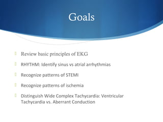
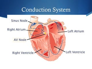
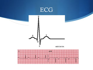
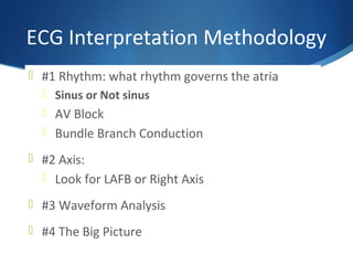
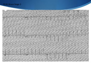
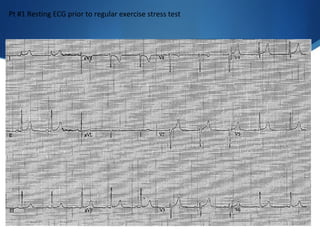
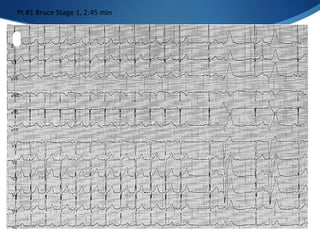
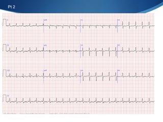
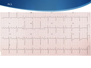
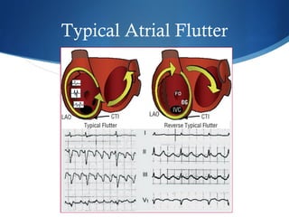
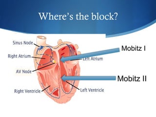
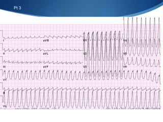
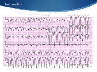
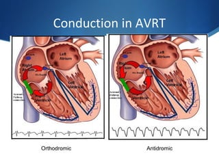
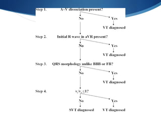
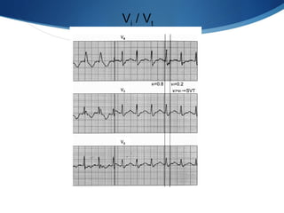
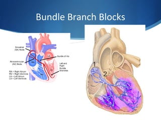
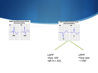
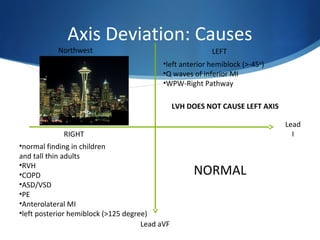
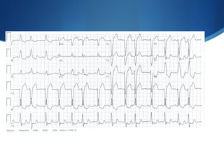
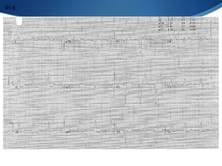
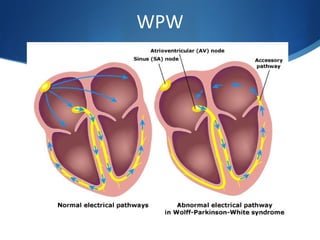
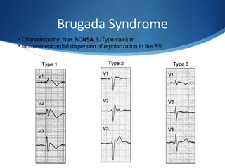
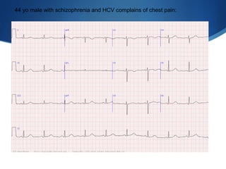
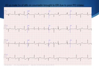
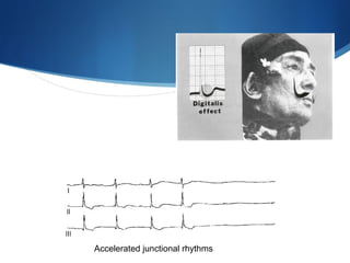
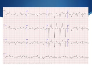
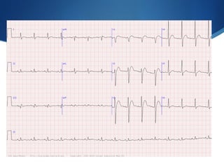
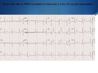
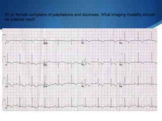
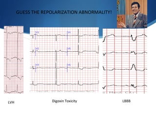
This document summarizes an ECG conference that reviewed basic ECG principles and interpretation methodology. It presented several patient cases to demonstrate sinus rhythm, atrial arrhythmias, STEMI patterns, ischemia, wide complex tachycardia, conduction blocks, axis deviations, AV nodal reentrant tachycardia, Brugada syndrome, digoxin toxicity, and more. The conference emphasized analyzing the ECG in terms of rhythm, axis, waveforms, and overall clinical picture.
8. ECG Interpretation Methodology
#1 Rhythm: what rhythm governs the atria
Sinus or Not sinus
AV Block
Bundle Branch Conduction
#2 Axis:
Look for LAFB or Right Axis
#3 Waveform Analysis
#4 The Big Picture
16. ECG Interpretation Methodology
#1 Rhythm: what rhythm governs the atria
Sinus or Not sinus
AV Block
Bundle Branch Conduction
#2 Axis:
Look for LAFB or Right Axis
#3 Waveform Analysis
#4 The Big Picture
17.
18. Mobitz I Wenckebach
• Although it is possible to syncopize due
to Mobitz I, it is generally considered
benign and a normal variant
• If you see it at night during sleep, not an
emergency
29. V TACH!!!
Axis Deviation: Causes
NORMAL
Northwest LEFT
RIGHT
Lead
I
Lead aVF
•normal finding in children
and tall thin adults
•RVH
•COPD
•ASD/VSD
•PE
•Anterolateral MI
•left posterior hemiblock (>125 degree)
•left anterior hemiblock (>-45o
)
•Q waves of inferior MI
•WPW-Right Pathway
LVH DOES NOT CAUSE LEFT AXIS
Antidromic AVRT
Axis is normal, VI/VT is >1
Ventricular Tachycardia
Sinus with APCs, nl axis, LBBB
WPW with LBBB, PVC is more narrow than sinus conducted beats!
Left Anterior Fascicular block
Note poor R wave progression,
Incomplete RBBB, RV strain pattern
Sinus tach
PE!
Inferior STEMI
Brugada Type I “Coved” Pattern
USE FLECAINIDE CHALLENGE (NA+ 1C)
Prolonged QT
Dig toxicity:
Not short QT, scooped ST, afib, ventricular ectopy
The increase in intracellular sodium causes an augmentation of the exchange of intracellular sodium for extracellular calcium. This shift can cause an increase in permeability to potassium; an increase in the outward current during the plateau phase, with a reduction in the duration of the action potential; and a short QT interval
Hyperkalemia
Hypercalcemia: 1) Digoxin toxicity 2) Hypercalcemia 3) Congenital Short QT
Short QT < 300
Hyperacute T wave with ST depession ->>STEMI
Arvd
Dig Toxicity:scooped” ST segment depression, most prominent in the inferolateral precordial leads and usually absent in the rightward leads; flattened T waves; increased U wave; and shortening of th QT interval